Case
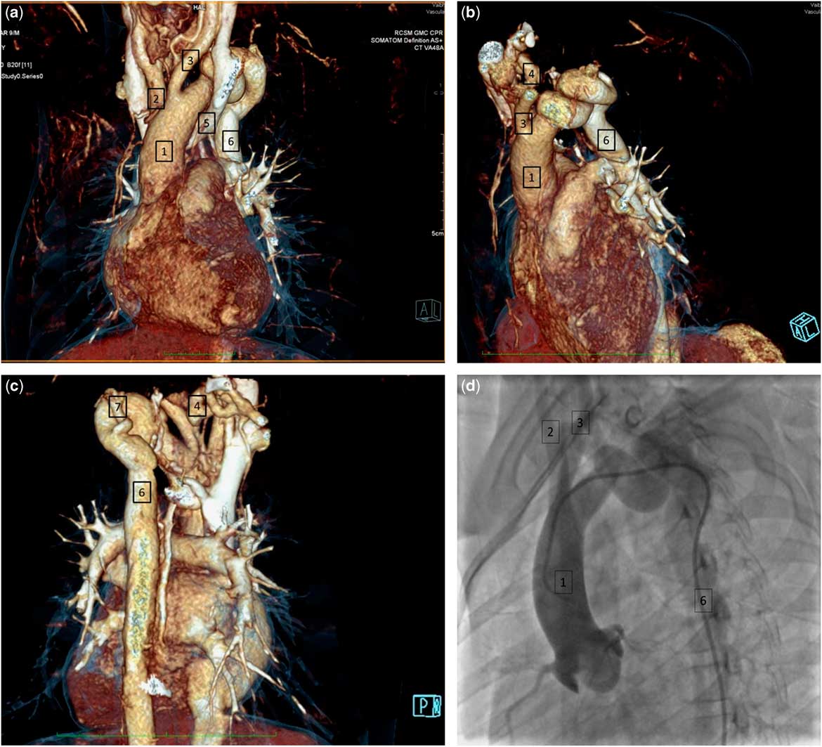
An asymptomatic 9-year-old boy, previously known to have a ventricular septal defect, presented for the first time at our hospital. We noted marked differences in the arm pulses as compared to the legs, with systolic blood pressures lower in the legs. There were no other unusual findings other than the previously reported ventricular septal defect having closed spontaneously. Contrast-enhanced computer tomography, however, revealed remarkable findings (Fig 1a-c). The left-sided aortic arch gave rise to the brachiocephalic artery, which bifurcated into the right common carotid and subclavian arteries. The right subclavian artery was tortuous proximally, but continued in normal fashion. The second branch, the left common carotid artery, had a normal origin and course. The arch then narrowed, and then took two remarkable turns, each through 360°. The left subclavian artery took a tortuous origin from the distal spiral. The arch then continued as the descending aorta, with cardiac catheterisation (Fig 1d) showing a narrowing just distal to the origin of the left common carotid artery. As far as we know, the arrangement of the arch, with two complete spirals, and coarctation proximal to the first spiral, is unique. The age and the nature of the anomaly suggest a congenital origin. Management is even more challenging than diagnosis. The traditional surgical options for repair of coarctation are hardly feasible. We deemed it less than optimal to subject an asymptomatic child to extensive surgery in an anomaly with an unknown progression. At present, therefore, our patient is on regular follow-up.
Fig. 1 (a, b, c) The computed tomographic anatomy of the aortic arch is shown in anterior (Fig. 1a), left sided (Fig. 1b), and posterior views (Fig. 1c). The images show the ascending aorta (1), the origin of the brachiocephalic artery (2), the left common carotid artery (3), the tortuous proximal parts of the right (4) and left (7) subclavian arteries, and the normal descending aorta (6). The retroaortic innominate vein (5) is seen coursing posterior to the ascending aorta; Fig 1 d - The aortic angiogram is shown in left anterior oblique orientation, at an angle of 60°, revealing the ascending aorta (1), the origins of the brachiocephalic (2) and left common carotid (3) arteries, followed by the two spiraling components of the arch and a normal descending aorta (6).

Acknowledgements
None.
Financial support
This research received no specific grant from any funding agency, commercial or not-for-profit sectors.
Conflicts of interest
None.