In the healthcare setting, contaminated room surfaces increase the likelihood of transmission of pathogens to patients via direct or indirect transmission. Reference Rutala and Weber1,Reference Rutala and Weber2 Healthcare-associated pathogens can persist on environmental surfaces for hours to days. Reference Weber, Anderson and Rutala3 Repeated exposure by patients to these contaminated surfaces can lead to acquisition of pathogens, which in turn may lead to healthcare-associated infections. Reference Rutala, Kanamori and Gergen4 For these reasons, it is important to ensure that touchable surfaces in patient rooms are free from potentially pathogenic microbes.
When indicated for research or epidemiologic investigation, to assess potential contamination of hospital surfaces, it is important to perform environmental cultures for specific pathogens of interest. However, standardized collection and processing methods for environmental sampling are lacking. Therefore, we compared 4 different sampling methods to assess comparative effectiveness for recovering pathogenic bacteria on 2 surfaces commonly found in patient rooms (ie, stainless steel and laminate).
Materials and methods
We tested 4 collection methods: (1) cotton swabs, (2) replicate organism detection and counting (RODAC) agar plates, (3) sponge sticks (Romer Labs, Newark, DE) using manual extraction and (4) sponge sticks using extraction with the Seward Stomacher (Seward, Davie, FL). Test surfaces included stainless-steel and laminate squares. We tested the following organisms: Klebsiella pneumoniae ATCC 700603, Staphylococcus aureus ATCC 43300, and Clostridioides difficile ATCC 9689. 5 We performed 9 replicates for each surface–method–organism combination.
Dilutions containing ∼10,000 CFU per milliliter (CFU/mL) of bacteria were made for each batch of testing. A viable colony count was obtained for each dilution by performing plate counts in triplicate. Colony counts were then averaged and used to calculate the estimated CFU/mL for each test dilution.
Known quantities of the test dilution were drawn into a pipette tip then dispensed as tiny droplets across each test surface. Applying known quantities allowed for the calculation of the estimated number of bacteria placed onto each test surface. Inoculated surfaces were allowed to air dry inside a biological safety cabinet ∼1 hour prior to sampling.
For Klebsiella pneumoniae and Staphylococcus aureus testing, Dey-Engley neutralizing agar was used for all methods. For Clostridioides difficile testing, Clostridium difficile selective agar containing 7% horse blood, sodium taurocholate, and lysozyme was used for all methods.
Swab samples were collected by rubbing a swab moistened with Remel DE-neutralizing broth over each 15.5-inch2 test surface, followed by a dry swab. Swabs were rotated as sampling was performed. Both swab tips were broken into a tube containing 1 mL phosphate-buffered saline. Tubes were mixed using a vortex, then an aliquot of each was plated to the appropriate agar plate. Each RODAC agar plate was pressed onto a 3.875-inch² round test surface and was held in place for 30 seconds. Sponge-stick samples were collected by rubbing a sponge premoistened with DE-neutralizing broth over each 15.5-inch² test surface. Each sponge head was ejected into a sterile bag containing phosphate-buffered saline. For manual agitation, each bag was kneaded by hand for 1 minute. For the stomacher method, bags were processed at 260 rpm for 1 minute. The contents of each bag were poured into individual tubes then centrifuged at 3,500 rpm for 15 minutes. The supernatant was removed and discarded from each tube. The remaining sample was mixed and measured, then an aliquot was plated to the appropriate agar plate.
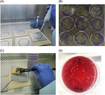
The K. pneumoniae and S. aureus test plates were incubated at 35°C for 24 hours and 48 hours, respectively. C. difficile test plates were sealed inside anaerobic jars immediately after inoculation then were incubated at 35°C for 48 hours. After incubation, a digital colony-counting device was used to enumerate the colonies growing on each of the agar plates. Figure 1 illustrates steps used to assess the different sampling methods. Colony-forming units recovered per square inch (CFU/inch²) were calculated for each replicate, and the average was obtained for each set of 9 replicates. For each category, 95% confidence intervals (CI) were calculated using the average and standard error of the replicate counts.
Illustration of steps used for assessing the effectiveness of 4 sampling methods. Photographs illustrating various stages of study: (A) applying bacterial dilution to test surface; (B) colonies of Staphylococcus aureus growing on agar plates from samples obtained by swabs; (C) collecting surface sample using sponge stick; and (D) colonies of Clostridioides difficile growing on a RODAC agar plate.

Results
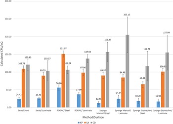
Figure 2 displays the CFU per square inch recovered for K. pneumoniae, S. aureus and C. difficile for each sampling method/surface combination. The overall recovery for K. pneumoniae was 26.88 CFU/inch² (95% CI, 23.15–30.62). Overall recovery for S. aureus was 98.53 CFU/inch² (95% CI, 91.68–105.38). Overall recovery for C. difficile was 137.72 CFU/inch² (95% CI, 124.35–151.08). When comparing methods, regardless of surface type, we obtained the following recovery results: swab, 78.73 CFU/inch² (95% CI, 66.66–90.81); RODAC, 97.71 CFU/inch² (95% CI, 86.05–109.36); sponge stick and manual agitation, 95.49 CFU/in² (95% CI, 74.82–116.15); and sponge stick and stomacher, 78.91 CFU/inch² (95% CI, 61.40–96.42). When comparing surfaces, regardless of method used, we obtained the following recovery results: stainless steel, 85.56 CFU/inch² (95% CI, 75.33–95.78), and laminate, 89.86 CFU/inch² (95% CI, 77.52–102.20).
Recovery of test organisms by four sampling methods. Average recovery in CFU/in² and 95% CI for Klebsiella pneumoniae, Staphylococcus aureus and Clostridioides difficile for each method and surface combination.

Discussion
We had initially hypothesized that the sponge sticks with stomacher extraction would yield the highest recovery. Sponge sticks have the largest surface area, and stomacher homogenization would likely provide the most vigorous extraction. However, our results did not support this hypothesis. In the setting of known concentration of inoculation and location of contamination, our study demonstrated that organism type was the most important factor in bacterial recovery. The sampling tool and surface type had less impact on bacterial recovery when applied to a test surface under these experimental conditions. The importance of organism type has been documented in similar environmental sampling studies. Reference Downey, Da Silva, Olson, Filliben and Morrow6,Reference Rawlinson, Ciric and Cloutman-Green7
Desiccation stress likely played a substantial factor in our results. Because K. pneumoniae exhibited the lowest percentage recoveries overall, this organism likely had the lowest tolerance to the effects of drying on the test surfaces. Recovery of C. difficile was highest overall, likely because the spores were able to better withstand the physical stress of drying and manipulation. The effects of desiccation in organism recovery have been detailed in similar reports. Reference Buttner, Cruz, Stetzenbach and Cronin8
Importantly, processing a swab or RODAC sample is much quicker than processing a sponge stick sample. Sponge samples must be homogenized, defoamed, centrifuged, decanted, and measured. The additional processing required for sponges allows more time for natural die-off of less hardy bacteria, or it could lead to bacterial cells that are viable but nonculturable. This effect could result in lower numbers of colonies recovered. Reference Buttner, Cruz, Stetzenbach and Cronin8,Reference Li, Mendis, Trigui, Oliver and Faucher9 In our study, this concept is reflected in the fact that both the highest (C. difficile) and lowest (K. pneumoniae) percent recoveries were seen when using sponge sticks with 2 different organisms. However, when used in actual hospital settings, sponges can sample an area 10–100× larger than swabs and RODAC; therefore, they could provide superior overall recovery.
Culture was the only detection method used in this study. If molecular methods had also been used, the recoveries of both viable and nonviable bacteria could have been determined. This methodology would have provided a more complete measure of the total quantity of all bacteria removed from the test surfaces, not just the ones that survived the sampling process. However, the use of molecular methods is less practical than culturing for assessing the role of the contaminated bacteria in outbreaks and for assessing the impact of cleaning and disinfection on the presence of viable bacteria. In addition, the presence of viable but nonculturable bacteria in the environment have not been shown to correlate to an increased risk of infection transmission. Reference Li, Mendis, Trigui, Oliver and Faucher9
Our results are reassuring for infection preventionists and environmental microbiologists because our experimental testing has shown that readily available tools and methods are able to detect viable bacteria on environmental surfaces.
Acknowledgments
The CDC Prevention Epicenters Program was not involved in the preparation, submission, or review of this manuscript.
Financial support
This study was supported by the CDC Prevention Epicenters Program (grant no. U54CK000483).
Conflicts of interest
All authors report no conflicts of interest relevant to this article.