Authors' reply
Dear Editors,
We are glad to hear about the interest in our work titled ‘Evaluation and comparison of type I tympanoplasty efficacy and histopathological changes to the tympanic membrane in dry and wet ear: a prospective study'. We would like to thank Dr Zhengcai Lou for reading our paper in depth. We have gone through the author's queries in detail.
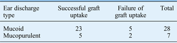
Firstly, Dr Lou raised the question of whether the degree and nature of the ear discharge had any bearing on the healing results following tympanoplasty. In our study (as mentioned in the article), of the 35 patients with wet ear, 28 patients had mucoid discharge and 7 patients had mucopurulent discharge at the time of surgery.Reference Shankar, Virk, Gupta, Gupta, Bal and Bansal1 On subsequent evaluation of the success of graft uptake between the two types of discharge, no statistically significant difference in success rate was found (p = 0.526) (Table I). Similarly, no difference in the graft uptake rate was found when the degree of the discharge was compared.
Success or failure of graft uptake by ear discharge type

Pearson chi-square (p = 0.526). Data represent numbers of ears.
Similar results have been shown by other authors. For example, a prospective audit study by Kotecha et al., which reviewed 1070 individuals, showed that patients with dry ear, or mucoid, serous or purulent discharge, had failure rates of 17.7 per cent, 17.1 per cent, 11.8 per cent and 17.2 per cent, respectively; these rates are comparable, irrespective of the nature of the discharge.Reference Kotecha, Fowler and Topham2
We agree with the author that the moist environment of a wet ear might accelerate the healing process of the ear drum.Reference Lou, Tang and Xiao3–Reference Caylan, Titiz, Falcioni, De Donato, Russo and Taibah7 In our study too, tympanic membrane vascularity was found to be higher in cases of wet ear (16 out of 25, 64 per cent) than in cases of dry ear (3 out of 21, 14.3 per cent) (p < 0.001). However, on subsequent comparison, no statistical difference was observed in the final graft uptake rates between the wet and dry ear based on the vascularity (p = 0.115).
The second comment queried whether the tympanosclerotic patches on the tympanic membrane were removed or not during the surgery. Nineteen (27.1 per cent) of our patients had tympanosclerosis at the time of surgery. Of these 19 patients, 13 were in the dry ear group and the remaining 6 were in the wet ear group. Five patients also had some middle-ear sclerosis along with the tympanosclerosis (four in the dry ear group and one in the wet ear group). During the over-underlay technique of tympanoplasty, all efforts were made to clear the tympanosclerotic patch before the grafting was done. In addition, the middle-ear sclerosis was cleared if present before grafting. Thus, no comparison between the presence and absence of tympanosclerosis on graft uptake rates was possible.