

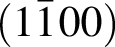
 plane and c(0001) plane of α–Al
 and
 , having an angle of about 43° with
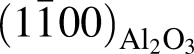 for the film deposited on m
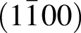 plane, and
 and
 for the film deposited on c(0001) plane though a Ca–Al–O amorphous layer formed between them. CoO seed layer near the interface and residual Co

Crossref Citations

This article has been cited by the following publications. This list is generated based on data provided by Crossref.
Sugiura, Kenji
Ohta, Hiromichi
Nakagawa, Shin-ichi
Huang, Rong
Ikuhara, Yuichi
Nomura, Kenji
Hosono, Hideo
and
Koumoto, Kunihito
2009.
Anisotropic carrier transport properties in layered cobaltate epitaxial films grown by reactive solid-phase epitaxy.
Applied Physics Letters,
Vol. 94,
Issue. 15,
Li, Qing
Feng, Yuzhang
Wang, Peng
and
Che, Renchao
2019.
Superior-capacity binder-free anode electrode for lithium-ion batteries: CoxMnyNizO nanosheets with metal/oxygen vacancies directly formed on Cu foil.
Nanoscale,
Vol. 11,
Issue. 11,
p.
5080.
Paul, Biplab
Lu, Jun
and
Eklund, Per
2019.
Growth of CaxCoO2 Thin Films by A Two-Stage Phase Transformation from CaO–CoO Thin Films Deposited by Rf-Magnetron Reactive Cosputtering.
Nanomaterials,
Vol. 9,
Issue. 3,
p.
443.
Zhang, Xi
Zhang, Yuqiao
Wu, Liao
Tsuruta, Akihiro
Mikami, Masashi
Cho, Hai Jun
and
Ohta, Hiromichi
2022.
Ba1/3CoO2: A Thermoelectric Oxide Showing a Reliable ZT of ∼0.55 at 600 °C in Air.
ACS Applied Materials & Interfaces,
Vol. 14,
Issue. 29,
p.
33355.
Zhang, Yuqiao
and
Ohta, Hiromichi
2023.
Recent progress in thermoelectric layered cobalt oxide thin films.
NPG Asia Materials,
Vol. 15,
Issue. 1,
Bhattacharjee, Jyoti
Roy, Subhasis
Shaikh, Abdul Aziz
and
Datta, Preetam
2025.
Efficient fabrication and characterization of doped nanocomposites for thermoelectric materials.
Nano Trends,
Vol. 10,
Issue. ,
p.
100109.