

1 Introduction
Judging by their frequent use, diagrams are important tools for biologists. They are used during the process of research, as biologists plan experiments with flow charts, and draw models to represent hypotheses for consideration. Diagrams are also frequent components of biologists’ key communication venues: they often appear in textbooks and research publications. There are now several studies available that address the use of diagrams in biologyFootnote 1. Griesemer (1991), for example, asks whether—and why—diagrams might be necessary for science. Others have discussed diagrams used to convey metaphors and facilitate analogical reasoning (Taylor & Blum, Reference Taylor and Blum1991b; Ruse, 1996)Footnote 2. Lynch (Reference Lynch1988), Maienschein (Reference Maienschein1991), and Abraham (Reference Abraham2003) are all concerned with the use of diagrams to convey theoretical content. Gilbert (1991) argues that Waddington presented genetic data in a diagrammatic format typical of embryology in an attempt to synthesize the two fields. This sample of papers suggests that diagrams play diverse roles in biological reasoning. We do not, however, understand what it is about diagrammatic representation that allows them to play these roles.
Larkin and Simon (Reference Larkin and Simon1987) compare linguistic and diagrammatic representation and show that while some diagrams are informationally equivalent to linguistic descriptions, this does not imply computational equivalence. Diagrams can be more efficient platforms for drawing inferences than informationally equivalent linguistic representations. Comparison between linguistic and diagrammatic representations, however, is limited in the extent to which it can clarify why biologists would use diagrams in biology. Biologists communicate with a variety of non-diagrammatic forms of visual representations, so understanding why biologists use diagrams requires understanding why they choose diagrams over non-diagrammatic visual representationsFootnote 3. Furthermore, since biologists typically use multiple kinds of representations when they communicate, understanding the role of a diagram often depends on analyzing the other representations involved; the most important ones to consider are often other kinds of visual representations. For these reasons, understanding what makes diagrams distinctive as representations is essential for understanding what roles they play in biological reasoning, and how they do so. In this paper, I characterize representational features of diagrams. Analysis of the relation between the forms and contents of diagrams will show there is significant diversity among the diagrams used by biologists, and that the features that distinguish these different types explain why biologists use particular kinds of diagrams for particular roles in biological reasoning.
2 Beyond simplicity: what diagrams have in common
Diagrams are visual representations, and it will be helpful to start by clarifying this broader class of representations. Visual representations are external representations in which some spatial relations in the picture are interpreted, and thus convey content. Spatial features of a photograph, for example, are interpreted as representing spatial features of a scene. Visual representations constitute a broad category with a lot of variety, because spatial relations in an image can refer to spatial properties (like shape) or to other kinds of properties—which need not be visible. In line graphs, for example, spatial relations are used to refer to relations between properties. Additional visible features in a picture, such as color, may also convey information. The variety of kinds of pictures is thus due to a variety in form–content relations. In order to understand a particular image, you must view its form, and also apply appropriate interpretive conventions to what you see, in order to relate the visible features of the picture to what it represents (Perini, Reference Perini2004). For example, you must interpret gray colors differently in a black and white photograph than in a color photograph.
Most pictures do not represent all aspects of their subject matter. There are some odd exceptions, such as an image representing itself, but most pictures remain non-committal about some properties of their subject matter (Lopes, Reference Lopes1995). For example, color photographs represent the features of an object visible from a particular angle, omitting information about the visible features on the opposite side, and information about the object's non-visible features. Similarly for black and white photos, except that colors are not represented. Diagrams are not distinctive among visual representations in that they are selective about the properties they represent.
However, there is something distinctive about the form–content relations of diagrams; they share a feature characterized by Goodman in Languages of Art. Diagrams are relatively non-replete, which means that compared with other pictures, relatively few visible features are used to convey content (Goodman, Reference Goodman1976). A typical line graph is relatively non-replete, because only the position of the line, and not its width or darkness, conveys content. A naturalistic pencil drawing is relatively replete, because the position, width, and darkness of pencil marks contribute to the content of the picture. Because diagrams are relatively non-replete, they represent fewer properties relative to non-diagrammatic images. However, diagrams are not less accurate as a result: Hall (Reference Hall1996) shows that they can be accurate, and that non-diagrammatic images can be inaccurate. For visual representations, as for other types of representations, omission and accuracy are separable.
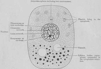
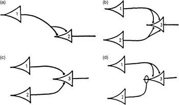
Low relative repleteness is a feature that all diagrams share. Since there is no sharp line between high and low relative repleteness, there will be some visual representations whose status is indeterminate. This does not interfere with the usefulness of characterizing diagrams as relatively non-replete. Identifying this feature provides a way to characterize what all the figures that have been identified and studied as diagrams have in common. Consider the diversity involved: Maienschein (Reference Maienschein1991) examines Wilson's cell diagrams, which represent the structures of various components of cells, while Abraham (2003) includes a discussion of McCullock and Pitts’ diagram of the functional connections involved in neural firing (Figures 1 and 2). Both the appearance and the subject matter of these images are quite different. One feature they share is a notable simplicity of visible form, compared with pictures like photographs and naturalistic renderings. The fact that figures identified as diagrams have a visibly simple and spare form might explain why all these particular figures were chosen as case studies of diagrammatic representation. Simplicity of appearance, however, offers no insight into diagrams as representations. Understanding diagrams as having form–content relations that are all relatively non-replete clarifies the problem at hand: explaining why biologists use diagrams requires understanding the advantages of relatively non-replete figures.
Diagram of a cell, reprinted in Maienschein (Reference Maienschein1991) from Wilson (Reference Wilson1896)

Diagrams of synaptic connections governing neural firing (detail), reprinted in Abraham (Reference Abraham2003) from McCulloch and Pitts (Reference McCulloch and Pitts1943)

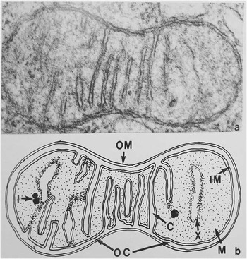
Prior work on diagrams shows that this project will require explanatory resources beyond relative repleteness. Michael Lynch studies figures that pair a diagram with a non-diagrammatic figure, an electron micrograph (Figure 3). The micrograph is an image of a particular sample, and represents the structure of an individual subcellular structure, a mitochondrion, in detail. The diagram conveys less information about the detail of the structure. For example, where the micrograph represents the width of membranes precisely as detected by the electron microscope, the diagram does not, making some areas that look different in the micrograph appear similar in the diagram. Lynch argues that the diagram does not merely leave out some information conveyed by the micrograph. Rather, the diagram is a means to convey content that is distinctively theoretical compared with the micrograph (Lynch, Reference Lynch1988). So the diagram is not used simply to select out some particular details from the full detail represented by the micrograph. It is not a matter of just ‘saying less’ with a figure of low relative repleteness. Lynch's discussion suggests that the two figures convey different kinds of content, and not just different amountsFootnote 4.
Paired figure: electron micrograph (above), diagram (below), reprinted in Lynch (Reference Lynch1988) from Threadgold (1967)

Thus, although the concept of repleteness allows for clarifying the questions we need to ask about why biologists use diagrams, the explanatory role low relative repleteness can play in this project is limited. It cannot explain differences among diagrams (as will be seen below, those go beyond differences in repleteness). Furthermore, on its own the fact that diagrams are relatively non-replete does not explain why biologists would use diagrams. Such figures convey less content compared with relatively replete figures, but there is no general advantage to communicating less content rather than more. Clarification of the feature diagrams share thus indicates that explaining reasoning with diagrams will depend on further analysis of diagrams. Below I describe three important types of diagrams in biology, and the distinctive features of those types explain their use in biologyFootnote 5.
3 Pictorial diagrams
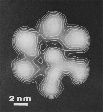
Consider Figure 4, a figure that combines two forms of representation. My aim is to characterize a particular type of diagram (superimposed on a non-diagrammatic image) and show how the features of that type of diagram explain why the diagrammatic form is combined with the other image in the figure.
Pictorial diagram (topo lines) superimposed on a non-diagrammatic image, representing the results of a study of the cross-sectional shape of the ATP synthase complex (Boekema et al., Reference Boekema, Berden and van Heel1986). Reprinted by permission from Elsevier Ltd

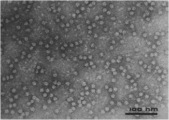
Figure 4 is the result of a study of the structure of a particularly important biological enzyme complex, the ATP synthase. This complex catalyzes the formation of ATP, the energy currency of the cell, in the last stage of oxidative phosphorylation. Because it plays such a key role in making energy available for cell processes, and because the structure of the enzyme would provide some insight into how the ATP synthase catalyzes the reaction, scientists were keen to get any information they could. One early step was the study by Boekema et al. (Reference Boekema, Berden and van Heel1986). The authors isolated the complex and used electron microscopy to generate an image of the isolated particles, which represents individual synthase particles as white areas on a dark background (Figure 5). However, the light spots corresponding to individual particles vary significantly in shape. This meant that the authors could not derive information about the shape of the particle from the appearance of individual spots. In order to get more precise and accurate information about particle shape, they oriented and scanned individual spots, then applied a mathematical analysis to the optical signals derived from multiple spots, in order to get an average of their shape.
Electron micrograph of isolated ATP synthase particles (Boekema et al., Reference Boekema, Berden and van Heel1986). Reprinted by permission from Elsevier Ltd

The results were presented in Figure 4, which involves two different kinds of images superimposed on one another. The first is the shading that ranges from light to dark, which represents the signal intensity of the averaged spots. Spatial relations of the figure are used to represent spatial relations of the signal intensities from the analysis, and the exact degree of lightness of the image correlates with the precise strength of the signal. The figure also includes a superimposed representation that is less replete than the first. Each topo line represents the spatial location of one particular value of signal intensity. The brightness and width of the topo lines are not informative. The positions of the topo lines convey significant detail, because they track precisely the location of specific signal intensities in two-dimensional space. Because each line represents a particular value, relative distance between the lines shows the steepness of the drop-off in signal toward the edge of the particle. The diagrammatic topo lines therefore carry less than the full information derived from the mathematical analysis.
Why did the authors superimpose a diagram over a more replete representation of the same structure? The topo lines are a means to express the results that were the ultimate aim of this study—information about the cross-sectional shape of the ATP synthase complex. That information is derived not from the particular intensity of the signal in any part of the complex, but from the drop-off of signal at the complex's boundaries. Human visual perception is not able to distinguish the kind of differences in light intensities needed to reliably pick out the outline shape of the complex from the full results, as represented in the gradual shading. However, the topo lines convey that drop-off very clearly—they are close together where the drop-off in signal intensity is rapid, which is at the edge of the complex. Thus, the diagrammatic representational scheme leaves out some information (the smooth gradient information about how the signal drops off) and conveys just that part of the mathematical output that is relevant to the goal of the study: it shows where the drop-off occurs, and thus represents the outline shape of the complex.
There are three things to note here. First, the topo lines drop out some information relative to the shaded form, but they still represent a specific shape: the precise location of the topo lines are used to represent the details of the complex's shape. The move to the diagram in this case is a move from one type of visual representation that conveys a great deal of detailed information to another type of visual representation that conveys less information, but the information it conveys is both detailed and limited to the content of interest: the conclusion of the study, and nothing more. Second, the value of the topo lines does not merely result from the fact that extraneous information is deleted (so the viewer's attention is focused on the information relevant to the study); it represents the drop-off in a way that is reliably comprehended by human interpreters of the visible form of the image, so it is a more effective means of conveying the results than the more replete representation.
Finally, in this case there is a visible similarity between the less and the more abstract images superimposed in the figure. This raises a question about whether reasoning involving visual representations in some way depends on visual matching—recognizing a straightforward visual similarity, such as similarity of outline shape—between the two figures. Later examples will show that visual matching is not required in reasoning with visual representationsFootnote 6.
4 Compositional diagrams
Some diagrams are compositional: they are built out of discrete atomic characters. Written sentences are compositional—composed of letters, spaces, and punctuation marks. In compositional visual representations, spatial relations among atomic characters are interpreted as representing relations among the referents of the atomic characters. Like words and numerals, however, the atomic characters of compositional diagrams need bear no resemblance to their referents, nor convey any information about them; they can function simply as labels. Simple chemical diagrams have this feature. Consider diagrams made with the following atomic characters: filled circles, which refer to hydrogen atoms; open circles, which refer to oxygen atoms, and straight lines, which refer to inter-atomic bonds. Spatial relations are interpreted in the following way: lines between two circles represent a bond between the two atoms denoted by the circle; angles between lines represent relative position of the atoms connected by those lines. This simple representational system will allow for representation of the structure of compounds like water and hydrogen peroxide, because the meaning of the diagrams produced are a function of the meaning of the atomic characters and the interpreted spatial relations among them.
For a more complex case that will allow for discussion of how compositional figures are involved in biological argumentation, consider Figure 6. This diagram is a representation of the mechanism by which the ATP synthase catalyzes the formation of ATP from precursors ADP and Pi. The complex includes three chemically identical subunits. The subunits can take on three different structural conformations. According to the binding-change model, at any given time each of the subunits is in a different one of the three conformations. The conformation of the subunit determines whether, and how tightly, it binds the precursors and products of the reaction. The loose conformation has a moderate affinity for the precursors ADP and Pi, so they will bind that site. The tight conformation binds ADP and ATP so strongly that neither is released; furthermore, when bound in this way ADP and Pi react to form ATP. ‘Open’ has such a low affinity for these compounds that neither bind. All three subunits transition at the same time, from one conformation to the next. Energy input drives the conformation changes, and the formation of ATP happens as a result of those changes. The subunit in the loose conformation binds the precursors, and when its conformation changes to tight, the precursors react to form ATP. After another transition to the open conformation, ATP is released, since in that conformation the subunit cannot bind anything.
Compositional diagram: the binding-change mechanism (Abrahams et al., 1994). Reprinted by permission from Macmillan Publishers Ltd: Nature, Copyright 1994

The diagram represents this model through the use of atomic characters, such as the wedge shapes, arrows, and linguistic characters. The three different wedge shapes refer to the loose, open, and tight conformations, respectively. Spatial relations among the atomic characters represent relations among the referents of those characters. Contiguity of the wedge shapes indicates that the enzyme complex has subunits in those particular conformations. Arrows between the different combined wedges refer to transitions between stages in the mechanism, in which chemicals are bound or released, energy is input, and the subunits change shape. Note that the atomic characters of this system could be used to make a different diagram, expressing a different model of the enzyme's function. For example, a diagram with three L wedges together, then three T wedges, then three O wedges would represent a mechanism in which all the subunits are in the same conformation, and they all transition together to the next conformation.
The diagram does not represent intrinsic features of the referents of the atomic characters. In this example, the wedge shapes do not represent the shape of the different subunit binding conformations. Rather, they simply denote the different conformations, which are named for their functional characteristics rather than spatial features. The fact that the forms of atomic characters do not map on to features of their referents means that they can function simply as labels. In such a system, atomic characters can refer to the elements of a system—even when those elements are very complex—without referring to their intrinsic properties.
The result is a figure that represents relations among the referents of the atomic characters, and is non-committal about many—or all—of the intrinsic properties of those referents. This is the point at which we should ask, why do biologists use a form of representation that is non-committal about such features? This type of selectivity in the content of the figure is relevant to biological reasoning in two important ways. First, compositional diagrams convey the kind of information that plays an important explanatory role in biology. Cummins (Reference Cummins1975) calls this functional analysis, in which a feature of a system is explained in terms of its component parts. Bechtel and Richardson's (Reference Bechtel and Richardson1992) discussion of research in biology make it clear that the relations among component parts of a biological system often are key to explaining that system. While both the identities of component parts and relations among those components are integral to the functional explanation, many properties of the system components are not relevant to explaining the capacity of the system. Compositional figures use atomic characters to refer to component parts (like subunits in particular conformations). Spatial relations among atomic characters are interpreted as referring to relations among the referents of the atomic characters, so that reference to the relations among the components is built into the representation. For this reason, the two kinds of information that make these models explanatorily relevant are presented in one figure: identification of important component parts of a system and of important relations among those components. The visual formatting of the diagram provides a way to refer to system components in such a way that their relationship to the system itself is also represented (Perini, Reference Perini2005a).
Because the form of the atomic characters need bear no information about the referents of those characters, information irrelevant to the functional analysis is not included in the diagram. This can be seen in the mechanism diagram, which identifies the important components of the model without representing properties irrelevant to the mechanism, and uses spatial relations (like contiguity) to represent key relations among system components. Thus, compositional visual representations make the explanatory aspects of the content especially salient.
The second advantage of the fact that compositional diagrams can omit information about the referents of atomic characters is that this type of selectivity can be critical to the strength of an argumentFootnote 7.
This can be demonstrated by considering the context in which the binding-change model was introduced. The binding-change model of ATP synthesis was proposed long before the structure of the ATP synthase complex was knownFootnote 8. The specific shape of the ATP synthase subunits, and the complex as a whole, is directly relevant to the binding-change mechanism, because protein function depends on three-dimensional conformation. Because of this dependency, confirmation of the binding-change model was ultimately dependent on showing that the complex had a structure that could function as the model described. However, the specific structure could not be predicted from the model. The binding-change model, along with background knowledge of proteins, does imply that the three subunits have different shapes from one another, but implies no specific structural features. So in presenting a model of the mechanism, it was important to remain non-committal about the specific shapes of the subunits. Since there was no support for any particular structure, the argument whose conclusion is a diagram that is non-committal about structure is stronger than would be the argument whose conclusion includes a representation of the subunits’ shapes.
There is one final lesson to be learned from this example. In the last section, I claimed that reasoning with diagrams need not involve visual matching. In many cases in biology, the conclusion of a paper is expressed through a diagram, and some—or all—of the evidence is presented through visual representations. In many of such cases, the forms of the visual representations offered as evidence do not have any obvious similarity to the form of the diagram they support, as was the case with Figure 6.
By the 1990s, Paul Boyer's binding-change model of the mechanism (Figure 6) had gained support, but as noted above, was not considered confirmed prior to publication of a model of the enzyme's three-dimensional structure. That occurred in with a paper on the results of a crystallographic study of the enzyme complex's structure (Abrahams et al., Reference Abrahams, Leslie, Lutter and Walker1994). Compared with Boekema's (Reference Boekema, Berden and van Heel1986) results on the cross-sectional shape of the complex, the structural model is both more comprehensive and much more precise. In addition to presenting their case for their model of the structure, Abrahams et al. argue for the binding-change model of the enzyme's mechanism, citing its structure as the crucial evidence needed to confirm the model of the mechanism. John Walker, the principle investigator for the structural study, and Paul Boyer, responsible for the binding-change model, shared half of the Nobel Prize in Chemistry for these contributions in 1997Footnote 9.
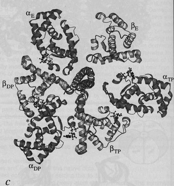
Figure 7 is a representation of the structure of the C-terminal ends of the proteins making up the subunits of the ATP synthase complex. The overall structure of the complex is sort of like a tangerine, with a central core and six protein subunits arranged around this core. The three catalytic β subunits, in gold, are distributed evenly around the core. The diagrams of the models for the ATP synthase structure (e.g., Figure 7) and the binding-change model (Figure 6) look completely different. Not only are they different in appearance, they involve quite different types of form–content relations. In Figure 7, ribbon-like shapes represent protein chains, and the spatial features of the image are used to represent spatial features among the protein chains of the complex. The diagram of the binding-change model, on the other hand, uses spatial relations to represent transitions over time, and mere co-location in the complex—not contiguity or other spatial relations. These two diagrams do not relate to each other on one coherent set of dimensions. The reasoning involved in taking images like Figure 7 as support for the model expressed by Figure 6must be a more complicated matter than perceptual comparison.
Visual representation of the structure of the ATP synthase complex (Abrahams et al., 1994). Reprinted by permission from Macmillan Publishers Ltd: Nature, Copyright 1994

So how does Figure 7 support the binding-change model? Recall that the binding-change model is non-committal about aspects of the ATP synthase mechanism, including the specific way that the structure of the complex instantiates that mechanism. The binding-change model does not imply a particular structure, but any enzyme that has the capacity to synthesize ATP in that way must have a structure. There are relevant theoretical commitments in the background: according to contemporary biochemistry, a protein's functional capacities are determined by its three-dimensional structure. The binding-change model, along with background knowledge about the relation between protein structure and function, has an important implication for the structure of the complex. According to the model, the three subunits will always be in different functional states. According to background knowledge, protein function is determined by three-dimensional structure. Together these imply that the three subunits will always be in different conformations from each other, and that in turn implies that the complex will by asymmetric in shape.
Figure 7 is a visual representation of the complex's structure, in which spatial relations in the figure represent spatial features of the complex. For this reason, Figure 7 can be used to evaluate the binding-change model, by looking for asymmetry in the diagram of the structure. This evaluation requires a visual abstraction on the structure diagram, because it is the generic property of asymmetry, rather than the specific shape properties, that is relevant to the binding-change model. The evidential reasoning involved in understanding Figure 7 as support for Figure 6 does not involve any perceptual matching between the two figures. Visible similarity is not necessary in reasoning with visual representations.
5 Schematic drawings
A third kind of diagram is distinct from pictorial and compositional diagrams. Schematic drawings do not represent specific properties due to interpretation of the details of their visible form, as is the case with pictorial diagrams. Unlike compositional diagrams, they are not composed of atomic characters, which can function like mere labels, simply denoting referents without representing the intrinsic properties of their referents. In schematic drawings, relatively generic visual features like contiguity, inside/outside, etc. are used to represent features that are themselves generic. As a result, schematic drawings abstract away from the particular properties of individuals, and instead represent more generic features.
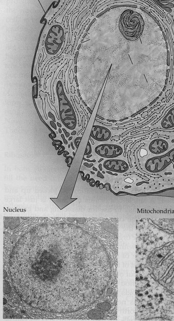
Figure 8 is a detail from a figure in a college-level biology textbook (Purves et al., Reference Purves, Orians and Heller1995). The arrow points from part of a diagram of an animal cell—that representing its nucleus—to an electron micrograph of a nucleus, directing students to relate the two different kinds of images. The images do not offer a simple visual match; the overall shape of the nucleus in each is visibly different, as is the relative position of the darker area within the nuclear envelope (representing the nucleolus). The pairing of the images thus prompts two questions: why are both included, and how do students comprehend the relation between the two?
Schematic diagram and electron micrograph composite figure (detail), of the cell and nucleus (Purves et al., Reference Purves, Orians and Heller1995). Reprinted by permission from Sinauer Associates

While the electron micrograph is much more replete than the diagram, the key difference between the two is in type of content each conveys. The electron micrograph is an image of a particular nucleus, produced through a detection technique that correlates the specific form of the image to the specific structure of the biological material that was scanned. A visual representation that uses specific visible details to convey content about specific detailed properties does convey content about the more generic features of nuclear structure, such as that it is bounded by a membrane, but the micrograph conveys this content in virtue of conveying information about the specific shape of the membrane. The diagram in this figure, on the other hand, does not represent the detailed shape of a particular nucleus. Although the diagram, as a visible form, has a particular shape, the diagram does not represent the nucleus as having this particular shape, because there is a distinctively different type of relation between its form and content compared with the micrograph. For the diagram, more general visible features, such as the gappy segments that enclose a space, and the relatively dark shape inside that area, are used to represent correspondingly general aspects of nuclear structure, such as the fact that it has an outer membrane, which has pores, and there is a structure within the nucleus—the nucleolus. In this way, the diagram represents nuclei as having a boundary, but not as having one with a particular shape; similarly the diagram represents nuclei as containing a nucleolus in their interior, but not as having a nucleolus that occupies a specific position within the nucleus.
In pairing the images in this way, the authors prompt students to understand the evidential relation between the two. That is not accomplished via recognizing a straightforward similarity between the forms of the two images. Comprehension of the evidential relation between the more detailed and specific micrograph and the more general content of the diagram requires a perceptual and cognitive accomplishment, a visual abstraction on the micrograph, by attending to its more generic visual properties rather than its visible details.
Why do biologists use schematic drawings? They not only are non-replete compared with non-diagrammatic visual representations, schematic drawings have reduced capacity for specificity compared with compositional diagrams, which identify system components and represent interrelations among them with precision. Schematic drawings also have a reduced capacity for detail compared with diagrammatic pictorial representations. Why, then, are they used so frequently in textbooks and research materials?
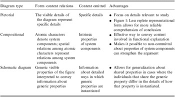
The schematic drawing provides a way to communicate about generic properties while not asserting that those properties are instantiated in a particular way. The ability to do that is crucial in a science like biology, which frequently requires communication about types of things that vary in their details, though they are the same in terms of more generic properties (Reference PeriniPerini, 2012). Biologists face this issue when communicating about almost any sort of biological structure, from organelles to individual organisms. Biologists often work with data in the form of relatively replete visual representations of biological forms—electron micrographs, photographs, etc. These images provide information about individual cases; they play an important evidential role. However, they are not effective means to express generalizations about structures that vary from individual to individual. The problem is that with images like electron micrographs and photographs, the visible details of the image convey content about the detailed properties of the particular individual pictured in the figure. Schematic drawings convey information about a class by conveying content about higher-order properties all its members share, while remaining silent about the detailed properties that vary among the members. Though compared with the other types of diagrams, schematic drawings lack capacity for detail and precision, this form of representation is well suited to convey information that generalizes over objects that differ in terms of specifics, but share higher-order features (Table 1).
Biological diagrams

6 Conclusion
These results show that any advantage that diagrammatic representational form might have for biologists is not due just to simplicity of form (even if that is the one feature all diagrams share), and that the relevant form–content relations distinctive of diagrams go beyond just ‘saying less’.
Diagrammatic pictorial representations derive from pictorial systems in which relatively few visible features matter for character identity, and only those are used to represent features of the referent. They can convey detailed information about a limited number of properties or relations, leaving out information about other properties that are not relevant to the issue at hand. This allows for focus on a limited number of specific properties. Schematic drawings are unlike pictorial representations, in that the visible features that are meaningful are not the specific visible features like the shape of a line, but generic visible features like contiguity, inside/outside relations, etc. These visible features are used to convey information about generic properties, without also implying that those are instantiated in any particular way. Compositional diagrams differ from both pictorial and schematic diagrams, because they are composed of atomic characters that need not convey any information beyond the identity of their referents. Since the arrangement of atomic characters is interpreted to represent relations among the referents of those characters, compositional diagrams highlight the relations holding among system components—often key information in biology.
In their introduction to a special volume in biological diagrams, Taylor and Blum (Reference Taylor and Blum1991) raise a question about whether there is something significant about the diagrammatic form in general that explains the use of diagrams, or whether only particular diagrammatic forms are relevant to understanding their use in biology. These results help explain why Taylor and Blum, with a volume of illuminating case studies in hand, would express puzzlement over whether diagrams share some feature that explains their use by biologists. It is not just that there are significant differences among types of diagrams. There is not a sharp line distinguishing diagrams (characterized as relatively non-replete visual representations) and non-diagrammatic figures. Although the line between relatively replete and non-replete images is obviously not sharp, this is not a trivial result. The reason is that for some diagrams—pictorial diagrams—the only difference from non-diagrammatic visual representations is in terms of degree of repleteness. They share the same capacity to represent specific, detailed properties. Other types of diagrams, however, are not just relatively non-replete compared with other non-diagrammatic figures; they have different kinds of form–content relations, and as a result convey different types of content. Schematic drawings, for example, represent generic properties rather than specific ones. Without understanding both what diagrams have in common, as well as the fact that there are significantly different types of diagrams, it is impossible to make generalizations about the use of diagrams in biology.
This study has shown that while all diagrams are relatively non-replete, and therefore ‘say less’ than non-diagrammatic figures, the kind of information left out, and what is conveyed, varies among the different kinds of diagrams. That in turn allows for explanation of why diagrams play certain roles in biological reasoning. This investigation of diagrams is merely another step towards a full account of why diagrams are important in biology. These results present one reason why that project is so challenging: in studying diagrams, we are not dealing with just one type of visual representation after all.
Acknowledgement
The author thanks Richard Burian for many helpful suggestions on earlier versions of this paper.