





Introduction
Lichens are a product of symbiosis between two or more microbial symbionts, chief among them a fungus (mycobiont) and a photosynthetic unicellular organism (photobiont). Together, lichen symbionts can create a single thallus with a stable and recognizable body plan (Honegger Reference Honegger1998; Spribille et al. Reference Spribille, Resl, Stanton and Tagirdzhanova2022). This tight integration between symbionts is arguably the most interesting feature of the lichen symbiosis. It is also a source of many methodological challenges. The lichen symbiosis is notoriously difficult to study, given its complexity and the recalcitrance that lichen symbionts, especially fungal mycobionts, show towards laboratory experimentation. Mycobionts are often challenging and time-consuming to isolate in pure cultures. Once isolated in cultures, they tend to resist genetic manipulation. Perhaps even more importantly, our ability to recreate the symbiosis from isolated symbionts is limited. These challenges have led to our understanding of lichen symbiosis lagging behind that of many other symbioses (Spribille et al. Reference Spribille, Resl, Stanton and Tagirdzhanova2022). The advent of high-throughput sequencing and omics methods has allowed us to interrogate intact lichen samples in situ. These methods have partially overcome the aforementioned challenges, leading to rapid advances in research on lichen symbiosis. Still, some technical challenges persist and many fundamental questions of lichen biology remain unanswered.
Here, we discuss the recent progress that omics has brought to lichen research. We focused on several areas that we identified as being of special interest to the field. We also highlight the opportunities, promising techniques, and potential next steps for deepening our understanding of the lichen symbiosis. We believe these steps could lead to lucrative paths of exploration in understanding how lichen symbiosis works (Fig. 1, Box 1).
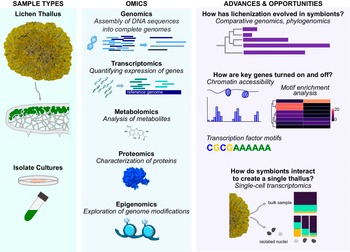
Overview of lichen research in the omics era. Lichen thallus and isolate cultures can be used in omics-based applications to advance our understanding of the evolution and molecular functions of these symbiotic associations. Moving forward with currently used approaches (genomics, transcriptomics, metabolomics and proteomics) in combination with novel approaches (epigenomics, single-cell and spatial transcriptomics) will allow the exploration of additional research frontiers including 1) the evolution of lichenization in symbionts, 2) how key genes involved in the symbiotic association are turned off and, 3) how individual symbionts interact to form the more complex thallus tissue. In colour online.

Figure 1. Long description
The infographic is divided into three columns.
Column 1: Sample Types.
At the top is a photo of a yellow Lichen Thallus. An arrow points down to a cross-section diagram showing green algal cells embedded in white fungal hyphae. Below this is Isolate Cultures, showing a petri dish and a test tube containing green liquid.
Column 2: Omics.
- Genomics: Assembly of D N A sequences into complete genomes, illustrated by blue horizontal bars and a double helix icon.
- Transcriptomics: Quantifying expression of genes, shown as small green segments aligning to a dark blue reference genome.
- Metabolomics: Analysis of metabolites, represented by a chemical molecular structure.
- Proteomics: Characterization of proteins, shown as a blue 3 D protein folding model.
- Epigenomics: Exploration of genome modifications, illustrated by blue circles representing histones wrapped in D N A with hexagonal tags.
Column 3: Advances and Opportunities.
- Evolution of lichenization: A phylogenetic tree paired with a horizontal bar chart.
- Gene regulation: A line graph showing chromatin accessibility and a heatmap for motif enrichment analysis. Below is a D N A sequence motif: C G C G A A A A A A.
- Symbiont interaction: A diagram comparing a bulk sample to isolated nuclei using single-cell transcriptomics, visualized with a stacked bar chart showing different cell population proportions.
Definitions for methods and subfields referenced in the text.
Omics: an umbrella term for a series of methods and disciplines based around analysis of large sets of data on biological molecules derived from a certain organism or sample. Omics includes genomics, transcriptomics, proteomics, and others (Dai & Shen Reference Dai and Shen2022).
Genomics: the study of entire genomes, or a complete set of genetic material of an organism, genome structure, evolution, and genetic elements present within. Typically involves sequencing of DNA molecules. Comparative genomics is a group of methods relying on comparing genomes from different species (Dai & Shen Reference Dai and Shen2022).
Transcriptomics: the study of transcriptomes, or the collections of RNA molecules present in a sample. Typically aims to study gene expression on the genome-wide level and involves sequencing of mRNA molecules (Dai & Shen Reference Dai and Shen2022).
Proteomics: the study of proteomes, or the collections of proteins present in a sample. Typically involves identification of proteins with mass spectrometry (Dai & Shen Reference Dai and Shen2022).
Metabolomics: the study of metabolomes, or the collections of small molecules present in a sample. Typically involves identification of metabolites with mass spectrometry (Dai & Shen Reference Dai and Shen2022).
Epigenomics: physical modifications of the genome (DNA and chromatin) which regulates gene expression. Distinct from epigenetics which refers to the heritable changes in gene expression which influence cell type differentiation (Struhl Reference Struhl2024).
Multi-omics, or integrative omics: a group of methods relying on integrating multiple types of omics data (e.g. genomic and transcriptomic) in one study. Not to be confused with meta-omics (Dai & Shen Reference Dai and Shen2022).
Meta-omics (metagenomics/metatranscriptomics/metaproteomics): application of omics techniques to mixed samples that contain multiple organisms. Metagenomics is distinct from metabarcoding and usually involves analysis of entire genomes of organisms extracted from the sample (Bharti & Grimm Reference Bharti and Grimm2021).
Metabarcoding: a group of methods for taxonomic profiling of mixed samples based on a single marker locus. Degenerate primers are used to amplify the same gene (e.g. 16S rRNA locus) from all genomes contained in a sample. Following PCR and sequencing, the data are used to identify taxonomic composition of a sample (Bharti & Grimm Reference Bharti and Grimm2021).
Shotgun sequencing: a method for sequencing a genome by breaking it into fragments at random, sequencing the fragments, and reconstructing genomic sequence through assembly (Bharti & Grimm Reference Bharti and Grimm2021).
Phylogenetics: a group of methods for reconstructing evolutionary relationships between taxa, often involves aligning sequences from different organisms and building phylogenetic trees as a way to represent how taxa are related to each other (Wiley & Lieberman Reference Wiley and Lieberman2011).
Phylogenomics: a group of methods that use genomic data for phylogenetics (Bourgeois & Warren Reference Bourgeois and Warren2021).
Population genomics: the study of variation in genomes within and between populations of a single species. Sometimes is defined as application of population genetics methods to whole-genome data (Bourgeois & Warren Reference Bourgeois and Warren2021).
Pangenomics: the study of pangenomes, or collection of genomic sequences from a single species. Pangenomic analysis often focuses on presence/absence variation and on identification of ‘core’ (present in the majority of individuals) and ‘accessory’ (present only occasionally) sets of genes (Secomandi et al. Reference Secomandi, Gallo, Rossi, Rodríguez Fernandes, Jarvis, Bonisoli-Alquati, Gianfranceschi and Formenti2025).
Sanger sequencing: high-precision low-throughput method of sequencing that relies on incorporation of chain-terminating dideoxynucleotides (van Dijk et al. Reference van Dijk, Jaszczyszyn, Naquin and Thermes2018).
Short-read sequencing: high-throughput method of sequencing based on the sequencing by synthesis technology, most notably implemented by Illumina. This technology is limited to short (50 to 400 base pairs) DNA fragments. Also known as next-generation sequencing or massively parallel sequencing (van Dijk et al. Reference van Dijk, Jaszczyszyn, Naquin and Thermes2018).
Long-read sequencing: high-throughput method of sequencing using the single-molecule real-time sequencing technology that can produce longer (over a million base pairs long) sequences, most notably implemented by PacBio and Oxford Nanopore. Also known as third-generation sequencing (van Dijk et al. Reference van Dijk, Jaszczyszyn, Naquin and Thermes2018).
Hi-C sequencing: high-throughput chromosome conformation capture to detect chromatin interactions (Belton et al. Reference Belton, McCord, Gibcus, Naumova, Zhan and Dekker2012). Informs 3D organization of genomes (Belton et al. Reference Belton, McCord, Gibcus, Naumova, Zhan and Dekker2012) and commonly used in genome assembly for generation of chromosome-level scaffolds (Ghurye et al. Reference Ghurye, Rhie, Walenz, Schmitt, Selvaraj, Pop, Phillippy and Koren2019).
RAD-seq: Restriction-site-associated DNA sequencing (Andrews et al. Reference Andrews, Good, Miller, Luikart and Hohenlohe2016). Genomic DNA is cut in fragments by a restriction enzyme and sequenced using short-read sequencing. The resulting data includes sequences of the genomic regions adjacent to the fragmentation sites. The method offers genome-wide data that can be used for phylogenetics at a smaller cost compared to whole-genome sequencing.
Target enrichment: a method for isolating specific DNA regions using complementary RNA or DNA probes (baits), designed and synthesized in vitro. Often used to create multi-locus datasets for phylogenetic studies. Also known as target capture (Andermann et al. Reference Andermann, Torres Jiménez, Matos-Maraví, Batista, Blanco-Pastor, Gustafsson, Kistler, Liberal, Oxelman and Bacon2019).
Genomic skimming: shallow (low-depth) sequencing method. Involves using genomic DNA to produce small amounts of data, which are not sufficient to recover the whole genome but allow the recovery of higher-coverage regions, including organelle genomes, repeat elements, and rRNA loci (Straub et al. Reference Straub, Parks, Weitemier, Fishbein, Cronn and Liston2012).
ATAC-seq: assay for transposase accessible chromatin with high-throughput sequencing (Buenrostro et al. Reference Buenrostro, Wu, Chang and Greenleaf2015) identifies regions in the genome that are open and not condensed. Provides information on the gene regulatory landscape for the entire genome and is often used for identifying regulatory elements within the genome, such as promoters.
ChIP-seq: chromatin immunoprecipitation sequencing. The method is used to study DNA-protein interactions, typically to identify genomic regions to which a specific transcription factor binds. This is achieved by fragmenting DNA and isolating the fragments bound to the protein of interest, using antibodies specific to the protein (Johnson et al. Reference Johnson, Mortazavi, Myers and Wold2007).
DAP-seq: DNA affinity purification sequencing (Bartlett et al. Reference Bartlett, O’Malley, Huang, Galli, Nery, Gallavotti A and Ecker2017). Similar to ChIP-seq, profiles potential transcription factor binding sites in a genome. Unlike ChIP-seq, DAP-seq relies on in vitro expression of the proteins of interest.
scRNA-seq/snRNA-seq: single-cell (sc)/single-nucleus (sn) RNA sequencing. Isolates individual cells/nuclei and generates transcriptomic profiles for each individually (Grones et al. Reference Grones, Eekhout, Shi, Neumann, Berg, Ke, Shahan, Cox, Gomez-Cano and Nelissen2024). The resulting data can be used to study different cell types or states present in a complex tissue.
Spatial transcriptomics: collection of approaches that profile gene expression in biological tissue while maintaining spatial context. Approaches commonly categorized as imaging-based (profiles pre-selected genes at cellular resolution) or sequencing-based (whole transcriptome spatial context at lower resolution) (Wang et al. Reference Wang, Liu, Zhao, Lee, Buzdin, Mu, Zhao, Chen and Li2023a).
Heterologous expression: expression of a gene from one organism in a different organism. Often achieved by molecular cloning (creating recombinant DNA molecules) followed by transformation or transfection (insertion of DNA molecules into a cell) (Frommer & Ninnemann Reference Frommer and Ninnemann1995).
AlphaFold: an algorithm for predicting 3D structure of a protein molecule based on the amino acid sequence (Jumper et al. Reference Jumper, Evans, Pritzel, Green, Figurnov, Ronneberger, Tunyasuvunakool, Bates, Žídek and Potapenko2021).
Progress and Advances in Lichen Research with Omics Technology
Generation of genomic resources
The first sequenced genome of a lichen symbiont, that of the mycobiont of Xanthoria parietina (L.) Th. Fr, was released (although not officially published) in 2011 (https://mycocosm.jgi.doe.gov/Xanpa2/Xanpa2.info.html). Over the following 15 years, many more, mostly mycobiont, genomes were produced. Currently, NCBI lists over 500 genomic assemblies of varying quality from the class Lecanoromycetes alone. Nevertheless, even for the better-studied mycobionts, the amount of genomic resources is lower than for their non-lichen relatives (compared to e.g. over 3000 genomic assemblies from a less species-rich class Eurotiomycetes).
Producing genomes for the lichen mycobiont was initially hindered by challenges related to the isolation of symbionts into pure cultures. Lichen mycobionts are difficult to isolate and, once in culture, they grow notoriously slowly. For example, the genome of X. parietina required two years of cultivation to amass enough culture-derived genomic DNA (Grube et al. Reference Grube, Berg, Andrésson, Vilhelmsson, Dyer, Miao and Martin2013). An alternative to laborious culture isolation is metagenomics, in which the total DNA of a lichen thallus is sequenced and used to produce genomic assemblies for individual symbionts. At its best, metagenomics allows challenges related to culture isolation to be bypassed and genomes of multiple symbionts to be obtained from one sequencing run. Metagenomics brings challenges of its own, which we address in detail below, yet it has proved to be an effective and efficient way to generate genomic resources for lichen symbionts.
The first photobiont genome, an Asterochloris from Cladonia grayi G. Merr. ex Sandst., was produced shortly after the first mycobiont genome (https://phycocosm.jgi.doe.gov/Astpho2/Astpho2.info.html). The amount of available genomic resources is lower compared to mycobionts, but currently, an increasing number of photobiont genomes are being produced, both from pure cultures and extracted from lichen metagenomes (Gazquez et al. Reference Gazquez, Bordenave, Montero-Pau, Pérez-Rodrigo, Marco, Martínez-Alberola, Muggia, Barreno and Carrasco2024; Poquita-Du et al. Reference Poquita-Du, Otte, Calchera and Schmitt2024; Puginier et al. Reference Puginier, Libourel, Otte, Skaloud, Haon, Grisel, Petersen, Berrin, Delaux and Dal Grande2024; Tagirdzhanova et al. Reference Tagirdzhanova, Saary, Cameron, Allen, Garber, Escandón, Cook, Goyette, Nogerius and Passo2024; Heuberger et al. Reference Heuberger, Wehrkamp, Pfammatter, Poretti, Graf, Herger, Isaksson, Schlagenhauf, Honegger and Wicker2025).
The majority of available lichen symbiont genome assemblies have been produced using short-read sequencing and consequently tend to be fragmented. Even for the lichen symbioses that had become commonly studied models, such as Lobaria pulmonaria (L.) Hoffm. or Umbilicaria pustulata (L.) Hoffm., only short-read genome assemblies were available until recently (Greshake Tzovaras et al. Reference Greshake Tzovaras, Segers, Bicker, Dal Grande, Otte, Anvar, Hankeln, Schmitt and Ebersberger2020; Cameron et al. Reference Cameron, Tremblay, Yahr, Blaxter and Finn2026). While genome fragmentation does not affect many studies, highly fragmented genomes are less useful for certain downstream applications, such as annotation of repetitive elements and studies of genome evolution. Since long-read sequencing and chromatin conformation capture sequencing (Hi-C) have become more robust and affordable, higher quality genomic assemblies are increasingly available (Greshake Tzovaras et al. Reference Greshake Tzovaras, Segers, Bicker, Dal Grande, Otte, Anvar, Hankeln, Schmitt and Ebersberger2020; McKenzie et al. Reference McKenzie, Walston and Allen2020; Singh et al. Reference Singh, Armaleo, Dal Grande and Schmitt2021b; Adams et al. Reference Adams, Escalona, Marimuthu, Fairbairn, Beraut, Seligmann, Nguyen, Chumchim and Stajich2023; Ahmad et al. Reference Ahmad, Ritz, Calchera, Otte, Schmitt, Brueck and Mehlmer2023; Llewellyn et al. Reference Llewellyn, Mian, Hill, Leitch and Gaya2023b; Yahr et al. Reference Yahr2024a). Long-read metagenomics allows the production of chromosome-scale genomic assemblies for mycobionts, as well as photobionts and other lichen-associated organisms, provided that enough sequencing data are produced (Heuberger et al. Reference Heuberger, Wehrkamp, Pfammatter, Poretti, Graf, Herger, Isaksson, Schlagenhauf, Honegger and Wicker2025). Recently, numerous sequencing research efforts, including the Aquatic Symbiosis Genomics (McKenna et al. Reference McKenna, Archibald, Beinart, Dawson, Hentschel, Keeling, Lopez, Martín-Durán, Petersen and Sigwart2021), Darwin Tree of Life (Darwin Tree of Life Project Consortium 2022), European Reference Genome Atlas (Mc Cartney et al. Reference Mc Cartney, Formenti, Mouton, De Panis, Marins, Leitão, Diedericks, Kirangwa, Morselli and Salces-Ortiz2024) and the Trebouxia Research Portal (https://trebouxia.net), are endeavouring to create high-quality genomes, including of lichen symbionts, using long-read metagenomics and Hi-C. Once released, the resulting data will be a highly valuable resource for lichen research.
Evolutionary history and microevolutionary patterns reflected in genomes
The availability of genomic information has allowed for a more precise reconstruction of the evolutionary history of lichen symbionts. Molecular phylogenetics of the mycobionts, already thriving in the era of Sanger sequencing, have benefited from the ability to construct phylogenetic trees based on hundreds or thousands of loci. Phylogenomics was used to resolve the backbone of Ascomycota and thus trace the evolution of fungal lifestyles including the lichen lifestyle (Díaz-Escandón et al. Reference Díaz-Escandón, Tagirdzhanova, Vanderpool, Allen, Aptroot, Češka, Hawksworth, Huereca, Knudsen and Kocourková2022), as well as to produce phylogenies of various clades of lichen mycobionts (Pizarro et al. Reference Pizarro, Divakar, Grewe, Leavitt, Huang, Dal Grande, Schmitt, Wedin, Crespo and Lumbsch2018; Grewe et al. Reference Grewe, Ametrano, Widhelm, Leavitt, Distefano, Polyiam, Pizarro, Wedin, Crespo and Divakar2020; Widhelm et al. Reference Widhelm, Grewe, Goffinet, Wedin, Goward, Coca, Distefano, Košuthová and Lumbsch2021).
Several strategies have been employed to generate data for phylogenomics of lichen symbionts. While some studies have relied on complete genomic assemblies generated either from cultures or with metagenomics (Divakar & Crespo Reference Divakar, Crespo, Upreti, Divakar, Shukla and Bajpai2015), others used various strategies to subsample genomic data and thus reduce costs. The strategies include 1) methods such as RAD-seq (Grewe et al. Reference Grewe, Huang, Leavitt and Lumbsch2017) and target enrichment (Grewe et al. Reference Grewe, Ametrano, Widhelm, Leavitt, Distefano, Polyiam, Pizarro, Wedin, Crespo and Divakar2020), which limit the sequencing effort to specific sites within the genome, and 2) genomic skimming (Meiser et al. Reference Meiser, Otte, Schmitt and Grande2017), which relies on shallow sequencing. Genomic skimming allows costs to be reduced, both in terms of computational resources and sequencing data by, for example, limiting the study to complete genomes of organelles. Mitochondrial genomes are both smaller and present in higher numbers of copies, which make them a cost-effective way to construct phylogenies (Xavier et al. Reference Xavier, Miao, Jónsson and Andrésson2012; Hoffman et al. Reference Hoffman, Karol, Ohmura, Pogoda, Keepers, McMullin and Lendemer2023).
Once the phylogenetic structure of a clade is revealed, genomic data can be used to answer follow-up questions. By comparing related organisms, we can see how the evolutionary transitions are reflected in their genomes. Comparative genomics can provide a powerful toolbox for lichen biology, in part owing to the diversity of lichen symbioses. Both for photobionts and mycobionts, the lichen lifestyle has evolved multiple times independently (Gargas et al. Reference Gargas, DePriest, Grube and Tehler1995; Spribille et al. Reference Spribille, Resl, Stanton and Tagirdzhanova2022), resulting in multiple evolutionary transitions that can be studied (Wang et al. Reference Wang, Liu, Zhang, Zhou, Zhang, Li, Yu, Zhang, Hao and Wang2014; Díaz-Escandón et al. Reference Díaz-Escandón, Tagirdzhanova, Vanderpool, Allen, Aptroot, Češka, Hawksworth, Huereca, Knudsen and Kocourková2022; Resl et al. Reference Resl, Bujold, Tagirdzhanova, Meidl, Freire Rallo, Kono, Fernández-Brime, Guðmundsson, Andrésson and Muggia2022; Song et al. Reference Song, Kim, Park, Lee, Choi, Jeon, Cheong, Choi, J-S and Lee2022; Puginier et al. Reference Puginier, Libourel, Otte, Skaloud, Haon, Grisel, Petersen, Berrin, Delaux and Dal Grande2024). The resulting symbioses have diverse phenotypes (e.g. body plans and metabolic profiles), and comparative genomics can provide clues on how a phenotype ‘works’ and what functional families of genes it requires (Pizarro et al. Reference Pizarro, Divakar, Grewe, Crespo, Dal Grande and Lumbsch2020). For example, comparative genomics has identified expansions of several groups of carbohydrate-active enzymes that are associated with the lichen lifestyle in green algae (Puginier et al. Reference Puginier, Libourel, Otte, Skaloud, Haon, Grisel, Petersen, Berrin, Delaux and Dal Grande2024).
Genomic data can also be used to study evolution on the sub-species scale. Population genomics is used to study microevolutionary patterns and processes affecting lichen symbionts (Werth et al. Reference Werth, Miao, Jónsson, Andrésson, Upreti, Divakar, Shukla and Bajpai2015). Studies of population structure, gene flow, and dispersal are especially informative in the case of endangered species, as they can inform conservation strategies (Allen et al. Reference Allen, McKenzie, Sleith and Alter2018; Yahr et al. Reference Yahr, Allen, Atienza, Burgartz, Chrismas, Dal Forno, Degtjarenko, Ohmura, Pérez-Ortega and Randlane2024b). When juxtaposed with information about geographical distributions of the studied populations and climatic conditions in different parts of the range, these data are used to study recent evolutionary history of lichen symbionts and their adaptations to the environment (Dal Grande et al. Reference Dal Grande, Sharma, Meiser, Rolshausen, Büdel, Mishra, Thines, Otte, Pfenninger and Schmitt2017; Alonso-García et al. Reference Alonso-García, Grewe, Payette and Villarreal2021; Merges et al. Reference Merges, Dal Grande, Valim, Singh and Schmitt2023; Wong et al. Reference Wong, Calchera, Otte and Schmitt2025). Population genomics can identify genes under purifying or diversifying selection, providing insight into selection pressures (Wong et al. Reference Wong, Calchera, Otte and Schmitt2025). Genomic data can also reflect the life cycle and reproduction strategy of the organism (Gazquez et al. Reference Gazquez, Bordenave, Montero-Pau, Pérez-Rodrigo, Marco, Martínez-Alberola, Muggia, Barreno and Carrasco2024), especially for the mycobionts, whose sexual reproduction is governed by the mating-type locus, which can be annotated and interpreted (Pizarro et al. Reference Pizarro, Dal Grande, Leavitt, Dyer, Schmitt, Crespo, Lumbsch and Divakar2019; Ament-Velásquez et al. Reference Ament-Velásquez, Tuovinen, Bergström, Spribille, Vanderpool, Nascimbene, Yamamoto, Thor and Johannesson2021).
Genome mining: an opportunity for natural product discovery
The increasing demand for novel bioactive products to combat antibiotic resistant pathogens and for disease treatment has resulted in the exploration of biosynthetic compounds produced by diverse taxa (Singh et al. Reference Singh, Dal Grande and Schmitt2022). The pharmacological properties of natural products include antimicrobial, antitumour and anti-inflammatory characteristics (Singh et al. Reference Singh, Dal Grande and Schmitt2022). Fungi have been commonly used for their bioactive secondary metabolites, with many drugs originating from fungal sources (Singh et al. Reference Singh, Xu, Zdouc, Pasinato, Egbert, Yu, Olafsdottir, Beltran-Sanz, Divakar and Pizarro2025). However, despite the quantity of drugs attributed to fungal sources, the taxonomic origins of these secondary metabolites have largely been limited to selected organisms that were easy to culture (Singh et al. Reference Singh, Xu, Zdouc, Pasinato, Egbert, Yu, Olafsdottir, Beltran-Sanz, Divakar and Pizarro2025). Novel secondary metabolite discovery can now be accomplished through identification of biosynthetic gene clusters (BGCs), with bioinformatic tools such as antiSMASH (Blin et al. Reference Blin, Shaw, Vader, Szenei, Reitz, Augustijn, Cediel-Becerra, de Crécy-Lagard, Koetsier and Williams2025) and databases such as MIBiG for experimentally validated BGCs (Zdouc et al. Reference Zdouc, Blin, Louwen, Navarro, Loureiro, Bader, Bailey, Barra, Booth and Bozhüyük2025).
Lichens are considered a treasure trove of secondary metabolites (Stocker-Wörgötter Reference Stocker-Wörgötter2008; Singh et al. Reference Singh, Xu, Zdouc, Pasinato, Egbert, Yu, Olafsdottir, Beltran-Sanz, Divakar and Pizarro2025). Annotation of mycobiont genomes has identified a high number of BGCs, including those potentially linked to novel secondary metabolites (Singh et al. Reference Singh, Dal Grande and Schmitt2022). As an increasing number of diverse taxa are included in the genome mining efforts (Abdel-Hameed et al. Reference Abdel-Hameed, Bertrand, Piercey-Normore and Sorensen2016b; Calchera et al. Reference Calchera, Dal Grande, Bode and Schmitt2019; Pizarro et al. Reference Pizarro, Divakar, Grewe, Crespo, Dal Grande and Lumbsch2020; Singh et al. Reference Singh, Armaleo, Dal Grande and Schmitt2021b, Reference Singh, Pasinato, Yriarte, Pizarro, Divakar, Schmitt and Dal Grande2024; Gerasimova et al. Reference Gerasimova, Beck, Werth and Resl2022, Reference Gerasimova, Beck, Scheunert and Kulkarni2024; Llewellyn et al. Reference Llewellyn, Nowell, Aptroot, Temina, Prescott, Barraclough and Gaya2023a; Pasinato & Singh Reference Pasinato and Singh2025), patterns are emerging, including a higher diversity of polyketide synthase BGCs in macrolichens (Kim et al. Reference Kim, Shishido, Pham, Kang, Liu, Wahlsten, Collemare, Park, Oh and Ha2025) or association of BGCs to climatic niches (Singh et al. Reference Singh, Calchera, Schulz, Drechsler, Bode, Schmitt and Dal Grande2021a).
Many lichen BGCs and their associated secondary metabolites remain unidentified (Singh et al. Reference Singh, Dal Grande and Schmitt2022). Following identification of BGCs, their function may still be unclear, since many are orphan (i.e. the structure/compound is unknown) or silent (i.e. not expressed naturally). Genomic resources alone cannot capture the bioactive potential and other approaches are necessary (Singh et al. Reference Singh, Xu, Zdouc, Pasinato, Egbert, Yu, Olafsdottir, Beltran-Sanz, Divakar and Pizarro2025). Following genomic identification of BGCs, metabolomics has been used to identify metabolites in lichenized fungi both at scale (Singh et al. Reference Singh, Xu, Zdouc, Pasinato, Egbert, Yu, Olafsdottir, Beltran-Sanz, Divakar and Pizarro2025) and in taxon-specific research (Abdel-Hameed et al. Reference Abdel-Hameed, Bertrand, Piercey-Normore and Sorensen2016a; Garg et al. Reference Garg, Zeng, Edlund, Melnik, Sanchez, Mohimani, Gurevich, Miao, Schiffler and Lim2016; Egbert et al. Reference Egbert, Hoffman, McMullin, Lendemer and Sorensen2022), allowing BGCs to be connected to their respective secondary metabolite product.
Within lichens, it is often assumed that the majority of secondary metabolites arise from the mycobiont (Bertrand et al. Reference Bertrand, Abdel-Hameed and Sorensen2018). Algae are known to produce diverse secondary metabolites (Kwon & Hovde Reference Kwon and Hovde2024), yet limited research is available on those found in lichen photobionts. Secondary metabolites from cyanobacterial photobionts have previously been identified (Kaasalainen et al. Reference Kaasalainen, Jokela, Fewer, Sivonen and Rikkinen2009, Reference Kaasalainen, Fewer, Jokela, Wahlsten, Sivonen and Rikkinen2012; Kampa et al. Reference Kampa, Gagunashvili, Gulder, Morinaka, Daolio, Godejohann, Miao, Piel and Andrésson2013) and some of these metabolites may be specialized for symbioses (Cameron et al. Reference Cameron, Sanchez, Goldman, Blaxter and Finn2024). The presence of such BGCs and secondary metabolites in cyanobacterial photobionts raises the question of whether chlorophyte photobionts might also contribute to secondary metabolite production.
Following the identification of diverse bacterial communities in lichens (part of the lichen microbiota (see below)), secondary metabolites and BGCs in the lichen microbiota have begun to be explored as an additional source of novel bioactive compounds. Biosynthetic gene clusters have been identified in Streptomyces isolated from a lichen (Schneider et al. Reference Schneider, Simic, Aachmann, Rückert, Kristiansen, Kalinowski, Jiang, Wang, Jiang and Lale2018) and in other Actinomycetes from the lichen-associated microbiota (Hou et al. Reference Hou, Nowak, Taylor, Calcott, Knight and Owen2023). The exploration of lichen-associated Actinomyces in comparison to other environmentally derived Streptomyces datasets suggested that lichen-based populations have a higher percentage of gene cluster families (Hou et al. Reference Hou, Nowak, Taylor, Calcott, Knight and Owen2023) and may be another source of novel secondary metabolites.
Organismal composition of lichen thalli
In addition to the mycobiont and photobiont, lichens contain other organisms, often termed lichen microbiota, whose role in the symbiosis and relationship to the myco- and photobionts remain largely mysterious (Grimm et al. Reference Grimm, Grube, Schiefelbein, Zühlke, Bernhardt and Riedel2021). Next-generation sequencing offered a way to profile the organismal composition of lichen thalli free of biases associated with culturing or the use of specific primers designed to detect only certain organisms. The two most common approaches to taxonomic profiling of lichen symbioses are metabarcoding and shotgun metagenomics (Grimm et al. Reference Grimm, Grube, Schiefelbein, Zühlke, Bernhardt and Riedel2021). With metabarcoding, lichen samples are profiled via sequencing of marker genes amplified with degenerate primers (Grube et al. Reference Grube, Cardinale, de Castro, Müller and Berg2009; Bates et al. Reference Bates, Cropsey, Caporaso, Knight and Fierer2011; Aschenbrenner et al. Reference Aschenbrenner, Cernava, Erlacher, Berg and Grube2017; Fernández-Mendoza et al. Reference Fernández-Mendoza, Fleischhacker, Kopun, Grube and Muggia2017). While metabarcoding is prone to primer bias and might miss certain lineages (Klindworth et al. Reference Klindworth, Pruesse, Schweer, Peplies, Quast, Horn and Glöckner2013), it is a cost-effective way to assess the taxonomic composition of a sample. Metabarcoding has been used extensively for studies with many samples, for example those focused on how lichen-associated bacteria are acquired and shared (Aschenbrenner et al. Reference Aschenbrenner, Cernava, Erlacher, Berg and Grube2017; Leiva et al. Reference Leiva, Fernández-Mendoza, Acevedo, Carú, Grube and Orlando2021). Metabarcoding using long-read sequencing technologies has emerged as an alternative to short-read metabarcoding because it can provide higher taxonomic resolution due to increased amplicon length (Jamy et al. Reference Jamy, Foster, Barbera, Czech, Kozlov, Stamatakis, Bending, Hilton, Bass and Burki2020).
Shotgun metagenomics is free of primer bias and has the additional benefit of providing functional information on lichen-associated organisms (Grube et al. Reference Grube, Cernava, Soh, Fuchs, Aschenbrenner, Lassek, Wegner, Becher, Riedel and Sensen2015; Sigurbjörnsdóttir et al. Reference Sigurbjörnsdóttir, Andrésson and Vilhelmsson2015; Greshake Tzovaras et al. Reference Greshake Tzovaras, Segers, Bicker, Dal Grande, Otte, Anvar, Hankeln, Schmitt and Ebersberger2020). Lichen-associated bacteria have been profiled using metagenomics, both across a diversity of lichen symbioses (Tagirdzhanova et al. Reference Tagirdzhanova, Saary, Cameron, Allen, Garber, Escandón, Cook, Goyette, Nogerius and Passo2024) and in more targeted studies. One lichen symbiosis, Lobaria pulmonaria, has emerged as a model for studies of bacterial communities in lichens (Grimm et al. Reference Grimm, Grube, Schiefelbein, Zühlke, Bernhardt and Riedel2021). Metagenomics have also been used to examine non-mycobiont lichen-associated fungi that are otherwise challenging to study (Tagirdzhanova et al. Reference Tagirdzhanova, Saary, Tingley, Díaz-Escandón, Abbott, Finn and Spribille2021; Keller et al. Reference Keller, Calchera, Otte and Schmitt2025). Similarly to metagenomics, metatranscriptomics and metaproteomics have been used to try to answer both the ‘who’s there’ and ‘what do they do’ questions (Eymann et al. Reference Eymann, Lassek, Wegner, Bernhardt, Fritsch, Fuchs, Otto, Albrecht, Schiefelbein and Cernava2017; Cernava et al. Reference Cernava, Aschenbrenner, Soh, Sensen, Grube and Berg2019). Notably, metatranscriptomic analysis revealed the presence of basidiomycete yeasts that are stably associated with some lichen groups, which set off a search for candidate symbionts that constitute the ‘core’ of lichen symbiosis in addition to the two main symbionts (Spribille et al. Reference Spribille, Tuovinen, Resl, Vanderpool, Wolinski, Aime, Schneider, Stabentheiner, Toome-Heller and Thor2016; Tuovinen et al. Reference Tuovinen, Ekman, Thor, Vanderpool, Spribille and Johannesson2019). While meta-omics has the advantage of profiling an entire community in one go, culture studies followed by genomic sequencing can also provide valuable information on individual members of lichen-associated organisms (Oh et al. Reference Oh, Han, Kang, Park and Kim2016; Pankratov et al. Reference Pankratov, Nikitin and Patutina2022).
Next generation sequencing has provided a plethora of new information about organisms in and on lichens, which has generated debate about the concept of the lichen itself (Hawksworth & Grube Reference Hawksworth and Grube2020; Sanders Reference Sanders2024). Many questions regarding the biology of lichen-associated microorganisms remain open. An additional biological component of these microbial communities, the protists and the viruses, remain largely underexplored with only a small number of reports existing (Merges et al. Reference Merges, Dal Grande, Greve, Otte and Schmitt2021; Ponsero et al. Reference Ponsero, Hurwitz, Magain, Miadlikowska, Lutzoni and U’Ren2021).
Lichen physiology through the omics lens
Omics has provided a powerful toolbox for studying the biology of lichen symbionts and is currently one of the most robust and straightforward ways to uncover molecular processes underlying the lichen symbiosis. Transcriptomics has been used extensively to compare the behaviour of lichen symbionts in different conditions and thus shed light on their physiological responses. To identify genes relevant for symbiosis, researchers have compared gene expression of lichen symbionts cultured separately to the same organisms in the symbiotic state (Tagirdzhanova et al. Reference Tagirdzhanova, Scharnagl, Sahu, Yan, Bucknell, Bentham, Jégousse, Ament-Velásquez, Onuț-Brännström and Johannesson2025, Reference Tagirdzhanova, Raistrick and Talbot2026a) or in co-culture with their partners (Armaleo et al. Reference Armaleo, Müller, Lutzoni, Andrésson, Blanc, Bode, Collart, Dal Grande, Dietrich and Grigoriev2019; Kono et al. Reference Kono, Kon, Ohmura, Satta and Terai2020). Several studies have attempted to identify gene expression patterns within a lichen body (Elshobary et al. Reference Elshobary, Becker, Kalichuk, Chan, Belmonte and Piercey-Normore2018; Tagirdzhanova et al. Reference Tagirdzhanova, Scharnagl, Sahu, Yan, Bucknell, Bentham, Jégousse, Ament-Velásquez, Onuț-Brännström and Johannesson2025).
Lichen stress tolerance has been studied by recording transcriptomic and proteomic responses to various stressors, such as desiccation (Junttila et al. Reference Junttila, Laiho, Gyenesei and Rudd2013; Carniel et al. Reference Carniel, Gerdol, Montagner, Banchi, De Moro, Manfrin, Muggia, Pallavicini and Tretiach2016; Del Campo et al. Reference Del Campo, Gasulla, Hell, González-Hourcade and Casano2023) and temperature shifts (Steinhäuser et al. Reference Steinhäuser, Andrésson, Pálsson and Werth2016), and exposure to nitrogen (Munzi et al. Reference Munzi, Sheppard, Leith, Cruz, Branquinho, Bini, Gagliardi, Cai and Parrotta2017), UV (Leksin et al. Reference Leksin, Shelyakin, Zakhozhiy, Kozlova, Beckett and Minibayeva2024), sea water (Chrismas et al. Reference Chrismas, Tindall-Jones, Jenkins, Harley, Bird and Cunliffe2024) and antibiotics (Chavarria-Pizarro et al. Reference Chavarria-Pizarro, Resl, Kuhl-Nagel, Janjic, Fernandez Mendoza and Werth2022b). These studies mostly used entire lichen thalli and not isolated cultures, which made it possible to contrast stress responses between the photobiont and the mycobiont (Chavarria-Pizarro et al. Reference Chavarria-Pizarro, Resl, Janjic and Werth2022a; Meyer et al. Reference Meyer, Koch, McDonald and Stanton2024; Valim et al. Reference Valim, Otte and Schmitt2025).
The interplay between the organismal composition and the physiology of resulting symbiosis can be studied by metatranscriptomics and metaproteomics. Several studies have shown how lichen-associated bacteria respond to shifting conditions (Eymann et al. Reference Eymann, Lassek, Wegner, Bernhardt, Fritsch, Fuchs, Otto, Albrecht, Schiefelbein and Cernava2017; Cernava et al. Reference Cernava, Vasfiu, Erlacher, Aschenbrenner, Francesconi, Grube and Berg2018, Reference Cernava, Aschenbrenner, Soh, Sensen, Grube and Berg2019). It was also possible to check how lichen symbionts behave depending on the other organisms around them, for example how the mycobiont gene expression is shaped by which photobiont it is partnered with (Almer et al. Reference Almer, Resl, Gudmundsson, Warshan, Andrésson and Werth2023), or how photo- and mycobionts respond to a lichen-associated fungus (Kraft et al. Reference Kraft, Scheidegger and Werth2022).
Lichen Metagenomics
Although metagenomics has rapidly advanced research outlooks in the field, it does not come without its own challenges. As the field of bioinformatics rapidly advances, decisions on best practices in the processing and handling of data are required. A metagenomics analysis workflow can be summarized simply into key steps including the following: 1) reads quality control; 2) assembly of reads into longer contiguous DNA sequences (referred to as contigs); 3) binning of contigs into groups representative of individual genomes; 4) quality scoring of genome completeness and contamination; 5) taxonomic classification and functional annotation of metagenome-assembled genomes (MAGs). For each of these steps, numerous tools are available and new ones are regularly developed implementing novel techniques including machine-learning approaches (see some examples in Fig. 2).
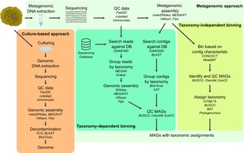
Genome acquisition pipelines used in lichen studies. The two main strategies include the culture-based approach, in which individual organisms are first isolated in culture and then subjected to standard genome acquisition, and the metagenomic approach, in which total DNA is isolated from lichen samples and analyzed in such a way as to produce metagenome-assembled genomes (MAGs). For the metagenomic approach, we show both taxonomy-dependent and taxonomy-independent strategies. For each step of the bioinformatic analysis, suitable software examples are given in italics. DB = database; QC = quality check. In colour online.
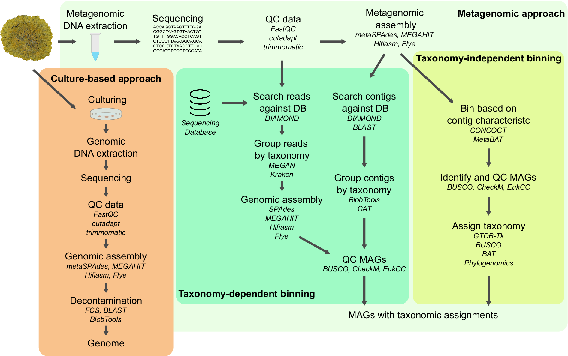
Figure 2. Long description
The flowchart begins with a lichen sample at the top left.
1. Culture-based approach (orange panel):
Arrows lead from the lichen to Culturing, then Genomic D N A extraction, Sequencing, Q C data (FastQC, cutadapt, trimmomatic), Genomic assembly (metaSPAdes, MEGAHIT, Hifiasm, Flye), Decontamination (F C S, BLAST, BlobTools), and finally a Genome.
2. Metagenomic approach (green and yellow panels):
Starting from the lichen, the path goes to Metagenomic D N A extraction, Sequencing, Q C data, and Metagenomic assembly (metaSPAdes, MEGAHIT, Hifiasm, Flye). From here, the process splits into two binning strategies:
- Taxonomy-dependent binning (center green panel):
Path A: Search reads against D B (DIAMOND) using a Sequencing Database, Group reads by taxonomy (MEGAN, Kraken), Genomic assembly, and Q C M A G s (BUSCO, CheckM, EukCC).
Path B: Search contigs against D B (DIAMOND, BLAST), Group contigs by taxonomy (BlobTools, CAT), and Q C M A G s.
- Taxonomy-independent binning (right yellow panel):
Bin based on contig characteristic (CONCOCT, MetaBAT), Identify and Q C M A G s (BUSCO, CheckM, EukCC), and Assign taxonomy (GTDB-Tk, BUSCO, BAT, Phylogenomics).
Both metagenomic paths terminate at M A G s with taxonomic assignments.
Binning is a key step in the generation of MAGs from metagenomic data, especially when short-read data are considered. Binning approaches can be broadly classified as taxonomy independent or dependent. The latter relies on taxonomic classification of sequences based on their similarity to sequences from reference databases (Mallawaarachchi et al. Reference Mallawaarachchi, Wickramarachchi, Xue, Papudeshi, Grigson, Bouras, Prahl, Kaphle, Verich and Talamantes-Becerra2024). While taxonomy dependent approaches can characterize known organisms from metagenomic samples, unknown organisms not found in reference databases might fail to be recovered (Mande et al. Reference Mande, Mohammed and Ghosh2012). Taxonomy independent approaches are better suited for more diverse environmental microbial communities. A popular approach is to group contigs based only on their intrinsic characteristics, such as k-mer composition and the abundance of reads which map to them within a metagenomic assembly (Mallawaarachchi et al. Reference Mallawaarachchi, Wickramarachchi, Xue, Papudeshi, Grigson, Bouras, Prahl, Kaphle, Verich and Talamantes-Becerra2024). Contigs thus grouped can be circumscribed as ‘bins’, among which researchers can detect MAGs of various organisms, including novel ones. The quality of the resulting assemblies in terms of their completeness and contiguity is often indistinguishable from the genomes produced from cultures. In the case of lichen metagenomes, such taxonomy independent approaches have been used to recover both bacterial and eukaryotic MAGs (Wicaksono et al. Reference Wicaksono, Cernava, Grube and Berg2020; Llewellyn et al. Reference Llewellyn, Nowell, Aptroot, Temina, Prescott, Barraclough and Gaya2023a; Tagirdzhanova et al. Reference Tagirdzhanova, Saary, Cameron, Allen, Garber, Escandón, Cook, Goyette, Nogerius and Passo2024).
Profiling eukaryotic diversity with metagenomics is generally more challenging than profiling prokaryotes. Bioinformatic tools tend to have a bias towards prokaryotes, which stems from eukaryotes having larger and more complex genomes filled with repeat elements that complicate assembly and binning (Escobar-Zepeda et al. Reference Escobar-Zepeda, de León A and Sanchez-Flores2015; Massana & López-Escardó Reference Massana and López-Escardó2022). This is exacerbated by the lower representation of non-model eukaryotes in reference genome databases (Saary et al. Reference Saary, Mitchell and Finn2020). Lichens represent complex samples that at a minimum contain one eukaryote, typically two and often many more, and are heavily affected by these limitations. However, eukaryotic metagenomics is gaining more traction and numerous tools are now available that can handle complex eukaryote-prokaryote communities (Saary et al. Reference Saary, Mitchell and Finn2020; Pronk & Medema Reference Pronk and Medema2022; Alexander et al. Reference Alexander, Hu, Krinos, Pachiadaki, Tully, Neely and Reiter2023).
Sequencing depth, or the total amount of sequencing data measured in base pairs, is a key consideration in planning a metagenomic study (Quince et al. Reference Quince, Walker, Simpson, Loman and Segata2017). Metagenomes sequenced too shallowly might fail to provide the desired output, yet deep sequencing for many samples can make metagenomic studies prohibitively expensive. Decisions should be made based on the research question. Higher sequencing depth translates into a higher quality of assembly and better chances of detecting organisms with lower abundance in the sample. When the goal is to generate a MAG of the mycobiont, even shallowly sequenced metagenomes might suffice, and over 90% of metagenomes sequenced at 2 Gbp sequencing depth have been found to yield a mycobiont MAG (Tagirdzhanova et al. Reference Tagirdzhanova, Saary, Cameron, Allen, Garber, Escandón, Cook, Goyette, Nogerius and Passo2024). To reliably obtain the photobiont MAG, 10 Gbp data can be required, and a comprehensive survey of MAGs from lichen-associated microbial communities might require even more data. Taxonomic profiling of shallowly sequenced metagenomes might lead to false absences, since organisms with lower abundance might fly under the radar (Tagirdzhanova et al. Reference Tagirdzhanova, Saary, Cameron, Allen, Garber, Escandón, Cook, Goyette, Nogerius and Passo2024). When faced with sequencing depth limitations, alternative approaches might be considered, such as metabarcoding or searching for rRNA or other functional genes of interest in the contigs. Occasionally, even deeply sequenced metagenomes fail to yield MAGs of sufficient quality. For instance, the presence of two closely related genomes within one sample can cause problems, such as when two photobiont strains co-exist in a thallus. In such cases, photobiont contigs are often highly fragmented and standard binning techniques cannot separate the two genomes, resulting in a highly duplicated assembly that will fail quality checks (Tagirdzhanova et al. Reference Tagirdzhanova, Saary, Cameron, Allen, Garber, Escandón, Cook, Goyette, Nogerius and Passo2024).
Metagenomic assembled genomes from lichen datasets continue to become increasingly available as more studies implement this approach (Greshake Tzovaras et al. Reference Greshake Tzovaras, Segers, Bicker, Dal Grande, Otte, Anvar, Hankeln, Schmitt and Ebersberger2020; Llewellyn et al. Reference Llewellyn, Nowell, Aptroot, Temina, Prescott, Barraclough and Gaya2023a; Tagirdzhanova et al. Reference Tagirdzhanova, Saary, Cameron, Allen, Garber, Escandón, Cook, Goyette, Nogerius and Passo2024; Heuberger et al. Reference Heuberger, Wehrkamp, Pfammatter, Poretti, Graf, Herger, Isaksson, Schlagenhauf, Honegger and Wicker2025). The generation of such data represents a valuable step forward in lichen research, creating opportunities for the exploration of evolutionary and ecological questions as described below. While metagenomics and new MAGs will not solve many of the remaining questions in the field, they can serve as a starting point for developing the next steps forward with new technology developments.
Frontiers and Opportunities
Pangenomics
In many organisms, including fungi, individuals within one species show considerable variation in gene content and genome structure; a single reference genome, therefore, cannot fully represent an entire species (Loegler et al. Reference Loegler, Friedrich and Schacherer2025). Pangenomics aims to map genomic variation within a species by studying numerous individually assembled genomes. Eukaryotic pangenomics is a rapidly growing field, powered by long-read sequencing and an increased ability to produce high-quality, highly contiguous assemblies (Loegler et al. Reference Loegler, Friedrich and Schacherer2025). While pangenomes for several non-lichen fungal species are already available (Mondo & Grigoriev Reference Mondo and Grigoriev2025), those of lichen symbionts remain unexplored.
Transposable elements and genome evolution
Transposable elements (TEs) contribute to eukaryotic genomic diversity and influence genome evolution (Pritham Reference Pritham2009; Castanera et al. Reference Castanera, López-Varas, Borgognone, LaButti, Lapidus, Schmutz, Grimwood, Pérez, Pisabarro and Grigoriev2016). In fungi in particular, TEs appear to drive the evolution of lifestyles in several ways (Muszewska et al. Reference Muszewska, Steczkiewicz, Stepniewska-Dziubinska and Ginalski2017; Sauters & Rokas Reference Sauters and Rokas2025). First, in plant pathogenic fungi, TE-rich regions may evolve faster and contain genes associated with pathogenicity (Torres et al. Reference Torres, Oggenfuss, Croll and Seidl2020; Fouché et al. Reference Fouché, Oggenfuss, Chanclud and Croll2022). Whether TEs affect mycobiont genome evolution in a similar way is yet to be investigated. Second, recently discovered giant transposon Starships are linked to numerous cases of horizontal gene transfer, including of genes that are beneficial to the fungus and linked to its lifestyle, such as virulence factors for pathogens or genes encoding antimicrobial proteins (Bucknell & McDonald Reference Bucknell and McDonald2023). A putative Starship has been reported from a lichen mycobiont (Tagirdzhanova et al. Reference Tagirdzhanova, Brown, Bucknell, Cameron, Finn, Blaxter, McDonald, Gluck-Thaler and Talbot2026b), but the true extent of the impact of Starships on lichen mycobionts remains unclear. Research is underway on the impact of TEs on the evolution of lichen mycobionts (Dal Grande et al. Reference Dal Grande, Jamilloux, Choisne, Calchera, Rolshausen, Petersen, Schulz, Nilsson and Schmitt2021; Paulsen et al. Reference Paulsen, Sharrett, Mumey, Larsen, Nguyen, Lendemer, Calabria, Hoffman, Magori and Allen2025; Cameron et al. Reference Cameron, Tremblay, Yahr, Blaxter and Finn2026), but those in lichen photobionts remain unexplored. Further study of the TE landscape, including Starships, requires 1) highly contiguous genome assemblies and 2) multiple genomes from the same species.
Photobionts through the omics lens
Many open questions about lichen photobionts can be addressed with omics-based techniques. These lines of research have been somewhat limited, since fewer genomic resources are available for lichen photobionts compared to their fungal partners. In the case of eukaryotic photobionts, this discrepancy comes in part from a methodological challenge: obtaining a photobiont genome from a lichen metagenome tends to require more data and is therefore more costly (Tagirdzhanova et al. Reference Tagirdzhanova, Saary, Cameron, Allen, Garber, Escandón, Cook, Goyette, Nogerius and Passo2024), leading to fewer photobiont MAGs being available. However, many more high-quality photobiont genomes are becoming available, which provides many new opportunities for research on lichen symbiosis. Comparative genomics of chlorophyte photobionts, pioneered by Puginier et al. (Reference Puginier, Libourel, Otte, Skaloud, Haon, Grisel, Petersen, Berrin, Delaux and Dal Grande2024), can serve as an example.
Gene regulatory landscape of lichen symbioses
By identifying specialized genes in newly generated genomic resources for lichens, we can make informed guesses about the key biological functions that shape the lichen symbiosis. The question remains as to how these genes are regulated in lichens. In other words, what are the mechanisms that change the behaviour of lichen symbionts based on environmental cues or interaction between symbionts? For example, what changes in gene expression accompany the transition from an aposymbiotic to symbiotic state, and how are these changes orchestrated on the cellular level? The gene regulatory landscape is influenced by a variety of mechanisms that can now be profiled following technical advances, including chromatin accessibility and transcription factor binding sites. Here, we propose key areas for further exploration focusing on techniques that may perform well with lichen samples while informing genome-wide regulatory landscapes.
A starting point in characterizing the gene regulatory landscape of lichens more broadly is chromatin accessibility. Chromatin accessibility refers to the physical access of DNA for transcription factor binding, which in turn influences which genes may be transcribed (Klemm et al. Reference Klemm, Shipony and Greenleaf2019). A variety of technologies exist which focus on quantifying chromatin susceptibility to enzymatic methylation or cleavage (Klemm et al. Reference Klemm, Shipony and Greenleaf2019). For instance, transposase-accessible chromatin using sequencing (ATAC-seq) provides a robust and rapid method for generating accessibility profiles (Klemm et al. Reference Klemm, Shipony and Greenleaf2019; Grandi et al. Reference Grandi, Modi, Kampman and Corces2022). ATAC-seq requires no a priori knowledge of the epigenetic mechanisms (Grandi et al. Reference Grandi, Modi, Kampman and Corces2022) and can generate genome-wide regulatory maps for novel species, including lichen symbionts. This data can be used as a starting point to understand how genes are regulated under different symbiotic states (e.g. lichen thallus vs isolate culture) or developmental stages, or in response to changes in environmental conditions.
Transcription factors (TFs) are critical in the regulation of gene expression. Despite the diversity in fungal and algal lineages, our knowledge of gene regulation is highly restricted to very few organisms. In silico characterization of TFs in fungi (Shelest Reference Shelest2017) and in Chlamydomonas reinhardtii, a model green alga (Riaño-Pachón et al. Reference Riaño-Pachón, Corrêa, Trejos-Espinosa and Mueller-Roeber2008; Virolainen & Chekunova Reference Virolainen and Chekunova2024), has previously relied on bioinformatic approaches and well-developed TF resources from other organisms. Now, we can leverage the increasing number of lichen genomes and systematically explore the TF landscape in these organisms using data-driven in silico approaches. These approaches would allow us to identify TFs and hypothesize which genes are regulated by them based on the predicted TF binding sites in the sequences of these genes, which can provide foundational insights into the gene regulation.
Different approaches are needed to move from generating hypotheses about which genes are regulated by which TFs, to validation. The identification of transcription factor binding sites (TFBS) can be accomplished with different techniques, including chromatin immunoprecipitation sequencing (ChIP-seq) or DNA affinity purification sequencing (DAP-seq), each offering their own benefits and limitations. ChIP-seq may present more technical challenges, since it requires gene-specific antibodies and a large quantity of cells for in vivo assay (Furey Reference Furey2012) but it can inform TF binding under specific conditions and treatments. Alternatively, DAP-seq provides the opportunity for high-throughput sequencing of all the potential TFBS in the entire genome identified in vitro (Bartlett et al. Reference Bartlett, O’Malley, Huang, Galli, Nery, Gallavotti A and Ecker2017), alleviating the challenges of antibody design and requirement for large amounts of biological material as needed for ChIP-seq. Both techniques have been used previously to explore circadian clock signalling (Smith et al. Reference Smith, Sancar, Dekhang, Sullivan, Li, Tag, Sancar, Bredeweg, Priest and McCormick2010), and nitrogen (Huberman et al. Reference Huberman, Wu, Kowbel, Lee, Daum, Grigoriev, O’Malley and Glass2021) and carbon utilization (Wu et al. Reference Wu, Thieme, Huberman, Dietschmann, Kowbel, Lee, Calhoun, Singan, Lipzen and Xiong2020) in the fungi Neurospora crassa Shear & B.O. Dodge, and stress responses in green algae (Wulf et al. Reference Wulf, Krüger, Klages, Viehöver, Jin, Wobbe, Eisenhut, Kruse, Blifernez-Klassen and Bräutigam2023; Kruse et al. Reference Kruse, Calhoun, Kamel, Edmundson, Holguin, Mach, McKie-Krisberg, Baumgart, Blaby and Bowen2025), demonstrating the potential for application in gene regulation research of lichen mycobionts and photobionts.
Functional diversity and spatial organization of individual symbionts in lichen
Lichens uniquely produce an anatomically complex structure which resembles neither mycobiont nor photobiont independently and instead resembles a multi-cellular organism. While the lichen looks and behaves like a multi-cellular organism with differentiated cell types, in reality it is stratified layers of morphologically simple fungal, algal and bacterial cells. What makes cells of the symbionts behave differently in different parts of a lichen thallus? Bulk genomic and transcriptomic approaches are unable to capture the differences between cells of individual symbionts within a thallus. Single-cell technologies have enabled the discovery and understanding of cell types and functions, first in humans and model organisms (Vandereyken et al. Reference Vandereyken, Sifrim, Thienpont and Voet2023) through research efforts such as the Biodiversity Cell Atlas (Sebé-Pedrós et al. Reference Sebé-Pedrós, Tanay, Lawniczak, Arendt, Aerts, Archibald, Arnone, Blaxter, Cleves and Coelho2025). Advancing our understanding of the cellular processes found in the lichen symbiosis requires expanding our toolkits to include single-cell and spatial transcriptomic approaches to generate high-quality cellular resolution data.
Single-cell (scRNA) and single-nucleus RNA (snRNA) sequencing profile the transcriptome of individual cells/nuclei allowing for the identification of transcriptomic differences between cells in the context of development, environmental conditions or genotype (Grones et al. Reference Grones, Eekhout, Shi, Neumann, Berg, Ke, Shahan, Cox, Gomez-Cano and Nelissen2024). For these methods, biological tissue must be dissociated into individual cells or nuclei. ScRNA sequencing is complicated in organisms that have cell walls, which need to be removed without damaging the plasma membrane in the process termed protoplasting (Adema et al. Reference Adema, Schon, Nodine and Kohlen2024). In contrast, snRNA sequencing relies on breaking cells and isolating nuclei (Guillotin et al. Reference Guillotin, Rahni, Passalacqua, Mohammed, Xu, Raju, Ramírez, Jackson, Groen and Gillis2023), removing the need for protoplast generation and presenting a promising technique for the generation of cellular resolution data in lichens.
Detecting different functional profiles in mycobiont and photobiont cells using single-cell transcriptomics represents a major step towards identifying the interactions between individual symbionts. However, single-cell transcriptomics cannot tell the whole story. This method requires dissociation of tissue prior to sequencing, meaning that the context of spatial organization is lost (Adema et al. Reference Adema, Schon, Nodine and Kohlen2024). We can identify groups of cells with different gene expression patterns, but we are left with the challenge of predicting how they are spatially distributed throughout the lichen thallus. Hypotheses can be generated connecting cellular functions to environmental context (e.g. photosynthetic activity of photobionts may be impacted by access to light) but may not reflect the true spatial organization of cell types in the lichen thallus. Spatial transcriptomic approaches can provide the missing spatial context. These technologies can be broadly classified into sequencing- or imaging-based approaches, each offering their own benefits and limitations (Wang et al. Reference Wang, Liu, Zhao, Lee, Buzdin, Mu, Zhao, Chen and Li2023a). Imaging-based approaches (e.g. 10X Xenium, Vizgen MERSCOPE, PHYTOMAP (Nobori et al. Reference Nobori, Oliva, Lister and Ecker2023)) can provide cellular resolution data but are limited in the number of genes that can be profiled (typically in the range of hundreds to thousands). This technology works best when researchers already have a list of genes that they want to profile. In contrast, sequencing-based approaches (e.g. 10X Visium, Takara Curio Bioscience Trekker) can profile the whole transcriptome but at a lower resolution (Wang et al. Reference Wang, Liu, Zhao, Lee, Buzdin, Mu, Zhao, Chen and Li2023a). When no a priori knowledge is available, as is the case for many lichen species, these methods can provide an initial exploration of spatial gene expression in the lichen thallus. Spatial metatranscriptomics has also been presented as a tool for characterizing host-microbiome interplay by allowing exploration of the spatial distribution of associated bacteria (Saarenpää et al. Reference Saarenpää, Shalev, Ashkenazy, Carlos, Lundberg, Weigel and Giacomello2024), providing further opportunity to study the lichen-associated microbiome in a spatial context.
Single-cell and spatial transcriptomics each independently hold value as techniques for the generation of cellular resolution data. However, in combination, these approaches have the potential for deeper characterization of spatial cellular dynamics in symbiotic systems (Liu et al. Reference Liu, Yang, Long, Zhang, Wang, Zhang, Dong, Zhao, Liu and Zhai2023; Serrano et al. Reference Serrano, Bezrutczyk, Goudeau, Dao, O’Malley, Malmstrom, Visel, Scheller and Cole2024; Ferreras-Garrucho et al. Reference Ferreras-Garrucho, Chancellor and Paszkowski2025). The combination of single-cell and spatial transcriptomics for lichens would allow the mapping of functionally distinct cell types in the lichen thallus, something which cannot be accomplished using single-cell transcriptomics alone. By combining single-cell transcriptomics and single-cell/single-nuclei ATAC-seq, we could simultaneously characterize transcriptional and regulatory profiles on the level of single cells (Flynn et al. Reference Flynn, Almonte-Loya and Fragiadakis2023; Nobori Reference Nobori2025). Single-cell multi-omic approaches have been applied in studies on plant stress (Liu et al. Reference Liu, Ma, Chen, Li, Wang, Wei, Hong, Sun, Cheng and Zhao2024b, Reference Liu, Kang, Du, Liu, Liang, Wang, He, Zhang, Wang and Hong2025; Sheng et al. Reference Sheng, Wang, Lin and Chen2025) and immune responses (Nobori Reference Nobori2025) and represent a new way forward to deeply profile cell types and cell type-specific gene regulation in lichens. By studying how gene expression differs across cells in a lichen thallus, we will get closer to understanding how lichens grow and develop and how symbionts interact with each other.
Linking omics to experiments
As a data-driven approach, omics is a powerful toolbox for generating hypotheses. The current challenge is how to progress towards testing these hypotheses experimentally. Our ability to explore functions of individual genes and proteins is limited by the lack of experimentally tractable models. Ideally, we would be able to 1) knock out or modify genes in a lichen symbiont and 2) recreate the symbiosis using the modified symbiont to see how the phenotype of the lichen is affected. However, neither step of this process is reliably achieved. Genetic manipulation remains challenging or impossible for the majority of lichen symbionts. An exception to this rule are some Umbilicaria mycobionts, which can be transformed and for which gene functional studies are possible (Park et al. Reference Park, Jeong, Wang, Kim, Yu, Kim, Cheong, Kang, Y-H and Hur2013; Wang et al. Reference Wang, Li, Wang, Qian, Bian, Wei, Wei and Xu2023b). However, it appears that these mycobionts lend themselves more readily to laboratory experimentation due to their ability to undergo dimorphic switch and grow as yeasts in culture (Wang et al. Reference Wang, Wei, Bian, Wei and Xu2020). It is currently unclear how transferable findings made in the Umbilicaria system are to lichens as a whole. The second step, reliable and tractable lichen resynthesis, so far remains out of reach (Belosokhov & Spribille Reference Belosokhov and Spribille2025). Solving these methodological challenges would revolutionize research on the lichen symbiosis.
Even in the absence of a tractable lichen model, other strategies can be used for gene functional studies on lichens. Genes from lichen symbionts can be profiled by expressing them in other organisms that are more amenable to laboratory experimentation. By subjecting the genes of interest to different assays, we can investigate different aspects of the gene function. For instance, Resl et al. (Reference Resl, Bujold, Tagirdzhanova, Meidl, Freire Rallo, Kono, Fernández-Brime, Guðmundsson, Andrésson and Muggia2022) profiled putative cellulases from a lichen mycobiont by expressing them in the non-lichen fungus Trichoderma and measuring their enzymatic activity. A similar strategy has been used to study BGCs from lichen mycobionts, and while challenging (Bertrand & Sorensen Reference Bertrand and Sorensen2019; Gill et al. Reference Gill, Sorensen, Collemare, Scott and Mesarich2023), it has yielded results (Harvey et al. Reference Harvey, Tang, Schlecht, Horecka, Fischer, Lin, Li, Naughton, Cherry and Miranda2018; Kim et al. Reference Kim, Liu, Woo, Kang, Park, Yu, Ha, Oh, Yang and Kim2021). Many more applications are possible. For example, if we are mapping cell-signalling pathways important for lichenization, we need to test whether two proteins interact. For this test, a variety of assays can be used including yeast two-hybrid (Y2H) or co-immunoprecipitation assays (Ito et al. Reference Ito, Ota, Kubota, Yamaguchi, Chiba, Sakuraba and Yoshida2002; Lin & Lai Reference Lin and E-M2017). To check whether a protein is secreted outside of the cell, a yeast secretion trap assay can be used (Lee et al. Reference Lee, B-D and Rose2006). Insights generated using heterologous expression are less direct compared to those provided by gene functional studies in their native organism. However, they can be crucial in testing omics-generated hypotheses on the role individual genes play in the symbiosis.
AlphaFold, an algorithm for predicting protein 3D structure from its sequence alone, provides another potent tool to translate genomic information into functional insight (Kovalevskiy et al. Reference Kovalevskiy, Mateos-Garcia and Tunyasuvunakool2024). When the goal is to understand the function of a gene, knowing the structure of the corresponding protein can be essential. AlphaFold-predicted 3D structures can be used to model protein-protein interactions, including in the context of interspecies communication (Homma et al. Reference Homma, Huang and van der Hoorn2023), or identify most likely ligands for a receptor protein (Sim et al. Reference Sim, Kim, Kim, Choi and Lee2025). In lichen biology, AlphaFold has been used to characterize mycobiont proteins identified as potentially secreted (Tagirdzhanova et al. Reference Tagirdzhanova, Scharnagl, Sahu, Yan, Bucknell, Bentham, Jégousse, Ament-Velásquez, Onuț-Brännström and Johannesson2025).
With AlphaFold and other machine learning tools, in silico experiments are possible. While these experiments alone cannot provide a definitive answer, they can be used as a way to bridge a gap between omics-derived hypotheses and experimental validation. Previously, researchers would have needed to laboriously screen numerous genes to, for example, identify which two proteins interact in a signalling pathway; now a high-throughput in silico screen can be used to narrow down the list and select candidates for further validation (Yu et al. Reference Yu, Chojnowski, Rosenthal and Kosinski2023). Many underexplored questions within lichen biology could benefit from this approach, including understanding how lichen symbionts sense each other and how they respond to their environment (Heinen et al. Reference Heinen, Rovenich and Altegoer2025).
Outlook
Omics-based techniques have transformed many areas of biology; lichen biology is not an exception. Meta-omics has allowed us to study lichens in situ and opened numerous avenues of research. Genomic, transcriptomic and proteomic data have simplified the move beyond taxonomic identification to functional characterization of organisms within lichen thalli. Multi-omics, or the integration of multiple types of omics data in one study, can be especially powerful. At the same time, high-quality genomic resources are the foundation of this type of work. Ongoing efforts in the generation of genomic resources will further advance studies in organismal composition, evolutionary relationships and functionality.
As new techniques continue to be developed, new doors will open. We will be able to address questions that were previously out of reach. Meta-omic approaches have been rapidly adopted into lichen research but the molecular toolbox does not end there. Adapting methods to such a complex model as lichens might be challenging. Yet it can be done, as we see novel molecular techniques applied to diverse plants (Du et al. Reference Du, Wang, Chen, Xu, Zhou, Shou and Chen2023; Liu et al. Reference Liu, Guo, Gangurde, Garg, Deng, Du, Lu, Chitikineni, Xiao and Wang2024a; Wu et al. Reference Wu, Morotti, Yang, Wang and Tatsis2024), algae (Zhang et al. Reference Zhang, Zeng, Xie, Zhang, Ji, Xu, Xie and Wang2024) and fungi (Chen et al. Reference Chen, Liu, Chen, Huang, Hsiang and Zheng2021; Lax et al. Reference Lax, Baumgart, Tahiri, Nicolás-Muñoz, Zhang, Blaby, Mondo, Kamel, O’Malley and Ng2025). We can explore these approaches through the dialogue between disciplines. Lichen researchers have the in-depth knowledge of the system and can identify the most promising and pressing questions, and most convenient lichen symbioses to use as models. Specialists working in other biological systems can provide the expertise necessary for troubleshooting the techniques. Such collaborative approaches would support an expanded toolkit and build upon resources and research in other fields.
Acknowledgements
The authors thank Rowena Hill, Ioana Onut Brännström and Toby Spribille for discussions and comments on earlier drafts of this manuscript. GT acknowledges financial support from the Wenner-Gren Foundations and Stockholm University. EC acknowledges the support of the Biotechnology and Biological Sciences Research Council (BBSRC), part of UK Research and Innovation, Core Capability Grant BB/CCG2220/1 at the Earlham Institute and the Earlham Institute Strategic Programme Grant Cellular Genomics BBX011070/1 (BBS/E/ER/230001A).
Author ORCIDs
Ellen S. Cameron, 0000-0003-3931-4245; Gulnara Tagirdzhanova, 0000-0002-8493-2387.
Competing Interests
The authors declare none.

 Open access

