

Introduction
The Gun Claim (sometimes also written Gunn Claim), Yukon Territory (130°0′51″W, 62°50′50″N), Canada is located ~4 km southeast of Wilson lake and 16 km southeast of Itsy Peak. It is the type locality for itsiite (Kampf et al., Reference Kampf, Peterson and Joy2014), meierite (Peterson et al., Reference Peterson, Färber, Evans, Groat, MacNeil, Joy, Lafuente and Witzke2016), pellyite (Montgomery et al., Reference Montgomery, Thompson and Meagher1972) and the co-type locality of titantaramellite (Alfors and Pabst, Reference Alfors and Pabst1984). The mineralisation is a barium-rich low-temperature skarn related to a porphyritic quartz monzonite stock. Skarn bodies have developed in limestone adjacent to the igneous contact. The major mineral assemblage includes andradite, baryte, cerchiaraite, gillespite, pellyite, quartz, sanbornite, taramellite and witherite (Montgomery et al., Reference Montgomery, Thompson and Meagher1972; MacNeil et al., Reference MacNeil, Peterson, Färber, Groat and Witzke2013; Kampf et al., Reference Kampf, Peterson and Joy2014).
Occurrence and physical properties
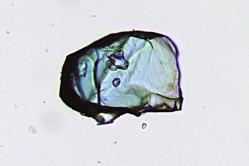
Bavsiite has been approved by the Commission on New Minerals, Nomenclature and Classification of the International Mineralogical Association (IMA2014-019, Bojar and Walter, Reference Bojar and Walter2014) and the type specimen is hosted in the mineralogical collection of the Universalmuseum Joanneum Graz, Austria (catalogue number 85.282). Bavsiite and suzukiite, which have nearly the same chemical composition, were found in a white, marble-like skarn-type rock. Bavsiite (Fig. 1) occurs as sky-blue, platy crystals up to 0.3 mm across with a thickness of a few hundredth of a millimetre and flattened on {001}. Bavsiite is transparent with a vitreous lustre and is non-fluorescent. The streak is white, the Mohs hardness is ~4 and cleavage parallel to (001) is good. Density was not measured directly due to frequent inclusions in bavsiite. Directly associated minerals are alstonite, baryte, celsian, diopside, fresnoite, mica, suzukiite, walstromite, witherite and minerals of the cerchiaraite group.
Transmission-light microscopy image of a single crystal of bavsiite (cat. nr. 85.282). Field of view is 0.2 mm. Photograph: F. Walter.

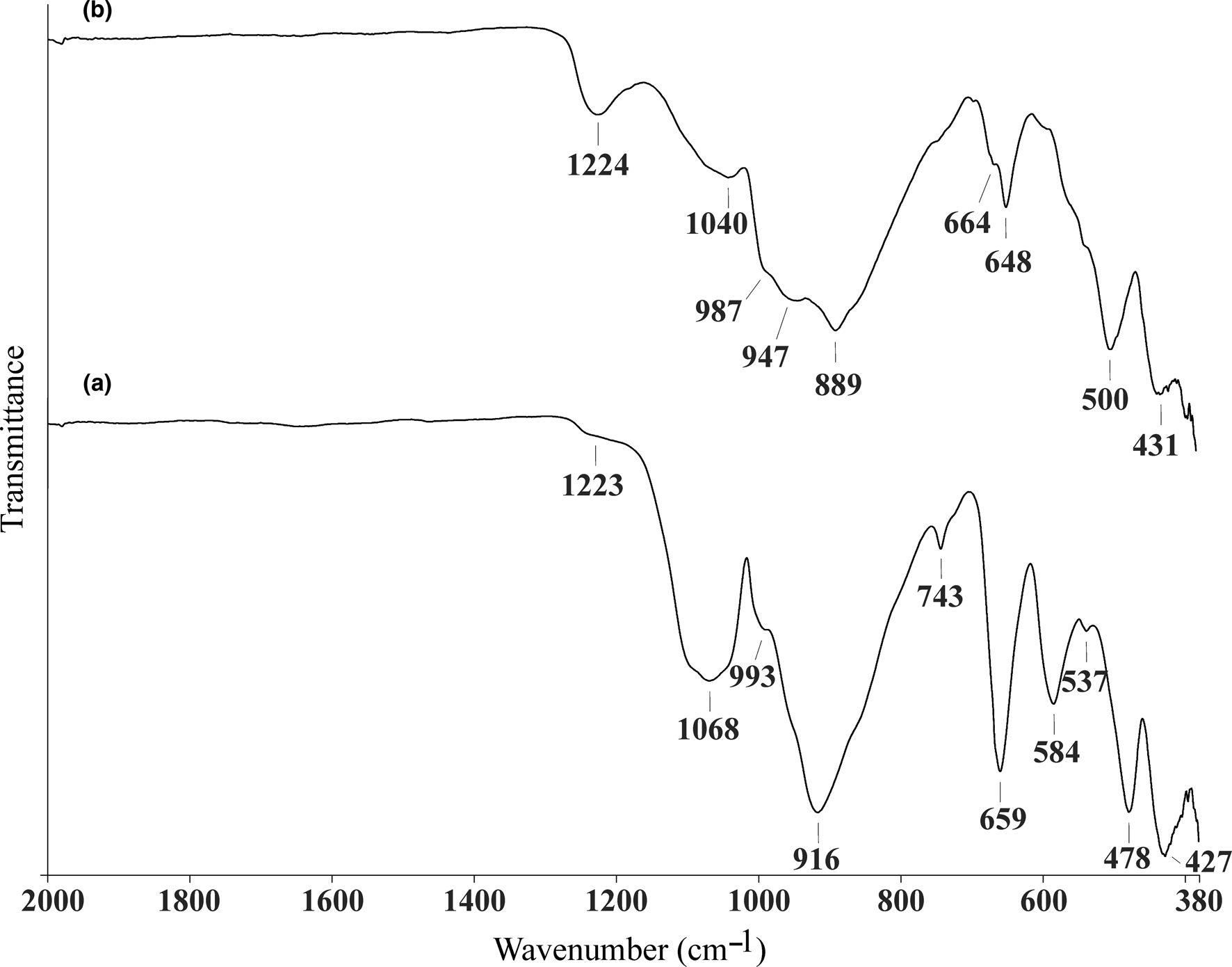
The Fourier-transform infrared spectrum was recorded using a Perkin Elmer Spectrum 100 spectrophotometer (ATR diamond cell, resolution 4 cm–1, 20 scans). The infrared spectra for bavsiite and suzukiite are overlaid for comparison and given in Fig. 2. The following bands for bavsiite were observed (in cm–1; s: strong, m: medium, w: weak, sh: shoulder): 1223w, 1068s, 993sh, 916s, 743w, 659s, 584m, 537w, 478s and 427m. The bands in the region from 1200 to 900 cm–1 are related to the stretching of Si–O in the SiO4 tetrahedron and V–O of the VO5 pyramid. The region of 740 to 400 cm–1 is related to the bending modes of the SiO4 and VO5 structural elements.
The Fourier-transform infrared ATR spectra of bavsiite (a) and of suzukiite (b).

Bavsiite is optical uniaxial (+); its optical constants measured with 589 nm on a spindle stage are ω = 1.725(3) and ε = 1.750(3), the dispersion is weak with r > v. Pleochriosm for ω is medium blue and for ɛ, pale yellow.
Composition
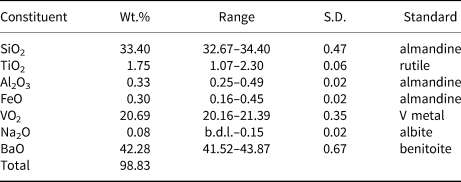
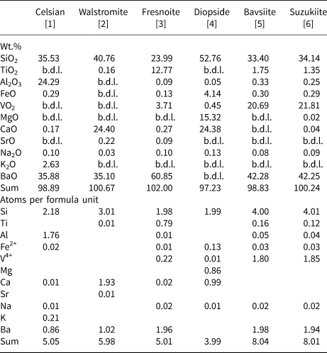
Electron microbeam wavelength dispersive analyses were performed on a Jeol 6610 LV electron microscope equipped with an OXFORD 50 mm2 energy dispersive and an OXFORD INCA WAVE spectrometer operated at an accelerating voltage of 20 kV and a beam current of 10 nA. Data reduction was done with the PAP routine (Pouchou and Pichoir, Reference Pouchou, Pichoir, Heinrich and Newbury1991) implemented in the INCA software of Oxford Instruments. Results of the analyses of bavsiite and minerals of its paragenesis are given in Tables 1 and 2. The empirical formula of bavsiite on the basis of 14 oxygen atoms is: Na0.02Ba1.98Ti0.16Fe2+0.03V4+1.80Al0.05Si4.00O14. The simplified chemical formula is Ba2V2Si4O14 which requires BaO 43.02, VO2 23.27 and SiO2 33.72 wt.%. The elements Ca, Mg, Mn and Sr were also measured, but found to be below the detection limits. The chemical composition of suzukiite and its empirical formula is very close to that of bavsiite: Na0.02Ba1.94Ti0.12Fe2+0.03V4+1.85Al0.04Si4.01O14.
Representative compositions from electron microprobe wavelength dispersive analyses of bavsiite. Average of 10 spot analyses from five crystal fragments.

S.D. – standard deviation; b.d.l. – below detection limit.
Representative electron microprobe results from wavelength-dispersive spectroscopy analyses and formula units for bavsiite and minerals of its paragenesis*.

* [1] celsian (on the basis of 8 oxygen atoms); [2] walstromite (9 oxygens); [3] fresnoite (8 oxygens); [4] diopside (6 oxygens); [5] bavsiite (14 oxygens, mean value of 10 analyses of 5 crystal fragments); and [6] suzukiite (14 oxygens, mean value of 5 analyses of one crystal fragment),
b.d.l. – below detection limit.
Associated feldspars are very low in Ca and high in Ba with higher Ba content in their cores. Fresnoite analyses (Na0.02Ca0.02Ba0.96Fe2+0.01V4+0.22Ti0.79Al0.01Si1.98O8) yielded considerable V4+ (tetravalence from stoichiometric considerations) values.
Slightly low analytical sums are caused most probably by a varying probe current.
Powder X-ray diffraction
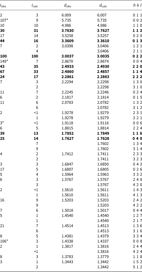
The powder X-ray diffraction data (Table 3) for bavsiite were obtained at 293 K using a Bruker-AXS D8 diffractometer with CuKα radiation. To minimise the background, the sample was placed on a low-background silicon wafer; an internal standard was not used. Unit-cell parameters, derived from whole pattern fitting (Pawley method) with fundamental parameters using TOPAS 4.2 software (Bruker-AXS, 2009) are: a = 7.051(1) Å, c = 11.470(1) Å and V = 570.2(1) Å3, which are in agreement with those obtained from the single-crystal study (see below). Due to the cleavage of bavsiite parallel to (001) and due to the sample preparation, the intensities for (002), (004), (006) and (008) of the observed pattern are too high in comparison to the (00l) intensities of the simulated powder pattern, calculated with data from the single-crystal structure refinement (Table 3). In the powder pattern of bavsiite impurities of feldspar, suzukiite and mica are visible (Fig. 3a). For comparison, the simulated powder patterns of bavsiite calculated with parameters of the single-crystal structure refinement at 100 K (Fig. 3b) and of β-BaVSi2O7 (Fig. 3c) calculated with parameters of the structure refinement from Liu and Greedan (Reference Liu and Greedan1994) are overlaid. The powder patterns confirm that bavsiite and β-BaVSi2O7 have the same crystal structure but are polymorphic to suzukiite (BaVSi2O7, orthorhombic) shown by overlaying the experimental powder pattern of suzukiite crystals from the type locality of bavsiite (Fig. 3d).
The powder X-ray patterns (CuKα, 40 kV and 40 mA) of: (a) bavsiite, measured with impurities of feldspar (Or), suzukiite (Su) and mica (Ph) – note the preferred orientation effects on the (00l) reflections in the experimental pattern of bavsiite; (b) calculated powder pattern of bavsiite with data from structure refinement; (c) calculated powder pattern of β-BaVSi2O7 with parameters of the structure refinement from Liu and Greedan (Reference Liu and Greedan1994); and (d) suzukiite, BaVSi2O7, experimental pattern with crystals from the type locality of bavsiite.

Powder X-ray diffraction data (d in Å) for bavsiite.

I obs and d obs from measurement at 293 K (Bruker D8 diffractometer, CuKα radiation); d calc calculated with profile fitting using TOPAS 4.2 and a = 7.051(1) Å, c = 11.470(1) Å; I calc calculated with atomic parameters from single-crystal structure refinement of bavsiite.
*I obs due to preferred orientation of the cleavage plane (001). Scaling is based on I obs (022) = 100.
The strongest lines are indicated in bold.
Single-crystal X-ray structure refinement
A pale blue platy crystal of bavsiite (Fig. 1) was used for X-ray single-crystal data collection on a Bruker AXS Smart Apex CCD diffractometer equipped with graphite-monochromatised MoKa radiation (0.71073 Å). The data were recorded with the program SMART, integrated with the program SAINT up to θmax = 26.3°, and corrected for absorption using the program SADABS (Bruker, Reference Bruker2008). The analyses of the intensity data indicate the two possible space groups I4/m and I4/mmm. The merged intensities for I4/m result in R int = 0.033, σ = 0.015 and for I4/mmm in R int = 0.062, σ = 0.018. For bavsiite we adopted as a starting point the space group I4/m and the atomic positions as reported for the structural identic synthetic phase β-BaVSi2O7 by Liu and Greedan (Reference Liu and Greedan1994). The small amounts of titanium and iron in the empirical formula of bavsiite (Table 2) were summarised with full occupancy of vanadium at the V site and all other sites were expected to be fully occupied by silicon, barium and oxygen, respectively. Least-squares structure refinement using anisotropic displacement parameters for all atoms was carried out with the program SHELXL-2013 (Sheldrick, Reference Sheldrick2015). It converged at satisfactory R values (R 1 = 0.0279) but the second parameter in the weighting scheme was unusually large (17.98). Refinement with space group I4/mmm gave nearly the same values for atomic positions and anisotropic displacement parameters but with slightly higher R values (R 1 = 0.0323) and also an unusually large second weighting parameter (24.32). These results are an indication of a twinning effect in the crystal structure of bavsiite. No splitting of reflections in the data collection was observed, thus a twinning by merohedry is predicted. In Laue group 4/m a mirror m parallel to the fourfold axis acts as a twin element and the apparent Laue group 4/mmm results. The refinement with space group I4/m using the twinning law 010/100/00 $\bar{1}$ confirmed the merohedral twinning effect by lowering R 1 to 0.0159 and the presence of twin domains with proportions 0.29 to 0.71. In addition, the weighting scheme shows acceptable values (WGHT 0.0179, 2.33) and a smooth difference-Fourier map shows that absorption effects are well corrected.
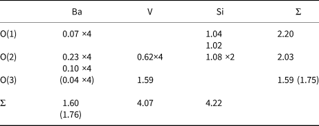
Details on data collection, crystallographic data and refinement are given in Table 4. Refined atomic coordinates and displacement parameters are given in Table 5, and selected interatomic distances and angles are listed in Table 6. Bond valences (Table 7) were calculated using the equation of Brown and Altermatt (Reference Brown and Altermatt1985) with the parameters of Gagné and Hawthorne (Reference Gagné and Hawthorne2015). The crystallographic information files have been deposited with the Principal Editor of Mineralogical Magazine and are available as Supplementary material (see below).
Crystal parameters, data collection and structure refinement details for bavsiite.

R 1 = Σ||F o|–|F c||/Σ|F o|, wR 2 = [Σ(w(F o2–F c2)2)/Σ(w(F o2)2)]½
w = 1/[σ2(F o2)+(0.0179P)2+2.33P], P=(Max(F o2,0) + 2F c2)/3
Atomic coordinates, equivalent isotropic and anisotropic displacement parameters (Å2) for bavsiite.

U eq is defined as one third of the trace of the orthogonalised U ij tensor.
The anisotropic displacement factor exponent takes the form: –2π2 [h 2a*2U 11+…+2hka*b*U 12]
Selected bond distances (Å) and angles (°) in bavsiite.

Bond-valence sums in valence units (vu) for bavsiite.

Bonds for O(1): 2 × Si + 2 × Ba; O(2): 1× Si + 1 × V + 2 × Ba; O(3): 1 × V + (4 × Ba)
Description of the crystal structure
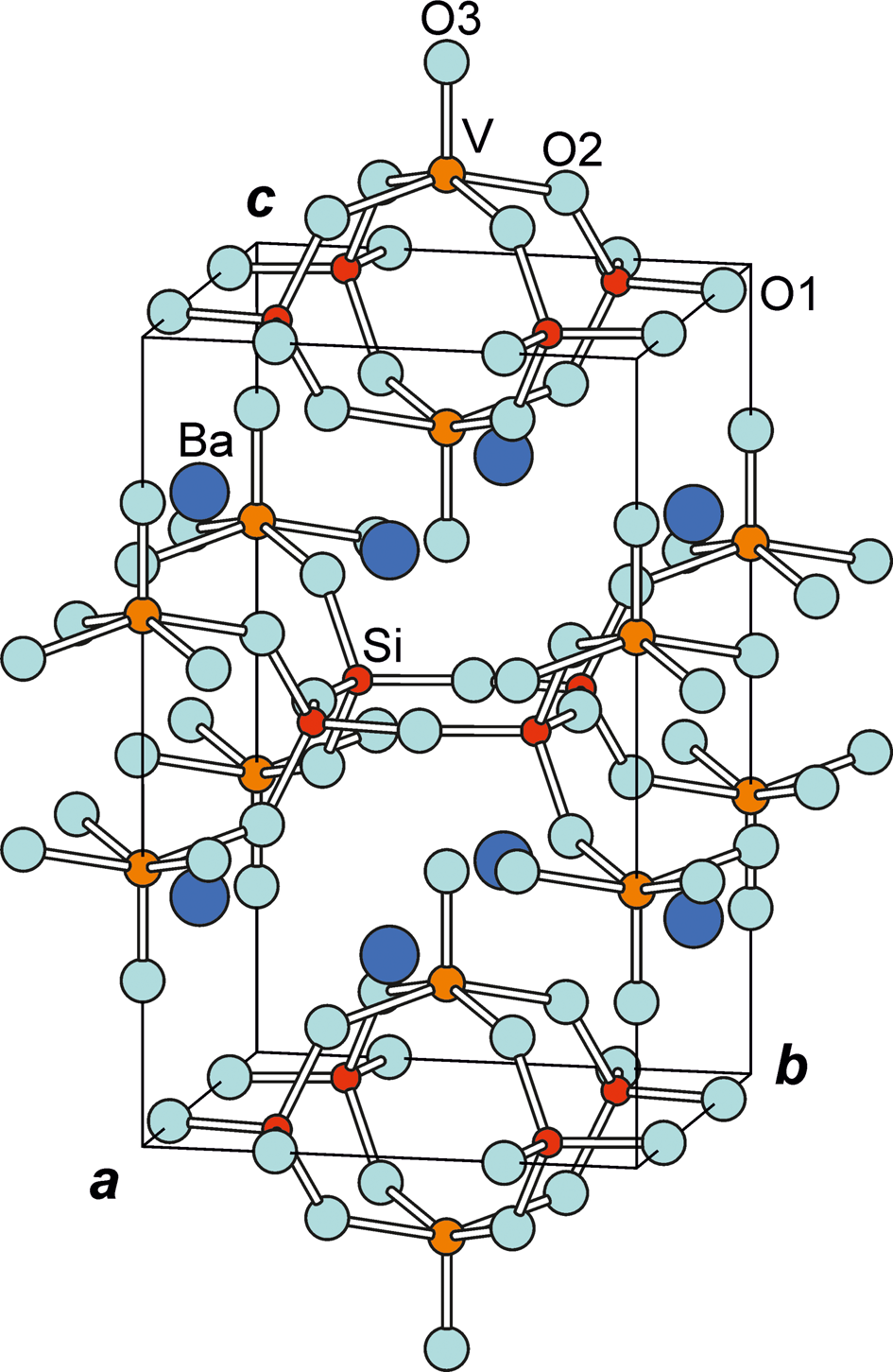
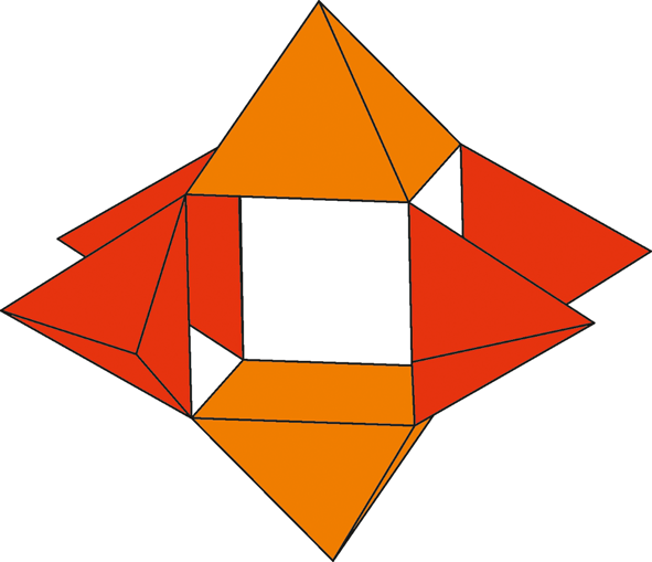
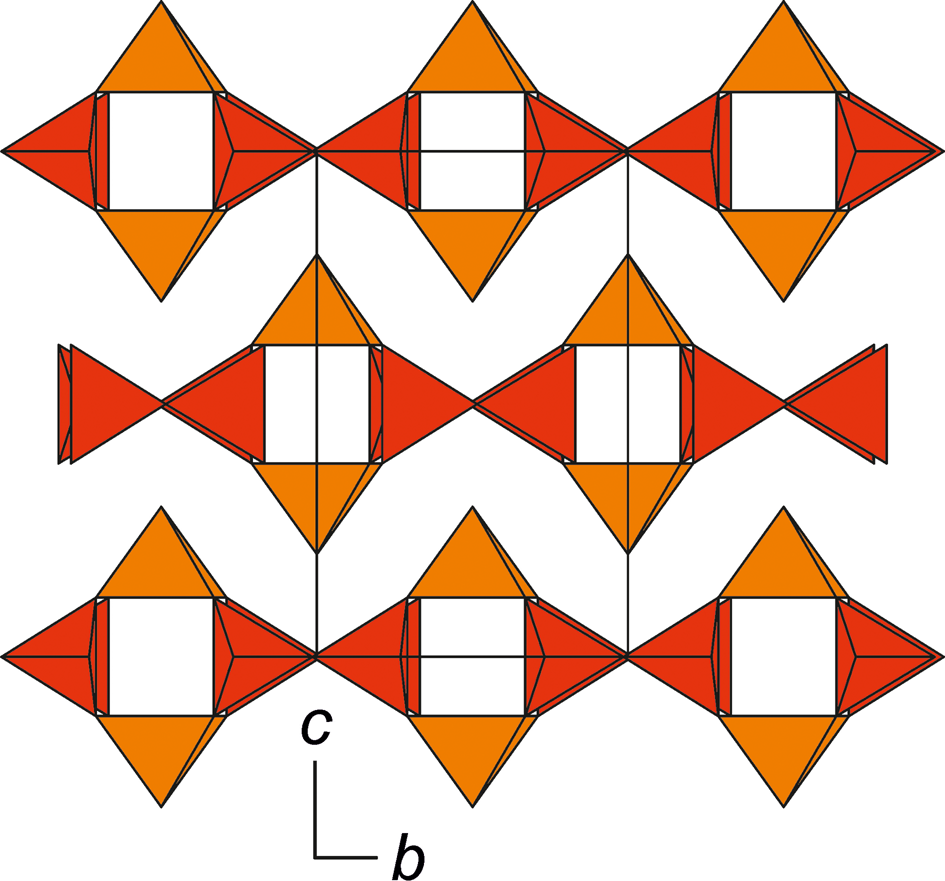
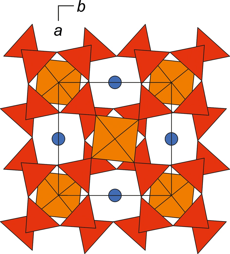
Bavsiite, Ba2V2O2[Si4O12], crystallises in a tetragonal unit cell of space group I4/m with one of the oxygen atoms in a general position whereas all other sites are located at special positions (Table 5). The structure can be considered as consisting of unbranched single [Si4O12]8– rings connected by [VO5]6– square pyramids (Fig. 4), or of cage–like [Si4V2O18]12– clusters built by four [SiO4]4– tetrahedra and two [VO5]6– square pyramids (Fig. 5). These clusters are cross-linked to form a pseudo-two-dimensional network (2D) parallel to (001) containing empty channels along the a axis as shown in Fig. 6. There is no connection between the [VO5]6– square pyramids by themselves and the V–V distance of 3.580 Å sets an upper limit for the channel height (Fig. 6). The 2D networks are held together by Ba2+ ions located in channels parallel to the c axis (Fig. 7).
Inclined projection of the crystal structure of bavsiite. Ba–O bonds are omitted for clarity.

The cage-like [Si4V2O18]12– cluster is a basic structural unit of bavsiite. The [SiO4] tetrahedra are coloured red and [VO5] square pyramids brown.

The cross-linked clusters form a pseudo-two-dimensional network parallel to (001) containing empty channels along the a axis. Ba atoms are omitted for clarity.

The crystal structure of bavsiite projected along [001] with Ba atoms (blue circles) in the channels along c.

The Si–O bond length ranges from 1.594(3) to 1.616(5) Å, average 1.603 Å, and the O–Si–O angles from 106.6(2)° to 114.6(2)°, average 109.5°, indicating a nearly regular tetrahedron. The bond angles of the unbranched [Si4O12]8– ring are 112.1(3)° for O(1)–Si–O(1)’ and 157.9(5)° (= 270° – 112.1°) for Si–O(1)–Si’.
Vanadium occurs in a distorted square pyramidal oxygen environment with equatorial bond length of 1.951(3) Å and a very short axial one of 1.608(5) Å. The displacement of the central V atom off the basal square plane towards the axial oxygen O(3) is indicated by the O(2)–V–O(3) basal to axial oxygen angle of 103.29(8)° and is also visible in Fig. 4.
Ions of Ba2+ occupy channels along c and can be considered as 12-coordinated with Ba–O bond lengths ranging from 2.812(4) to 3.295(2) Å and an average of 3.085 Å. A further but very long Ba–O distance of 3.562(5) Å would sum to a 16-coordination with an even longer average length of 3.205 Å.
Discussion
In the unbranched single [Si4O12]8– ring the anisotropic parameter U 33 of the bridging oxygen O(1) is significantly higher than U 11 and U 22 generated by the strong bonding to two Si (Si–O(1) = 1.609(5) Å and Si–O(1)’ = 1.616(5) Å) in the plane Si–O(1)–Si’ parallel to (001) and loosely bonding to two Ba ions (Ba–O(1) = 3.295(2) Å) with c-axis–Ba–O(1) = 29.75° with respect to U 33. The silicon tetrahedron in bavsiite is more regular than in β-BaVSi2O7 [Si–O(1) = 1.622(5) Å, Si–O(1)’ = 1.597(5) Å and 2× Si–O(2) = 1.603 Å] with Si–O averages of 1.603 Å (bavsiite) and 1.606 Å, respectively. The bond-valence sum in bavsiite for the tetrahedrally coordinated O(1) is high (V = 2.20) whereas the value for O(2) is nearly ideal (V = 2.03). O(2) connects the silicon tetrahedron with the [VO5]6– square pyramid and has an additional two bonds with Ba. Such square pyramids are important structure elements in similar silicates such as β-BaVSi2O7, (Liu and Greedan, Reference Liu and Greedan1994), suzukiite, BaVSi2O7 (Ito et al., Reference Ito, Matsubara, Yokoyama, Momma, Miyawaki, Nakai and Kato2014), haradaite, SrVSi2O7 (Basso et al., Reference Basso, Lucchetti, Palenzona and Zefiro1995) and synthetic SrVSi2O7 (Berger and Range, Reference Berger and Range1996). In the [VO5]6– square pyramid two types of bonds are visible by their V–O distances: (1) a very short vanadyl bond (shorter than 1.74 Å) and (2) longer equatorial bonds in (V4+On) polyhedra in the range 1.90 to 2.12 Å (Schindler et al., Reference Schindler, Hawthorne and Baur2000). In bavsiite the vanadyl bond V–(O3) = 1.608(5) Å is longer than in β–BaVSi2O7, suzukiite and haradaite but nearly equal as in synthetic SrVSi2O7: 1.592(8) Å, 1.593(5) Å, 1.593(9) Å and 1.61(1) Å, respectively. The equatorial bonds in bavsiite and β–BaVSi2O7, (1.951(3) Å and 1.936(5) Å, respectively) are shorter than in suzukiite, haradaite and synthetic SrVSi2O7 (1.9986(19) Å, 1.990(4) Å and 1.980(5) Å, respectively). In bavsiite the bond-valence sum 1.59 valence units (vu) for O(3) reflects the double bond nature of the vanadyl bond. The nearest further cation bond is to Ba with 3.562(5) Å and would increase the valence value to 1.75 vu for O(3).
The tight bonds in the [Si4V2O18]12– cluster result in high valence values for Si and V (4.22 and 4.07 vu, respectively) whereas the Ba atoms in the channels along the c axis are only loosely bonded with valence sums of 1.60 vu (12-coordinated) or 1.76 vu (16-coordinated). The good cleavage in bavsiite parallel to (001), also visible by preferred orientation reflexes (00l) in the powder X-ray pattern (Fig. 3), is mainly due to the lengthened Ba–O bonds, which hold together the 2D-network.
According to Strunz and Nickel (Reference Strunz and Nickel2001) bavsiite is a cyclosilicate with 4-membered single rings (9.CF.). Bavsiite is isotypic to synthetic β-BaVSi2O7 (Liu and Greedan, Reference Liu and Greedan1994) and polymorphic to suzukiite, BaVSi2O7. In the crystal structure of suzukiite (Ito et al., Reference Ito, Matsubara, Yokoyama, Momma, Miyawaki, Nakai and Kato2014) SiO4 tetrahedra are connected to corrugated Si4O12 chains along the c axis and VO5 square pyramids link the chains to form layers parallel to the ac plane, which are connected by barium atoms.
Supplementary material
To view supplementary material for this article, please visit https://doi.org/10.1180/mgm.2019.59
Acknowledgements
We are grateful to Stuart Mills for editorial handling and helpful comments and Gunnar Färber, Germany, who provided the bavsiite-bearing samples. The comments of Peter Leverett and two anonymous reviewers improved the manuscript substantially.