Sensing, stimulating, and growing neurons in a dish
In neuroscience, cells or tissues are often used under in vitro or ex vivo conditions as model systems to study the brain. Neurobiologists are able to separate cells or a piece of intact tissue from specific regions of the brain and grow them in a dish (cell culture).Reference Banker and Goslin1 Depending on the neurobiological field in question, the biological model systems are highly specialized. To study the signaling mechanism in learning and memory, brain slices or embryonic neurons isolated from the areas implicated in these processes, such as the hippocampus, are used.Reference Bliss and Collingridge2 To understand the regulation of circadian rhythm in the mammalian brain, a group of neurons from the biological clock region known as the suprachiasmatic nucleus are used.Reference Welsh, Logothetis, Meister and Reppert3 In case of applied neuroscience research such as the restoration of vision, retinal slices or retinal ganglion cells are used as a tissue model.Reference Sekirnjak, Hottowy, Sher, Dabrowski, Litke and Chichilnisky4 Establishing working biological model systems is an important driving force in neurobiology, and interdisciplinary approaches are empowering advances in neuroscience and engineering.
There has been a strong need for innovative technologies to improve existing in vitro neural tissue culture platforms. The major issues for current platforms are the quality of measured neural signals, spatial precision of neural stimulation, and more accurately reproducing in vivo cellular environments in a dish. Innovations can emerge from three different aspects, namely sensing, stimulating, and growing neurons, and materials play a key role in providing these innovations. Novel sensor materials are used to measure neural signals, while high-performance transducers are used to modulate or control neural activities. Combining both sensing and stimulation would provide better experimental solutions for the study of neural circuits by allowing reliable access to individual neurons. Biomimetic materials can provide live neurons with in vivo-like environments in a dish to obtain more realistic neural tissue models.
There are several in vitro neural interface technologies that have been actively developed in the past decades for the purpose of design and analysis of cultured neurons and circuits. One of the most well-known technologies is a planar-type microelectrode array (MEA) (see the Ordonez et al. article in this issue), which is a cell culture platform with electrical interfaces for recording and stimulating neurons.Reference Taketani and Baudry5 In this platform, sensors (microelectrodes) are embedded in a surface where neurons can grow, and multiple neurons can be electrically interfaced at the same time so that network studies are possible. There are a few commercially available MEA platforms for electrophysiological research: 60- or 64-channel MEAs with metal sensors are most widely used, while ultrahigh density MEAs with thousands of active complementary metal-oxide-semiconductor sensors are also available.Reference Jones, Livi, Lewandowska, Fiscella, Roscic and Hierlemann6 Another example is the development of engineered cell culture platforms for the manipulation of neuronal growth under precisely controlled conditions. Microfluidic interfaces with separated culture compartments were developed to control extracellular fluidic environments and guide the outgrowth of new neuron branches known as neurites.Reference Taylor and Jeon7 A surface micropatterning method that prints surface-bound micropatterns of proteins on cell culture devices made it possible to control neuronal adhesion between the surface and cell membrane, the direction of a long, slender neurite known as an axon, and the form of neural networks.Reference Wheeler and Brewer8
These technologies can serve as powerful and innovative tools for experimental neuroscientists. In the pharmaceutical industry, an efficient drug-screening assay system can replace costly and labor-intensive screening processes for the nervous system. In biotechnology, cell-based biosensors can be applied to delicate tasks such as environmental monitoring or biological warfare. Furthermore, a practical in vitro model system can be built by manipulating neuronal cultures to understand clinical neural engineering problems in brain-machine interfacing or neural tissue engineering. For more details on advances in these technologies, the reader is referred to recent review articles.Reference Taketani and Baudry5, Reference Wheeler and Brewer8, Reference Nam and Wheeler9
Biological properties: Cytotoxicity or biocompatibility
Cytotoxicity and biocompatibility are key issues for materials that are used in in vitro neural interfaces, and their toxicity and biological effects on neuronal growth should be carefully assayed before further progress is made. Sensors, stimulators, or cell culture substrates that make close physical contact with live neurons might have adverse effects on the cultivation of neurons by interfering with cellular characteristics such as neuronal adhesion, cell body (soma) size, length of neuron branches (neurites), and the degree of branching. Common cytotoxicity assays are MTT [3-(4,5-dimethylthiazol-2-yl)-2,5-diphenyltetrazolium bromide] assays, and calcein AM. An MTT assay measures enzymatic activity, while the calcein AM assay stains live cells with green fluorescence signals. In these assays, glass coverslips or plastic tissue culture dishes coated with cell-adhesive coatings (poly-D-lysine, laminin, or poly-D-lysine/laminin mixtures) are used as positive control groups, and neuronal growth is compared quantitatively to evaluate toxicity effects. Since neurons are different from other cell types in terms of their unique compartmentalized structures (dendrites, soma, and axon), morphological analysis is also routinely performed. Morphological features such as the length of neurites, size of the cell body, and number of neurites are used for the characterization of early neuronal growth.
Long-term biocompatibility is another important concern when neurons are cultivated for a few weeks. To study neural circuits in vitro, neurons are often cultured for such periods so that biological maturation processes such as axon/dendrite branching, an increase of electrical excitability, and synapse formation can occur. During long-term cultivation, materials that comprise neuronal growth environments can perturb the maturing neurons by physical contact or biochemical reactions. Eventually, long-term chronic exposure would lead to unintended biological damage to neuronal health and maturation processes. In the case of polymeric materials, unreacted monomers should be completely removed to ensure that there is no cytotoxicity. For example, polydimethylsiloxane—a well-known soft-lithographic material for cell culture devices—had to be treated with organic solvents to extract uncross-linked oligomers before usage.Reference Millet, Stewart, Sweedler, Nuzzo and Gillette10 In the case of nanomaterials, nanoparticles containing heavy metals such as cadmium lead to programmed cell death, less metabolic activity, and abnormal cellular morphology, which implies their potential cytotoxicity for long-term usages.Reference Kotov, Winter, Clements, Jan, Timko, Campidelli, Pathak, Mazzatenta, Lieber, Prato, Bellamkonda, Silva, Kam, Patolsky and Ballerini11
Electrical properties
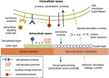
Electrical interfaces are used to sense neural signals (action potentials) or to stimulate neurons to evoke electrical activity. Both tasks require the transduction of electrical charges through an electrode-electrolyte interface, and this interface is characterized by its electrical impedance and the charge injection limit of the interfacial double-layer capacitances (Figure 1).
For sensing neural signals, membrane currents (ionic and capacitive components) and the resulting voltage drops across the tissue are the main signal sources. When an action potential is generated by a neuron, both ionic (Na+ and K+) and capacitive currents are formed near the membrane, and electrical voltage drops are generated along the current path. Although the original action potentials are typically 100 mV when measured across the membrane (intracellular recording), voltage drops measured from outside the membrane are 1000 times smaller (extracellular recording). The amplitude of the extracellular signals ranges from tens to hundreds of microvolts, which is extremely small from an electrical measurement viewpoint. To measure such weak signals, sensors (electrodes) should have high sensitivity and low intrinsic thermal noise. Both requirements can be met by choosing materials that can provide low impedances (high interfacial capacitances) in the frequency range of action potentials (300–10,000 Hz). Electrical impedance for 1 kHz sinusoids is typically used as a figure of merit of recording electrodes.
Electrical stimulation for neural responses requires the passage of electrical charges through the electrode-electrolyte interface. Both voltage and current pulses can be used to transport charge, and the interfacial double-layer capacitance determines the efficiency of the charge injection. The charge transfer from electrode to electrolyte takes place by a faradaic or non-faradaic (or capacitive) mechanism. The current through non-faradaic charge transfer refers to capacitive current that flows through the double-layer capacitor, while faradaic currents accompany various electrochemical reactions on the electrode surface. Ideally, the charge injection process should specifically target the electrical activity of the neuron. In reality, it is likely to trigger surface redox reactions such as the reduction and oxidation of water, metal oxide formation, valency changes within an oxide, corrosion, or gas evolution. As some of the reactions can directly or indirectly damage neurons by altering extracellular or intracellular environments, the maximum amount of charge that an electrode can store without generating harmful chemical byproducts is an important figure of merit for the stimulating electrode.Reference Merrill, Bikson and Jefferys12
Noble metals
Gold and platinum are common materials used for neural electrodes. They have been preferred to other metals, as they are known to be inert under biological environments. To reduce the interfacial impedances for a given micrometer-sized electrode, its surface area can be increased by electrochemical deposition of these metals. A highly porous electrode surface can be fabricated by depositing platinum in this way, which forms a dendritic structure (platinum black) when a current density of 100 nA/μm2 is applied, increasing electrode sensitivity by a factor of 1000.Reference Novak and Wheeler13 The electrode impedance was reduced from 1.4 MΩ to 10 kΩ for a 10 μm-diameter microelectrode using this method. Although its mechanical properties are not suitable for long-term use or implantation, platinum black has served as a standard material that enhances the sensitivity of neural sensors for extracellular recordings. Careful engineering of electrodeposition conditions has resulted in a stable and robust nanoporous platinum structure, with an impedance of 2.4 kΩ and a charge injection limit of 3 mC/cm2 for a 45 μm-diameter microelectrode.Reference Park, Song, Boo and Chung14
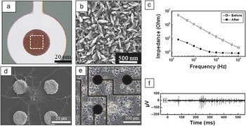
Gold nanostructures have also been fabricated on microelectrodes to enhance their sensitivity by increasing the surface area. Nano-sized thin gold pieces (“nanoflakes”) were formed on micro-sized flat gold electrodes by electrodeposition with an impedance of 11 kΩ for a 50 μm-diameter microelectrode (Figure 2),Reference Kim, Kang, Nam and Choi15 and gold nanopillar electrodes were fabricated using template-based methods.Reference Bruggemann, Wolfrum, Maybeck, Mourzina, Jansen and Offenhausser16 Other than engineering the surface area of the electrode, three-dimensional (3D) protruding microelectrodes were introduced by the Spira group at the Hebrew University of Jerusalem for the purpose of directly measuring action potentials (in-cell recording).Reference Hai, Shappir and Spira17 They used the electroplating process to fabricate arrays of rounded protruding gold microelectrodes (“gold spines”) that could be engulfed by neurons and coated their surface with polypeptides that bind to the cell membrane receptor proteins. In this way, they induced tight sealing between the cell membrane and electrode surface. They were able to record extraordinarily large neural signals on the order of a few mV, as well as being able to detect subthreshold membrane activity that is generally not measurable by extracellular recording techniques.
Carbon nanotubes
Carbon nanotubes (CNTs) have been shown to have several distinct effects on electrical interfaces. First, CNTs have been shown to dramatically enhance the electrode performance by decreasing the electrode impedance and increasing the charge injection limit.Reference Gabay, Ben-David, Kalifa, Sorkin, Abrams, Ben-Jacob and Hanein18–Reference Wang, Fishman, Dai and Harris20 Reported impedance values were 1∼10 kΩ, and the charge injection limit was 3 mC/cm2.Reference Gabay, Ben-David, Kalifa, Sorkin, Abrams, Ben-Jacob and Hanein18, Reference Wang, Fishman, Dai and Harris20 Second, it was reported that larger neural signals were recorded from CNT electrodes. In addition to the effect of decreased impedance, it was found that some CNTs produced a stronger mechanical bond with cells. Neurons showed higher affinity to chemical vapor deposited CNTs than silicon nitride surfaces by spontaneously migrating and adhering to pristine CNT surfaces for two weeks.Reference Gabay, Ben-David, Kalifa, Sorkin, Abrams, Ben-Jacob and Hanein18 This high physical affinity to the electrode surface was correlated with the recording of large signals on the order of a few hundred microvolts, or 10 times larger than conventional microelectrodes.Reference Shoval, Adams, David-Pur, Shein, Hanein and Sernagor21 Third, neurons cultured on CNTs had altered electrophysiological properties due to the penetration of the cell membrane by CNT fibers.Reference Cellot, Cilia, Cipollone, Rancic, Sucapane, Giordani, Gambazzi, Markram, Grandolfo, Scaini, Gelain, Casalis, Prato, Giugliano and Ballerini22, Reference Cellot, Toma, Varley, Laishram, Villari, Quintana, Cipollone, Prato and Ballerini23 This example implies that nanoscale interfacing could significantly alter the intrinsic cell properties by electrically short-circuiting neuronal compartments.
Silicon nanowires
Silicon nanowires have been implemented with either field-effect transistor (FET)-type active sensors or metal nanoelectrodes for in vitro neural interface platforms. The Lieber group at Harvard University has reported silicon nanowire field-effect transistors (NW-FETs) arrays with active sensing areas ranging from 0.01∼0.06 μm2. They showed that simultaneous recordings from the axon and dendrites of a single neuron were possible with NW-FET arrays.Reference Patolsky, Timko, Yu, Fang, Greytak, Zheng and Lieber24 In addition, neural signals ranging from 0.3–3 mV were recorded from neural circuits in brain slices using a NW-FET array (device sensitivity 31.1 nS/mV) on a flexible transparent substrate.Reference Qing, Pal, Tian, Duan, Timko, Cohen-Karni, Murthy and Lieber25 These works showed that the NW-FET is a promising sensor that can provide sufficient sensitivity with unprecedented spatial selectivity (∼0.06 μm2). In the case of metal nanoelectrodes, the Park group at Harvard University developed a vertical silicon nanowire array with individual nanowires 150 nm thick and 3 μm high.Reference Robinson, Jorgolli, Shalek, Yoon, Gertner and Park26 Several nanowires were grouped (2 μm spacing) to cover a single neuron, and an array of grouped nanowires were used to interrogate a small neural circuit. A high signal-to-noise ratio on the order of 100 was achieved with the measured signal amplitude on the order of a few mV.
Conductive polymers
Conductive polymers have emerged as versatile multifunctional neural interface materials for recording and stimulation. The conductivity of conductive polymers such as polypyrrole (PPy) and poly(3,4-ethylenedioxythiophene) (PEDOT) can be controlled by an electrochemical polymerization process, and this can be exploited to lower the interfacial impedance for neural sensors.Reference Green, Lovell, Wallace and Poole-Warren27 A typical electrode impedance range achieved by a conductive polymer was 10–100 kΩ for 1250 μm2 electrodes. The electrical polymerization process was also shown to be effective for entrapping biological materials such as nerve growth factors or peptides on electrode surfaces.Reference Cui and Martin28, Reference Kim, Richardson-Burns, Hendricks, Sequera and Martin29 Recently, nanotubes were fabricated with PPy or PEDOT, and microelectrodes with these nanotubes had lower impedances and higher charge injection limits than those with thin-film PPy or PEDOT. It was also observed that the cultured neurons had longer neurites on nanotubes.Reference Abidian, Corey, Kipke and Martin30
Insulation materials for electrical interfaces
Long-lasting electrical insulators are needed to ensure good signal quality by minimizing any parasitic signal paths that shunt the neuronal signals. The degradation of the insulation layer would lead to a decrease in shunt resistance, which in turn reduces the magnitude of measured signals and degrades signal-to-noise ratios. Unlike probe-type in vivo neural interfaces, insulating materials in vitro serve as culture substrates, and the biocompatibility of the insulator becomes the foremost issue for successful experiments. Several materials have been reliably used for insulation and growing neurons. A low-residual stress silicon nitride film that is compatible with a standard semiconductor fabrication process is a good choice due to its pinhole-free high film quality,Reference Borkholder, Bao, Maluf, Perl and Kovacs31 and sandwich structures made of SiO2/Si3N4/SiO2 have been used to ensure the film quality.Reference Ecken, Ingebrandt, Krause, Richter, Hara and Offenhäusser32 Polymers such as polysiloxane,Reference Gross, Wen and Lin33 SU-8,Reference Heuschkel, Fejtl, Raggenbass, Bertrand and Renaud34 parylene,Reference Erickson, Tooker, Tai and Pine35 or polyimideReference Novak and Wheeler13 have been used as non-cytotoxic insulation materials in vitro.
Optical properties
Optical properties are a particularly important consideration for the investigation of neural interfaces using optical imaging or stimulation. In the case of optical imaging, transparency and fluorescent properties of the materials used should be considered. When materials are optically transparent, transmitted light microscopy under phase-contrast or differential interference contrast (DIC) mode can be easily used to characterize live neural cells and tissues in vitro. These are common cell imaging methods in biological laboratories. Neurons grown on opaque surfaces need to be analyzed by fluorescence or reflected DIC microscopy. Fluorescence microscopy has become a powerful and routine technique to investigate subcellular events owing to the development of DNA recombinant methods that allow various fluorescent proteins to be expressed in a cell. To take full advantage of this imaging technique, fluorescent characteristics of the material such as auto-fluorescence or quenching effects are important factors to be taken into account for successful experiments.
Optoelectronic properties can be utilized in sensing and stimulating neurons (see the Chernov et al. article in this issue). As action potentials are electrical signals in nature, it is possible to detect a modulated neural signal by light. For example, planar gold nanoparticle arrays were designed to measure action potentials indirectly through optical signals originating from surface plasmon resonance effects.Reference Zhang, Atay and Nurmikko36 This label-free optical detection was possible due to electrostatic field-induced plasmon modulation in gold nanoparticles. Meanwhile, photons can also induce electrical currents to stimulate neurons. Localized electrical currents were generated by applying a laser pulse to a reverse biased boron doped Si wafer, and these transient currents then passed through nearby neurons to induce neuronal activity.Reference Starovoytov, Choi and Seung37 This method suggests that lead-free optical stimulation is possible for cultured neuronal networks. Recently, neurons that have light-sensitive ion channels or pumps on their cell membranes have been produced by genetic engineering. This makes it possible to excite or inhibit neural activity by irradiation with different wavelengths. This technique, which is called optogenetics, has become an emerging technology that offers a highly efficient way of stimulating a subset of neurons with similar biological properties in a heterogeneous neural circuit.Reference Fenno, Yizhar and Deisseroth38 Other than sensing and stimulation, amorphous silicon was used as a photoconductor to fabricate ultrahigh-density neural sensors with 3,600 electrodes.Reference Bucher, Brunner, Leibrock, Schubert and Nisch39
Chemical properties
Neural interfaces can provide biological signals or cues to control neuronal growth, which can be realized by functionalizing the interface with biological substances. To this end, it is important to investigate chemical properties of the material with the following questions in mind: Is the surface hydrophilic or hydrophobic? What kind of functional groups are present on the surface? Is it possible to functionalize the surface to have certain chemical properties that will be favorable for cell adhesion and growth? What kind of protein linking chemistry or immobilization schemes should be used?
Cell adhesive versus repulsive materials
To grow neurons in cell culture conditions, cell adhesion and neurite growth should be facilitated by the culture substrates (Figure 1). The surface of neural interfaces can be converted into neuron-friendly surfaces by using various biomimetic or biological materials. First, positively charged polymers can promote neuronal adhesion and induce neuronal outgrowth. As the outside of a cell membrane is known to be negatively charged, cell membranes will be attracted to positively charged surfaces. Synthetic polymers such as poly-D-lysine, polyelectrolyte (e.g., polyethyleneimine), or aminosilane are used as coating materials to form positively charged surfaces. Second, proteins can be directly used to promote cell adhesion and growth. This involves extracellular matrix (ECM) proteins (e.g., laminin, fibronectin, and collagen) (see the Chen and Allen article in this issue), NgCAM (neuron-glial cell adhesion molecule), N-cadherin, and RGD (arginine-glycine-aspartic acid) peptides. These proteins stimulate the receptor proteins on cell membranes and trigger biochemical signaling mechanisms inside the cell to recruit cell adhesion and neurite outgrowth. In some cases, a mixture of synthetic polymers and ECMs is used.
In vitro neural interfaces for sensing, stimulating, and growing neurons. To grow neurons on the substrate, substrate surfaces are functionalized with cell adhesive or repulsive materials by a linker layer. Neurons adhere to the surface by recognizing cell-adhesive proteins or electrostatic binding forces. Cell-repulsive polymers inhibit neuronal adhesion and growth by resisting protein adsorptions. In the case of electrical interfaces, microelectrodes can measure minute extracellular action potentials generated by membrane ion channels. Electrical charges are delivered to neuronal membrane by injecting currents through the double layer and by electrochemical reactions. Chemical stimulation is also possible by delivering extracellular signaling molecules using microfluidic interfaces.

Gold nanoflake microelectrodes for neural recording and stimulation. (a) Optical image of electrochemically deposited gold nanoflake microelectrode. (b) Scanning electron microscopy (SEM) image of nanoflake structures. (c) AC frequency sweep before and after the deposition of gold nanoflakes. (d) SEM image of fixed rat hippocampal neurons on four nanoflake microelectrodes. (e) Phase-contrast image of live hippocampal neuronal networks on a gold nanoflake microelectrode array. (f) High-quality neural recording of extracellular action potentials 16 days after the cell culture. Reproduced with permission from Reference 15. ©2010, IOP Science.

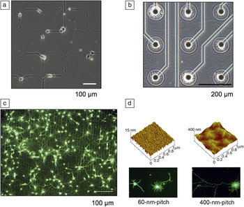
In contrast to promoting neuronal adhesion, it is also useful to develop methods to repel cell attachment and growth. Consequently, several types of cell-repulsive materials that inhibit neuronal adhesion and growth have been used. These materials are especially useful for micropatterning neuronal cultures.Reference Sekirnjak, Hottowy, Sher, Dabrowski, Litke and Chichilnisky4 The most well-known such material is polyethyleneglycol, which has been shown to be effective in maintaining patterned neuronal outgrowth for a long-term period (2∼4 weeks) by resisting protein adsorption (Figure 3a).Reference Branch, Wheeler, Brewer and Leckband40, Reference Kang, Kang, Lee, Choi and Nam41 Agarose hydrogel layers inhibited the attachment of neurons and glial cells (non-neuronal cells in nervous tissue) (Figure 3b).Reference Kang, Lee, Lee and Nam42 Hydrophobic surfaces made of fluorosilane have also been shown to be effective in inhibiting neuronal attachments.Reference Wyart, Ybert, Bourdieu, Herr, Prinz and Chatenay43
Interfacing various growth substrates with live neurons. (a) Optical image of confined neuronal growth on a surface initiated film of cell-repulsive poly(oligo[ethylene glycol] methacrylate) on glass. Poly-L-lysine patterns were printed by microcontact printing for selective neuronal adhesion. Reproduced with permission from Reference 41. ©2010, Wiley. (b) Agarose-hydrogel micropatterning for interfacing microelectrodes with micro-neural circuits (optical image). Reproduced with permission from Reference 42. ©2009, Royal Society of Chemistry. (c) Fluorescence image of calcein AM staining of cultured neurons on a patterned carbon nanotube substrate at seven days after cell seeding. Reproduced with permission from Reference 50. ©2010, IOP Science. (d) Nanotopographic surfaces (anodized aluminum oxide) with 400 nm pitch (right) induced accelerated neurite outgrowth more than those with 60 nm pitch (left). Atomic force microscopy images (top) show bare AAO surfaces, and fluorescence microscopy images (bottom) show neurons cultured on each surface. Reproduced with permission from Reference 53. ©2010, Wiley.

Biofunctionalization materials
In order to immobilize biological materials or proteins on neural interfaces with defined chemical bonds, interfaces need to be converted into chemically active (or functional) surfaces. Self-assembled monolayers with known functional groups have been used for this activation. If the original surface is rich in hydroxyl groups (-OH), organosilane chemistry can be readily applied to functionalize the surface. Organosilanes with various functional groups such as amines (-NH3), carboxyls (-COOH), thiols (-SH), or epoxides are commercially available. If metal surfaces (e.g., gold or platinum) are available, surfaces can be treated with alkanethiol derivatives. These groups can be linked with the amine group of proteins through appropriate cross-linkers.Reference Branch, Wheeler, Brewer and Leckband40, Reference Kang, Kang, Lee, Choi and Nam41 Recently, polydopamine, a protein inspired by adhesion proteins in mussels, has been introduced as a universal coating material for biological interfaces. It can adhere to any surface and serves as a chemical platform for further chemical reactions. Polydopamine has been successfully applied to functionalize various neural interface materials, including gold, platinum, indium tin oxide, glass, silicon nitride, and liquid crystal polymers.Reference Kang, Choi and Nam44
Mechanical properties
Recent progress in cell biology has revealed that mechanical properties of extracellular environments modulate many cellular behaviors such as cell adhesion, growth, proliferation, and death.Reference Wang, Tytell and Ingber45 In the case of interfacing with neural tissues in vitro, mechanical stimulation can be embedded in the interface by choosing materials with different roughness, topography, elasticity, or stiffness.Reference Hoffman-Kim, Mitchel and Bellamkonda46 Moreover, for the design of 3D neural interfaces, it is important to select a material that can be micro-machined into 3D structures. Materials that are compatible with conventional microfabrication techniques or 3D printing are chosen for this purpose.
Nanomaterials for topographical cues
Nanomaterials that can provide nanoscale topographical features have become popular materials, as culture substrates with nanoscale features have significantly different effects on neuronal adhesion and growth. Vertical nanowires were shown to selectively promote neuronal adhesion and guide neurite outgrowth even without any cell-adhesive coating.Reference Shalek, Robinson, Karp, Lee, Ahn, Yoon, Sutton, Jorgolli, Gertner, Gujral, MacBeath, Yang and Park47, Reference Hallstrom, Martensson, Prinz, Gustavsson, Montelius, Samuelson and Kanje48 Micropatterned islands of tangled CNTs also showed similar spontaneous adhesion and growth effects.Reference Sorkin, Greenbaum, David-Pur, Anava, Ayali, Ben-Jacob and Hanein49 Guided neuronal growth was reported on various nanotopographical substrates made of nanomesh CNTs (Figure 3c),Reference Jang, Namgung, Hong and Nam50 electrospun nanofibers,Reference Gertz, Leach, Birrell, Martin, Feldman and Corey51 or patterned polyurethane acrylate.Reference Jang, Kim, Feltrin, Jeon, Suh and Pertz52 Anodized aluminum oxide with periodic nanotopography (pitch 400 nm) showed accelerated neuronal growth at the early stage of in vitro neural developments, while a smaller pitch (60 nm) did not affect the neuronal growth (Figure 3d).Reference Cho, Kang, Kang, Jang, Nam and Choi53
Materials for 3D neural interfaces
As the brain is mainly a soft tissue and 3D in nature, culture substrates that provide such environments would be useful for constructing more realistic tissue models. 3D neural interfaces require a mechanical construct (scaffold) for neuronal growth and electrical or fluidic access in 3D spaces. A photoresist, SU-8, which is a base material for various microelectromechanical system devices, has been used to construct a 3D neural interface with fluidic and electrical connections.Reference Rowe, Almasri, Lee, Fogleman, Brewer, Nam, Wheeler, Vukasinovic, Glezer and Frazier54, Reference Musick, Khatami and Wheeler55 A photopolymerizable hydrogel (poly[2-hydroxyethylmethacrylate]) has been used to construct 3D scaffolds by direct-write assembly.Reference Hanson Shepherd, Parker, Shepherd, Gillette, Lewis and Nuzzo56 A mixture of silica beads and neurons were assembled using the colloidal technique to construct 3D neuronal circuits in vitro.Reference Pautot, Wyart and Isacoff57 Hydrogels such as collagen or fibrin gel and cell suspension were directly printed on a substrate to construct 3D neural tissues using a 3D printing technique.Reference Boland, Xu, Damon and Cui58
Summary
This article overviewed different aspects of materials that need to be considered in vitro neural interface technology. Physical, chemical, and biological properties were considered for sensing, stimulating, and growing neurons in vitro. In the near term, materials that can provide solutions for more precise, sensitive, and non-invasive recording and stimulation will be sought. Also, newly discovered materials such as graphene will be actively investigated for their interfacial properties (cytotoxicity, impedance, charge injection limits, and measurable signals). In the longer term, alternative sensing and stimulation interfaces, such as the emerging use of optical interfaces, will be pursued to replace traditional electrical interfaces. With advances in these technologies, we will be able to manipulate neural circuits more reliably and precisely and obtain information that is relevant to the functioning of the brain. Future in vitro neurotechnologies will allow us to develop powerful and novel diagnostic tools for detecting neural diseases in clinic and to discover new drugs.
Acknowledgments
The author acknowledges support from the National Research Foundation (NRF) (No. 2009–0080081) and the Brain Research Center of the 21st Century Frontier Research Program funded by the Korean government (MEST).