Introduction
In the last decade, the development of advanced robotic limbs has revolutionized the simple hook prosthesis powered by body movements currently used by many amputees, with multi-fingered lightweight devices capable of up to 22 degrees of freedom and populated with force and movement sensors that more closely resemble the operation of the human hand.Reference Carpaneto, Micera, Zaccone, Vecchi and Dario1, Reference Cipriani, Controzzi and Carrozza2 Despite such progress in prosthetic devices, providing the user of the robotic limb with natural control and feel remains a formidable challenge.
Most robotic prostheses are controlled through command signals recorded from remaining limb muscles (electromyogenic signals) that are measured by surface electrodes, amplified, and converted to mechanical signals.Reference Uellendahl3, Reference Farrell and Weir4 In order to provide a more natural control and feel of the prosthetic hand, Kuiken and collaborators developed a surgical strategy that transfers residual arm nerves to pectoral muscles (a method known as targeted muscle reinnervation), from where electromyogenic activity can be recorded and used to control the robotic hand.Reference Kuiken, Li, Lock, Lipschutz, Miller, Stubblefield and Englehart5 In addition, recent data from animal studies indicate that transferred sensory nerves can also reinnervate the skin over the pectoral area, suggesting that this technique can provide sensory feedback to amputees and contribute toward the development of closed-loop control systems.Reference Marasco and Kuiken6 Alternatively, prosthetic devices can be controlled by signals obtained through microelectrode arrays implanted in the brain premotor cortex from where neural activity is known to correlate with intention of movement.Reference Chapin, Moxon, Markowitz and Nicolelis7, Reference Schwartz, Cui, Weber and Moran8 In human volunteers, this so-called brain-machine interface (BMI) technology has proven successful in allowing paralyzed patients to control a hand prosthesis by thought,Reference Simeral, Kim, Black, Donoghue and Hochberg9, Reference Hochberg, Serruya, Friehs, Mukand, Saleh, Caplan, Branner, Chen, Penn and Donoghue10 and recent animal studies suggest that sensation might be conveyed to the user via electrical micro-stimulation of the sensory cortex.Reference Fitzsimmons, Drake, Hanson, Lebedev and Nicolelis11 However, the invasive craniotomy surgery required for cortical interface placement and the lack of modulation from other areas in the brain or spinal cord needed for context-dependent control and sensory discrimination limit their use.Reference Brown, Koerber and Millecchia12, Reference Lee, Carvell and Simons13
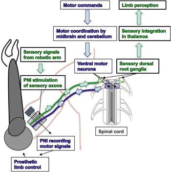
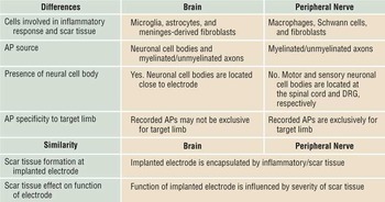
In contrast to BMI, placing electrodes in the peripheral nerves of amputees offers a readily accessible portal to the bidirectional flow of information between the nervous system of the user and smart robotic prosthetic devices. Motor commands initiated by the user are transmitted from the motor cortex in the brain to the midbrain, cerebellum, and ventral motor neurons in the spinal cord for integration and coordination, ultimately traveling through the peripheral nerve where they can be recorded by peripheral nerve interfaces (PNIs) (Figure 1). Conversely, sensations from the limb such as motion, pressure, and temperature can be detected by sensors populating the robotic device and potentially can be conveyed to the user by direct stimulation of specific sensory fibers at the PNI. Such information will follow the natural sensory pathway, which is normally carried from the peripheral nerves to the spinal cord via neurons located on both sides of the vertebral column known as the dorsal root ganglia (DRG), from there to the thalamus for integration, and ultimately reaching the sensory cortex in the brain for limb awareness and movement coordination (Figure 1). Indeed, interfacing neuronal cell bodies in the DRG or their axons in the peripheral nerve provides arguably the most appropriate site for conveying sensory feedback information to the amputee, as it ensures that such sensory information is modulated by the spinal cord and regions of the brain prior to being transferred to the somatosensory cortex for more accurate limb awareness. Despite the fact that both BMIs and PNIs exchange information between the nervous system and prosthetic devices, tissue response by the brain or the peripheral nerves to the electrodes varies significantly. A major difference that affects the sensitivity of the recorded signals lies in the fact that BMI electrodes primarily record from neuron cell bodies and dendrites (branched projections of the cell body) in the cerebral cortex, while PNIs record and stimulate only the long neuronal projections (neurites) known as axons. A comparison between BMI and PNI is summarized in Table I. While comprehensive reviews are available on the BMIs,Reference Schwartz, Cui, Weber and Moran8, Reference Grill, Norman and Bellamkonda14–Reference Gilja, Chestek, Diester, Henderson, Deisseroth and Shenoy16 here we focus our review on materials designed to increase the sensitivity, selectivity, reliability, and safety of the PNI electrodes.
Bi-directional signal processing through peripheral nerve interfaces (PNIs). Motor commands initiated in the brain follow a downstream pathway to the ventral spinal cord from where they activate motor signals in the peripheral nerve that can be recorded to control a robotic limb. Sensory signals from the robotic arm follow an opposite route and can be conveyed to the user through selective electrical stimulation of the amputated nerve. The sensory stimulus travels to the spinal cord, the thalamus, and sensory cortex in the brain for limb awareness and feedback control.Reference Brown, Koerber and Millecchia12, Reference Lee, Carvell and Simons13

Comparison of brain and peripheral nerve-machine interface characteristics.

AP, action potential; DRG, dorsal root ganglia.
PNI designs: Selectivity versus invasiveness
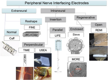
Peripheral nerve interfacing has been accomplished either through extraneural electrodes, such as cuff electrodes, or intraneural electrodes (i.e., intrafascicular, intraneuronal, or regenerative electrodes). The most current electrodes in each type are included.
Cuff electrodes
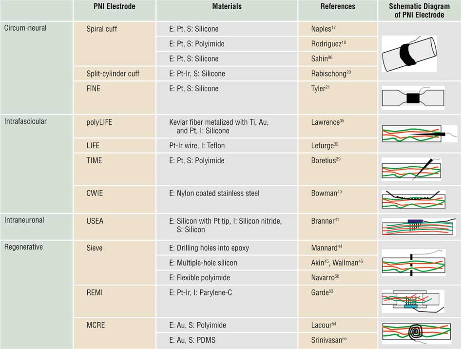
Cuff electrodes are implanted internal to the skin and outside the nerve (i.e., extraneural) and consist of two or more metal electrodes embedded on an insulating tubular substrateReference Naples, Mortimer, Scheiner and Sweeney17–Reference Tyler and Durand21 (Figure 2). Platinum and platinum-iridium are normally used; either stimulating the enclosed nerve or to record the compound action potential. The insulating tubular substrate completely encircles the nerve with flexible and self-sizing silicone or polyimide cuff materials that avoid stretching or compression damage of the enclosed nerve (Table II). Cuff electrodes have an established history for long-term recording of neural activity—up to 63 weeks after implantation in human volunteers.Reference Fisher, Tyler, Anderson and Triolo22 While this approach is successful for nerve recording and stimulation, the activity it records is a weighted average from the entire nerve rather than units from single axons. The signals are relatively small and from a limited number of electrodes, and complications such as nerve inflammation and tissue damage curtail the reproducibility of recordings.Reference Vince, Brelen, Delbeke and Colin23, Reference Wodlinger and Durand24 Despite these limitations, cuff electrodes are minimally invasive and provide multiple opportunities for correct placement. Progression in cuff electrode design continues, as recent “FLAT” electrodes are now capable of more selective stimulation of individual nerve bundles, or fascicles, within the nerve through careful electrode pairing within the cuff (Figure 2).Reference Durand, Park and Wodlinger25
Classification of the different types of peripheral nerve interface electrodes. Extraneural electrodes are either normal (cuff)Reference Naples, Mortimer, Scheiner and Sweeney17–Reference Tyler and Durand21 or deform the nerve (FLAT),Reference Durand, Park and Wodlinger25 while minimally invasive. Penetrating electrodes are inserted either perpendicular (USEA)Reference Branner and Normann41, Reference Branner, Stein, Fernandez, Aoyagi and Normann42 or parallel (TIME)Reference Boretius, Badia, Pascual-Font, Schuettler, Navarro, Yoshida and Stieglitz39 to the nerve axons. Those that require complete transection of the nerve have either enclosed electrodes (Sieve)Reference Edell, Churchill and Gourley43–Reference Wallman, Zhang, Laurell and Danielsen46 or open architecture (REMI).Reference Garde, Keefer, Botterman, Galvan and Romero53

Various types of peripheral nerve interface electrodes.

Red lines (sensory axons) and green lines (motor axons); E, electrode; I, insulator; S, substrate; FINE, flat-interface nerve electrode; LIFE, longitudinally implanted intrafascicular electrode; TIME, transverse intrafascicular multichannel electrode; CWIE, coiled wire intrafascicular electrode; USEA, Utah slanted electrode array; REMI, regenerative multielectrode array; MCRE, micro-/multichannel roll electrode; PDMS, poly(dimethylsiloxane).
Electrodes in the DRG
Insertion electrodes have been placed directly into the sensory DRG where they can record directly from the neuron cell bodies, thus obtaining larger signals, and where both recording and stimulation have been achieved.Reference Weber, Stein, Everaert and Prochazka26, Reference Gaunt, Hokanson and Weber27 However, these sensory ganglia lie along the vertebral column and are covered by bone, thus access for implantation requires invasive surgery. Furthermore, since the DRG only contains sensory neurons, no information from motor intent can be recorded at this site.
Intraneural (LIFE, TIME, CWIE, and USEA) electrodes
Intraneural electrodes are placed directly in the peripheral nerves, and their close proximity to the individual axons provides increased neuronal selectivity and sensitivity. They are able to access both motor and sensory axons, which allows the electrodes to record command signals for control and to stimulate sensory axons, thus conveying modality-specific sensation to amputees.Reference Dhillon, Lawrence, Hutchinson and Horch28–Reference Rossini, Micera, Benvenuto, Carpaneto, Cavallo, Citi, Cipriani, Denaro, Denaro, Di Pino, Ferreri, Guglielmelli, Hoffmann, Raspopovic, Rigosa, Rossini, Tombini and Dario30 These observations have stimulated the study and development of a number of different intraneural electrodes for PNIs.Reference Navarro, Krueger, Lago, Micera, Stieglitz and Dario31
According to the specific mode of intraneural electrode placement within the nerve tissue, four major electrode designs can be recognized: longitudinally implanted intrafascicular electrodes (LIFE), transverse intrafascicular multi-channel electrodes (TIME), coiled wire intrafascicular electrodes (CWIE), and the Utah slanted electrode array (USEA) (Table II, Figure 2).
The LIFE is a Teflon insulated, bipolar 25 μm diameter platinum-iridium electrode with a platinized 2 cm active tip and an estimated Young’s modulus of 202 GPa. It has been used to record activity from the radial nerve of cats for up to six months.Reference Lefurge, Goodall, Horch, Stensaas and Schoenberg32 Limitations of the LIFE include drift in the population of cells being recorded and a decrease in signal-to-noise ratio over time.Reference Malmstrom, McNaughton and Horch33 More recent versions of the LIFE have been tested but are not commercially available, including the polymer-based LIFE (poly-LIFE) and the the thin-film LIFE (tf-LIFE). The poly-LIFE is a silicone insulated 12 μm diameter Kevlar (poly-paraphenylene terephthalamide) fiber coated with titanium, gold, and platinum over a 1 cm active area.Reference McNaughton and Horch34 This electrode has an average impedance of 14.4 kΩ, signal-to-noise ratio of 3.6, single spike amplitudes as high as 120 μV peak to peak, and background noise of 10–20 μV, with an average of 30 μm encapsulation layer thickness.Reference Malmstrom, McNaughton and Horch33 The poly-LIFE was successfully used in the short term to stimulate severed nerves proximally to the stump of subjects with upper limb amputation, where it elicited graded sensations of touch, joint movement, and position.Reference Dhillon, Lawrence, Hutchinson and Horch28, Reference Lawrence, Dhillon, Jensen, Yoshida and Horch35 In turn, the tf-LIFE consists of a flat ribbon of polyimide 10 μm thick and 50 mm in length with a Young’s modulus of 8.3 GPa, with four 40 μm diameter circular platinized electrode contacts.Reference Lago, Yoshida, Koch and Navarro36 The tf-LIFE has been shown to develop an axon free scar layer, presumably caused by mechanical motion and compression injury induced by the electrode inside the nerve.Reference Lago and Navarro37, Reference Lago, Udina, Ramachandran and Navarro38
The TIME transversally penetrates the peripheral nerve and is designed to selectively activate subsets of axons in different fascicles within the nerve. For these electrodes, platinum electrodes are embedded on a flexible polyimide substrate, and selective stimulation of different fascicles has been demonstrated in short-term implantations.Reference Boretius, Badia, Pascual-Font, Schuettler, Navarro, Yoshida and Stieglitz39
The CWIE is made from nylon coated stainless steel wire, which is wound into helical flexible coils to allow for expansion and contraction within the surrounding nerve tissue. The CWIE has been implanted into rabbit motor nerve fascicles and demonstrated its use in neuromuscular electrical stimulation, but much remains to be done to fully ascertain the capabilities of this electrode design.Reference Bowman and Erickson40
The Utah slanted electrode array (USEA) consists of 100 needle electrodes of varying lengths (0.5 to 1.5 mm with 0.1 mm difference in length between rows of neighboring electrodes) and is designed to access most fascicles within the nerve. The needle electrodes are made from conductive doped silicon with platinum-plated tips that are electrically insulated with silicon nitride. This electrode array is perpendicularly inserted into peripheral nerves using a pneumatic impulse insertion technique. Since these needle electrodes directly contact individual nerve fibers, the USEA has been demonstrated to have more selective nerve stimulation at much lower current intensities compared to conventional cuff electrodes.Reference Branner and Normann41, Reference Branner, Stein, Fernandez, Aoyagi and Normann42
Regenerative (SIEVE, REMI, and MCRE) electrodes
An alternative design to penetrating electrodes was proposed more than 36 years ago based on the innate capacity of peripheral nerves to regrow spontaneously after injury.Reference Edell, Churchill and Gourley43–Reference Wallman, Zhang, Laurell and Danielsen46 In this method, the nerve, which in amputees is already severed and serves no specific function, is re-cut and encouraged to grow through electrodes that can be either ring or needle shaped. Ring electrodes are gold traces around holes in a flat circular “sieve” that is placed in between two ends of the cut nerve. The axons then grow through the ring electrodes, which have both recording and electrical stimulation capabilities. Silicon sieve electrode arrays have been used to obtain neural recordings up to 13 months post-implantation.Reference Bradley, Cao, Akin and Najafi47–Reference Ramachandran, Schuettler, Lago, Doerge, Koch, Navarro, Hoffmann and Stieglitz49 More recently, a polyimide sieve electrode with seven integrated recording-stimulating ring electrodes was developed.Reference Navarro, Calvet, Rodriguez, Stieglitz, Blau, Buti, Valderrama and Meyer50 Unfortunately, when these electrodes are used, neural activity can only be recorded from a fraction of the animals tested, and then only from a low proportion of the electrodes on the sieve, due to compression injury of the regenerated nerve.Reference Lago, Ceballos, Rodriguez, Stieglitz and Navarro51 This in turn is caused by the inability of the ring electrodes to accommodate the increase in fiber diameter over time that normally occurs as axons mature and undergo remyelination (regrowth of the insulating sheath surrounding axons).Reference Castro, Negredo and Avendano52
We recently developed a regenerative multielectrode interface (REMI) that is placed between the transected ends of an end-to-end repaired nerve (Figure 2), and we were successful in obtaining single and multicellular recordings.Reference Garde, Keefer, Botterman, Galvan and Romero53 In the REMI, 18 parylene-C insulated platinum-iridium electrodes are placed in the center of a polyurethane tube, the cut ends of the peripheral nerve are then placed at both ends of the tube, encouraging the nerve to regenerate through the needle-shaped multielectrode array. Since the needle electrodes do not restrict the growth of the nerve, nerve injury by compression does not occur. The REMI allows the recording of action potentials as early as 8 days post-implantation, with high signal-to-noise ratio, and as long as 120 days in some animals, with minimal inflammation at the electrode implantation site.
Also a relatively new design, the microchannel roll electrode (MCRE) records from axons guided to regrow through microchannels with embedded electrodes and aims at maximizing the contact between regenerating axons and the embedded metal electrodes. In a particular format, gold microelectrodes have been patterned on a polyimide substrate and microchannels created using photosensitive polyimide.Reference Lacour, Atta, Fitzgerald, Blamire, Tarte and Fawcett54 In a separate design, gold electrodes have been patterned on a poly(dimethylsiloxane) (PDMS) elastomer, and microchannels were created by placing a SU-8 photoresist on top of the PDMS layer.Reference Srinivasan, Guo and Bellamkonda55 These two-dimensional electrode microchannel arrays are rolled into a three-dimensional channel bundle (i.e., similar to a Swiss roll) designed to fit the transected peripheral nerve at both ends of the roll (Table II).
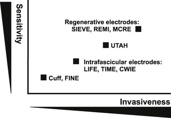
In general, the sensitivity of regenerative electrodes is superior to that of extraneural electrodes, as they are closer to the individual axons, able to record single action potentials, and able to elicit specific sensations. Their implantation requires invasive surgery, however, this might be justified in most amputee cases in which the transected nerve in the limb stump remains functional but serves no other function.
Material designs to enhance electrode safety and sensitivity
While a number of PNIs have been described, they all seem to suffer from limitations similar to those reported for the BMIs, particularly the lack of a reliably safe and sensitive electrode (see Figure 3). Several lines of research currently are directed toward the development of advanced electrode materials that can overcome these limitations. Biocompatible, corrosion-resistive materials for electrical recording and stimulation include gold, titanium, tungsten, platinum, iridium oxide, stainless steel, alloys of these metals, highly doped semiconductors such as silicon, and conductive polymers.Reference Merrill, Bikson and Jefferys56 Most common electrodes are made of platinum, which has charge injection capacities of 300–350 mC/cm2, and iridium oxide, with a capacity of 2–3 mC/cm2, which is safer as it prevents tissue electrolytic damage.Reference Merrill, Bikson and Jefferys56, Reference Grill and Reichert57 It has been recently demonstrated that the performance of extracellular microelectrodes can be improved by surface chemistry modifications, such as with conductive polymers or carbon nanotubes (CNTs). Gold electrodes coated with conducting polymers, such as polypyrrole (PPy) and poly(3,4-ethylenedioxythiophene) (PEDOT), decrease the impedance of the bare electrodes thereby increasing their charge transfer capacity by two and three orders of magnitude, respectively.Reference Jan, Hendricks, Husaini, Richardson-Burns, Sereno, Martin and Kotov58 Microelectrode arrays implanted in rat cortex revealed that PEDOT-coated electrodes have higher signal-to-noise recordings and an injection limit 15 times higher than platinum-iridium electrodes and electroplated iridium oxide electrodes.Reference Venkatraman, Elkabany, Long, Yao and Carmena59
Distribution of the different types of peripheral nerve interface electrodes according to sensitivity and invasiveness. Sensitivity refers to the ability of the electrode to record distinct action potentials from one or few axons that can be used as individual channels for control of the robotic device. Invasiveness relates to damage to the nerve while placing the electrode, from minimal damage by the cuff electrodes, which are placed over the nerve, to maximal damage by the regenerative electrodes that require complete transection of the peripheral nerve for implantation. REMI, regenerative multielectrode interface; MCRE, microchannel roll electrode; LIFE, longitudinally implanted intrafascicular electrodes; TIME, transverse intrafascicular electrodes; CWIE, coiled wire intrafascicular electrodes; FINE, flat-interface nerve electrode.

The strength, electrical conductivity, and high surface area of CNTs make them excellent candidates for interfacing with neural systems. CNTs were shown to offer a permissive substrate for neuron attachment and growth.Reference Mattson, Haddon and Rao60, Reference Galvan-Garcia, Keefer, Yang, Zhang, Fang, Zakhidov, Baughman and Romero61 We recently showed that coating with electroplated CNT/gold composites reduced the impedance of indium-tin oxide and tungsten wire electrodes an average of 23-fold, with a 45-fold increase in charge transfer, and also demonstrated a further increase in charge transfer if the electrodes are coated with a CNT/PPy composite.Reference Keefer, Botterman, Romero, Rossi and Gross62 Similar studies have shown that CNTs combined with polyelectrolyte out perform iridium oxide or PEDOT-coated electrodes.Reference Jan, Hendricks, Husaini, Richardson-Burns, Sereno, Martin and Kotov58 In addition to increasing surface area and conductivity, the enhanced capacitance of CNT coated electrodes might be due to tight contact with the neuron cell membranes, as observed in vitro.Reference Cellot, Toma, Varley, Laishram, Villari, Quintana, Cipollone, Prato and Ballerini63 Despite the dramatic improvement in electrode performance with CNTs, chronic recording or stimulation data are incomplete, and thus the benefit of CNT-coating in PNI remains to be fully investigated.
Preventing signal decay in PNIs
In the peripheral nerves, metal electrodes elicit an immune response characterized by the recruitment of macrophage cells that merge into foreign body giant cells of variable thickness, that eventually separate the electrode from the nerve cells and account for a rapid increase in electrode impedance.Reference Williams, Hippensteel, Dilgen, Shain and Kipke64, Reference Mercanzini, Colin, Bensadoun, Bertsch and Renaud65 In cuff electrodes, the immune response also recruits fibroblasts to the active sites, which form fibrous tissue (fibrosis) and contribute to the limited reproducibility of recordings and variability of stimulation.Reference Vince, Brelen, Delbeke and Colin66 Electrodes such as the USEA produce a larger inflammatory response due to tissue laceration, compression, and vascular injuries that occur during electrode insertion into the peripheral nerve. This immune response is similar to that elicited by electrodes inserted into the brain.Reference Winslow and Tresco67 Tissue damage is further exaggerated in regenerative electrodes, where the nerve is completely transected, and a prolonged repair inflammatory response is elicited.Reference Lago, Yoshida, Koch and Navarro36, Reference Garde, Keefer, Botterman, Galvan and Romero53 This inflammatory response peaks 7–14 days post-implantation, reducing the ability to record from low amplitude signals and contributing toward the progressive reduction in the number of active sites over time.Reference Williams, Hippensteel, Dilgen, Shain and Kipke64, Reference Leung, Biran, Underwood and Tresco68
In the brain, in addition to the initial inflammatory response to cortical electrode insertion, microscale movements of the tethered electrode arrays and electrode-tissue elasticity mismatch progressively exacerbate the chronic foreign body response, creating further damage, including neural and dendritic loss, which eventually results in a neuron “kill” zone around the electrode.Reference Biran, Martin and Tresco69 The fibrotic insulation barrier or scarring that separates the electrode from the neural tissue is accepted as the dominant failure mechanism in most cortical neural interfaces.Reference Williams, Hippensteel, Dilgen, Shain and Kipke64, Reference Leung, Biran, Underwood and Tresco68 However, this response seems to vary within single electrode arrays where a range of impedance values has been observed, even after years of implantation.Reference Simeral, Kim, Black, Donoghue and Hochberg9
The foreign body response is substantially different in the peripheral nerve compared to that in the CNS, and while signal decay and a foreign body response have been clearly documented in the peripheral nerve in both penetrating and regenerative electrodes,Reference Branner, Stein, Fernandez, Aoyagi and Normann42, Reference Garde, Keefer, Botterman, Galvan and Romero53 the specific cellular and molecular response to these electrodes needs to be further evaluated to define the extent to which such a barrier participates in PNI signal decay and to determine whether micromotion in the PNS creates an “axon death” zone.
In addition to material surface modifications, biological coatings were proposed a decade ago to modulate the tissue response to electrodes, following a report in which gold electrodes coated with polypyrrole and laminin peptide promoted the attachment of neural tumor cells in vitro Reference Cui, Lee, Raphael, Wiler, Hetke, Anderson and Martin70 and reduced the early inflammatory response in vivo, albeit ineffective in the long-term.Reference Cui, Wiler, Dzaman, Altschuler and Martin71 Subsequently, it was reported that systemic administration of the anti-inflammatory synthetic glucocorticoid dexamethasone reduced the number of scar forming glial cells (reactive gliosis) around inserted cortical electrodes,Reference Spataro, Dilgen, Retterer, Spence, Isaacson, Turner and Shain72 which in turn motivated the search for localized delivery of such agents to improve electrode recording and stimulation. Indeed, in the brain, dexamethasone-eluting nitrocellulose coated electrodes reduced the reactive gliosis caused by cortical silicon probes,Reference Zhong and Bellamkonda73 and coatings of dexamethasone-encapsulated polypropylene sulfide nanoparticles were also able to reduce the electrode impedance caused by the glial scar by 25% during 46 days in vivo.Reference Mercanzini, Reddy, Velluto, Colin, Maillard, Bensadoun, Hubbell and Renaud74 While dexamethasone in the peripheral nerve has been associated with inducing the expression of regeneration associated genes such as GAP-43,Reference Yao and Kiyama75 the use of anti-inflammatory molecules in PNIs has not been investigated.
Coatings to attract and guide neurons to the electrode interface
Adhesion molecules and nerve growth factors have also been proposed as candidates to enhance neuron-electrode interactions. The extracellular matrix molecules, such as collagen and laminin (see the Chen and Allen article in this issue), are recognized by integrin receptors present in the cell membrane, and polyethyleneimine, chitosan, laminin, fibronectin, and collagen IV are known to increase cell attachment and growth onto tungsten, platinum, gold, iridium, silicon, and thin-film polyimide/platinum LIFE electrodes in vitro.Reference Ignatius, Sawhney, Gupta, Thibadeau, Monteiro and Brown76–Reference Bossi, Benvenuto, Wieringa, Di Pino, Guglielmelli, Boretius, Stieglitz, Navarro and Micera78 In addition, neural specific cell adhesion molecules such as axonin-1 and NgCAM, which are normally expressed in neurons and glia (non-neuronal cells in nervous tissue) during development, can enhance neuron attachment and neurite outgrowth when coated onto a silicon oxide substrate.Reference Sorribas, Braun, Leder, Sonderegger and Tiefenauer79 Furthermore, electrodes coated with the extracellular protein L1 show enhanced neural growth and minimal glial attachment to the electrode compared to those coated with laminin.Reference Azemi, Lagenaur and Cui80 These findings support the use of biological coatings to regulate the neuronal response to the electrodes.
Neurotrophic growth factors coated onto metal electodes are well-known modulators of neuron survival and axonal growth. Specifically, nerve growth factor (NGF) has been shown to attract neurites into electrodes in the brain, extending neural recordings for as long as 15 months following cortical implantation;Reference Kennedy, Mirra and Bakay81 NGF-eluting hydrogels have also been used to coat multielectrode arrays;Reference Winter, Cogan and Rizzo82 and coating electrodes with collagen and brain-derived neurotrophic factor seems to support neuron attachment even after growth factor withdrawal.Reference Winter, Han, Jensen, Cogan and Rizzo83 Due to the reported benefit in the use of conductive polymers and neutrophic factors, some have tried to incorporate NGF with polypyrrole or PEDOT, and while the release of biologically active neurotrophins was confirmed over time, coating of these biomolecules seems to increase electrode impedance.Reference Green, Lovell and Poole-Warren84, Reference Huang, Hu, Lu, Ye, Zhang and Luo85
Despite this promising evidence, much remains to be done to fully demonstrate whether neurotrophin-coating electrodes can attract neurons, reduce electrode impedance, and ultimately improve long-term recordings/stimulation in vivo. In addition to attracting axon growth, conclusive evidence has demonstrated that growth factors can be used to guide neuron growth both in the CNS and PNS.Reference Romero, Rangappa, Garry and Smith86, Reference Hu, Cai, Yang and Smith87 This characteristic opens the possibility of using specific growth factors to biologically control the growth of defined neuron subtypes to desired electrode sites in regenerative PNIs.
Signal specificity: Recording motor intent and stimulating modality-specific sensations
The human hand is populated with an estimated 17,000 touch sensing receptors in the skin that provide information about small slips, skin deformation, and limb position. In sharp contrast, the most advanced hand prostheses rely on a small number (commonly less than five) of vibrotactile and electrotactile sensors for surrogate feedback sensation,Reference Phillips88 and most users operate the prosthetic limbs under visual control. The lack of a natural and intuitive bidirectional interface remains a formidable challenge in the development of advanced prosthetics.Reference Egermann, Kasten and Thomsen89 It has been shown that electrical stimulation of the transected peripheral nerve in amputees allows them to judge and set grip force and joint position in an artificial arm in the absence of visual input.Reference Dhillon and Horch29 However, most amputations at the level of the shoulder and above the elbow have transected radial, medial, and ulnar nerves, which at those levels are composed of multiple fascicles and some degree of mixed motor-sensory axon modalities in each fascicle.Reference Stewart90 In addition, it is well known that motor and sensory axon mixing increases in regenerative peripheral interfaces.Reference Brushart, Mathur, Sood and Koschorke91 Therefore, recording exclusively from motor axons and selectively stimulating specific sensory modalities is an extremely challenging task from a mixed nerve.
Such lack of neuron-type specificity can be seen during electrical stimulation on the peripheral nerve, where it is known that large myelinated axons (i.e., motor and limb position sensory nerves) are depolarized with smaller currents, while smaller diameter neurons (i.e., pain fibers) require larger stimuli.Reference Lertmanorat and Durand92 Thus, when stimulating the small caliber fibers, large-size axons will be stimulated as well, particularly with cuff electrodes. This limitation is partially obviated by the use of intraneural electrode arrays within single fascicles as axon mixing is reduced within single nerve bundles. Indeed, if such electrodes are placed on separate fascicles of nerves attached to the gastrocnemius (calf) muscle in cats, they are able to elicit selective electrical stimulation.Reference Yoshida and Horch93 However, chronically implanted intrafascicular electrodes result in an immune response that includes fibrosis and tissue swelling, utimately resulting in loss of nerve fibers and shifts in activation threshold due to the corresponding increase in electrode impedance.Reference Lefurge, Goodall, Horch, Stensaas and Schoenberg32, Reference Goodall, Lefurge and Horch94
In regenerative sieve multielectrode arrays, only a small fraction of regenerating axons succeed in crossing the sieve electrode, and most of them correspond to sensory and not motor neurons,Reference Castro, Negredo and Avendano52 while both sensory and motor axons are in close electrode proximity in the REMI.Reference Garde, Keefer, Botterman, Galvan and Romero53 However, in sharp contrast to cuff and penetrating electrodes, regenerative peripheral interfaces can exploit growth factors and guidance molecules to control axonal regeneration in a way that modality axons can be potentially guided to separate compartmentalized electrodes. We recently reported supportive evidence to that argument by showing that compartmentalized diffusion delivery of NGF and neurotrophin-3 growth factors in a “Y”-shaped nerve guide preferentially entice the growth of TrkA+ pain fibers and TrkC+ limb position subsets of neurons, respectively.Reference Lotfi, Garde, Chouhan, Bengali and Romero-Ortega95 However, whether this strategy can be translated into selective recording from motor axons and modality-specific sensory stimulation remains to be determined.
Summary
Electrical signals have been recorded from the human brain through microelectrode arrays for a time period of up to 2.5 years, whereas animal experimental recordings from the dorsal root ganglia and peripheral nerve signals have been limited to a few months. While such reports are encouraging, neural recordings are not stable over time and fail to provide long-term safety and reliability, which limits their clinical application. Despite this challenge, our understanding of the mechanisms of failure has been increasing in recent years, and a number of strategies aimed at improving sensitivity have been recently reported. In that light, a number of new electrode material designs have been developed that bear promise in achieving better control at the neuron-electrode interface and, in so doing, increase the possibilities of developing long-lasting, safe, and sensitive peripheral neuron interfaces capable of providing voluntary thought-control of advanced robotic prosthetic limbs with multiple degrees of freedom. Such interfaces will eventually allow the possibility of selectively stimulating sensory-specific modalities, providing feedback information, and conveying a natural feel to the user of such advanced bionic limbs.