
Introduction
Hexagonal boron nitride (h-BN) has long been used in applications requiring dry lubricants. In addition, its excellent electrical resistivity (with a dielectric strength of 80 kV.mm−1), high thermal conductivity (71 W.m−1.K−1), and low thermal expansion (<1 × 10⁻⁶ K−1) have made it a material of choice in industrial processes, such as for supports and cells used in chambers for high-temperature materials growth[Reference Solozhenko, Lazarenko, Petitet and Kanaev1] or for large-scale exothermic reactions.[Reference Tian, Lin, Xu, Wan, Lin and Wang2] Remarkably, a recent body of work overhauls the conventional outlook on h-BN by suggesting that atomic arrangements in individual layers can be engineered to introduce a new generation of versatile catalysts.[Reference Nash, Restrepo, Parra, Giesler, Penabade, Aminpour, Le, Li, Farha and Harper3–Reference Grant, McDermott, Venegas, Burt, Micka, Phivilay, Carrero and Hermans7] This application has long been overlooked, partly due to the high chemical and thermal stability[Reference Solozhenko, Lazarenko, Petitet and Kanaev1] together with the wide band gap of h-BN, estimated to be between 3.6[Reference Fomichev, Rudnev and Nemnonov8] and 7.1 eV[Reference Carpenter and Kirby9]. Instead, immediate applications of h-BN monolayers following the advent of two-dimensional (2D) materials such as graphene and transition metal dichalcogenides, have been in providing atomically thin and flexible insulating layers for nanoelectronics[Reference Akinwande, Petrone and Hone10] and far-ultraviolet sources for optoelectronics.[Reference Watanabe, Taniguchi, Niiyama, Miya and Taniguchi11] At this time, all fields are converging toward the need for a deeper understanding of defects,[Reference Zhong, Bruno, Ethan, Ruitao, Rahul, Humberto, Marcos and Mauricio12–Reference Sajid, Reimers and Ford14] their formation and their effects on 2D materials properties, to reach more prevailing rules for material design in the foreseeable future.
Boron and nitrogen vacancies, together with Stone–Wales (SW) defects have been shown to be the most common defects found in individual h-BN layers,[Reference Nash, Restrepo, Parra, Giesler, Penabade, Aminpour, Le, Li, Farha and Harper3, Reference McDougall, Partridge, Nicholls, Russo and McCulloch15] beside edge defects. In multi-layered materials or heterostructures, stacking becomes another distinction that has to be taken into account.[Reference Henck, Pierucci, Aziza, Silly, Gil, Sirotti, Cassabois and Ouerghi16] All can significantly impact the optical,[Reference Berzina, Korsaks, Trinkler, Sarakovskis, Grube and Bellucci17] electrical and magnetic properties of h-BN.[Reference Sajid, Reimers and Ford14] In addition, doping or impurities in place of the defects have led to interesting properties.[Reference McDougall, Partridge, Nicholls, Russo and McCulloch15] However, characterizing the nature of the defects remains challenging.[Reference McDougall, Partridge, Nicholls, Russo and McCulloch15] Secondary ion mass spectrometry can provide some insight on the carbon and oxygen content but the association with a particular defect is not straightforward.[Reference McDougall, Partridge, Nicholls, Russo and McCulloch15] Nanoscale imaging techniques including scanning probe microscopy and transmission electron microscopy (TEM) have revealed the presence of defects, in particular vacancies, but the effect of the incident beam such as in TEM could itself affect the nature of the defects. X-ray spectroscopies, electron energy loss spectroscopy, and electron-paramagnetic resonance[Reference Sajid, Reimers and Ford14] can also provide complementary information regarding the nature of the defects and the bond energies resulting from the presence of defects or impurities. However, this exercise remains challenging.
Hence, the effect of introducing defects in h-BN to create a new metal-free heterogeneous catalytic candidate taking the form of defect-laden hexagonal boron nitride (dh-BN), should be investigated further. It has now been confirmed that dh-BN can facilitate hydrogenation and olefin bond activation.[Reference Nash, Restrepo, Parra, Giesler, Penabade, Aminpour, Le, Li, Farha and Harper3, Reference Kroes, Fasolino and Katsnelson18] The experimental findings and conceptual predictions have been supplemented by density function theory (DFT) calculations. The results show that the olefin bond will preferentially be activated over four types of defect sites in h-BN: boron substitution for nitrogen (BN), nitrogen (VN) or boron (VB) vacancies, and SW defects.[Reference Nash, Restrepo, Parra, Giesler, Penabade, Aminpour, Le, Li, Farha and Harper3] Subsequent solid-state nuclear magnetic resonance (NMR) experiments identified nitrogen vacancies as the most probable active sites for olefin hydrogenation.[Reference Nash, Restrepo, Parra, Giesler, Penabade, Aminpour, Le, Li, Farha and Harper3]
Ball milling was the treatment used to produce dh-BN in the seminal study by Nash et al.[Reference Nash, Restrepo, Parra, Giesler, Penabade, Aminpour, Le, Li, Farha and Harper3] It is a common top-down approach to produce nanomaterials or to facilitate chemical reactions in the absence of solvents, potentially improving reaction efficiency.[Reference Immohr, Felderhoff, Weidenthaler and Schüth19, Reference Rodriguez, Bruckmann, Rantanen and Bolm20] This has been described for numerous applications including the realization of heterogeneous catalytic CO oxidation over metal oxide catalysts, such as NiO and Cr2O3,[Reference Immohr, Felderhoff, Weidenthaler and Schüth19, Reference Eckert, Felderhoff and Schüth21] and now for the hydrogenation and dehydrogenation on non-metal catalysts.[Reference Nash, Restrepo, Parra, Giesler, Penabade, Aminpour, Le, Li, Farha and Harper3] During ball milling, the flakes are mechanically exfoliated into thinner layers and reduced to smaller flakes through comminution, therefore increasing the surface area of the catalytic material.[Reference Nash, Restrepo, Parra, Giesler, Penabade, Aminpour, Le, Li, Farha and Harper3, Reference Molchanov, Byanov and Goidin22, Reference Zazhigalov, Haber, Stoch, Bogutskaya and Bacherikova23] Furthermore, it has been demonstrated that ball milling can introduce different types of defects, including edge defects and stacking disorder into h-BN flakes.[Reference Ghosh, Mazumdar, Das, Ghatak and Basu24, Reference Huang, Jia, Yasuda and Mori25] The nature and the density of defects may be controlled by varying operating parameters, i.e., milling intensity, duration and media composition. Nash et al.[Reference Nash, Restrepo, Parra, Giesler, Penabade, Aminpour, Le, Li, Farha and Harper3] further demonstrated that dh-BN produced through high energy ball milling can be utilized as a catalyst suitable for the hydrogenation of olefins in bulk-scale chemical experiments. Other processes of industrial relevance for producing defects, such as sintering or plasma etching, have not yet been carefully investigated for this application.
To date, the specific defects created and their role in the reactions have mostly been examined locally through gas chromatography—mass spectrometry analysis, solid-state NMR, and supported by quantitative measurements of binding energies such as with x-ray Photoelectron Spectra and DFT calculations.[Reference Nash, Restrepo, Parra, Giesler, Penabade, Aminpour, Le, Li, Farha and Harper3] For other 2D materials, Raman spectroscopy has been a powerful tool for identifying defects, particularly in graphene due to the disorder related D band.[Reference Graf, Molitor, Ensslin, Stampfer, Jungen, Hierold and Wirtz26] In MoS2, defects have been identified using the LA(M) band at ~227 cm−1. LA(M) originates from the first-order Raman mode due to the LA phonons at the M point in the Brillouin zone.[Reference Mignuzzi, Pollard, Bonini, Brennan, Gilmore, Pimenta, Richards and Roy27, Reference Kang, Paudel, Leuenberger, Tetard and Khondaker28] The apparition of oxidation signs in the form of MoO3 has also been used as an indicator of defect creation.[Reference Kang, Paudel, Leuenberger, Tetard and Khondaker28]
In h-BN, identifying defect-induced properties is more challenging due to the absence of the D band indicative of defects present in graphene.[Reference Li and Chen29] While scanning tunneling microscopy (STM) and TEM can offer high-resolution images of the atomic defects in 2D materials,[Reference Kobayashi, Fukui, Enoki, Kusakabe and Kaburagi30, Reference Hashimoto, Suenaga, Gloter, Urita and Iijima31] the requirement to deposit h-BN on a non-metallic substrate to study its metal-free catalytic properties makes it impractical for STM imaging. TEM is also challenging due to the thickness of the flakes in the powders used for ball-milling (sometimes greater than 50 layers). Thus, identifying defects in dh-BN to establish correlations with its catalytic properties continues to be quite challenging. However, comparing the effect of these defects on the reactivity of the formed dh-BN powders can provide valuable information for process optimization.
Motivated by the potential application of h-BN as a heterogeneous catalyst and the role defects play during the catalytic reactions, the present work investigates the effect of defects introduced in pristine h-BN through ball milling and heat treatment. A comparative study of the microstructure, physical and chemical properties of the dh-BN obtained under the various conditions is carried out to establish some connections with catalytic processes. Lastly, an initial assessment of the catalytic performance of the dh-BN powders is discussed.
Experimental methods
Sample preparation
Ball milling
The pristine h-BN powder was obtained from Saint-Gobain Ceramic Materials (Grade PCTF5). To remove moisture, the powder was dried at 180 °C in vacuum overnight before transferring to a glovebox filled with argon for ball milling. Two types of milling media and vials were used for milling, zirconia (ZrO2, 12.7 mm in diameter, approximately 5.97 g in weight) and tungsten carbide (WC, 12.7 mm in diameter, approximately 16.5 g in weight). For each treatment, 3 g of dry h-BN powder were sealed in a 65 mL milling vial and four milling balls were employed to supply kinetic energy. After sealing the vial, the powder was processed with a mixer/mill (8000 M SPEX CertiPrep). When using the ZrO2 balls, the powders were milled for 30, 60, 90, and 120 min. When using the WC balls, the powder was milled for 120 min. After milling, the dh-BN powder was stored in the glovebox until characterization. Upon transfer for spectroscopy measurements, the powder was sealed in a custom compartment to minimize exposure to air before characterization.
Heat treatment
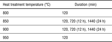
Heat treatments were initially conducted in the air in a Thermolyne Small Benchtop Muffle Furnace (Type 1400, Thermo Scientific) for optimization. The parameters identified for defect creation were used to recreate the treatment in a controlled environment under glovebox conditions filled with argon. In both cases, pristine h-BN flakes from Saint-Gobain Ceramic Materials (Grade PCTF5) were transferred into the furnace, after the temperature was stabilized. At the end of the treatment, the samples were rapidly taken out from the furnace for fast cooling (or quenching) to room temperature. The detailed parameters used for heat treatment are listed in Table I.
Summary of the heat treatment conditions used for optimization of defect creation in pristine h-BN.

Characterization
Scanning electron microscopy (SEM)
The morphology of h-BN and dh-BN powders, including shape and size, were studied using SEM (Zeiss ULTRA-55 FEG).
Raman spectroscopy
Raman spectra were acquired by using a confocal Raman microscope (WITec Alpha 300RA) with a 532 nm laser excitation. Raman spectra were collected with a 50 × objective lens (Zeiss, numerical aperture of 0.7) and a spectrometer with an 1800 lines/mm grating, evaluated to provide a 0.9 cm−1 spectral resolution. For all spectra acquired, settings were maintained at a laser power of 5.7 mW and integration time of 1 s. Raman spectra from pristine h-BN powder and heat-treated h-BN powder prepared outside of the glovebox were acquired in air. Ball-milled and heat treated dh-BN powders prepared in the glovebox were sealed in custom-made chambers with a sapphire window (compatible for Raman measurements) inside the glovebox to prevent exposure to ambient conditions (H2O, O2, N2, and CO2).
Fourier transformed Infrared spectroscopy (FTIR)
FTIR spectra were acquired in the air, immediately after opening the sealed powder container, using a PerkinElmer FTIR spectrometer (Perkin Elmer Corp, Spectrum 100 series) equipped with an attenuated total reflection (ATR) sampling accessory unit with a single reflection diamond/ZnSe crystal plate. Because of the properties of the ATR windows, the wavelength range was restricted to 650–4000 cm−1. The spectral resolution was set to 4 cm−1 with iris aperture of 8.9 mm for all measurements. Four scans were collected and averaged for each powder sample.
Results and discussion
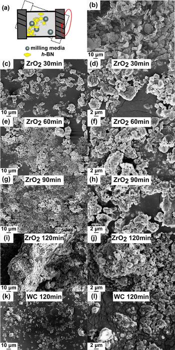
We first investigated the effect of ball milling on the properties of h-BN. The morphology of the BN powders was studied using SEM to understand the effectiveness of ball milling in exfoliation and size reduction of the flakes. The morphology of the pristine h-BN flakes and the resulting dh-BN after ball milling are presented in Figs. 1(b) and 1(c–l), respectively. Higher resolution images are also provided in Fig. S1 (supplementary material). Analysis of the SEM images suggests that before treatment, the flakes were ~6–10 µm in size, in agreement with the product specification sheet (mean value of diameter: ~7 µm). The pristine flakes exhibited irregular edges but smooth surfaces and few folds or dislocations. After 30 min of milling with the ZrO2 media, SEM images reveal flakes with smaller dimensions though large and intact flakes could still be observed [Figs. 1(c) and 1(d)]. This is attributed to the high-energy impact between the flakes, the milling balls and the vial walls during the treatment. As can be seen in Table S1 (supplementary material), after the first 30 min, the large pristine flakes were mostly fragmented with flakes lateral dimensions in the 2–5 µm range. The number of 2–5 µm flakes increased until 60 min of treatment [Figs. 1(e) and 1(f)]. After 90 and 120 min, the dimension of the flakes decreased further, with a large number falling below 2 µm [Figs. 1(g)–(j)]. The SEM images also show that after 120 min of ball milling, the lateral dimensions and thickness of the flakes decreased dramatically to nano-sized flakes and a significant agglomeration of the smaller flakes could be observed, which is in agreement with prior findings by Torii et al.[Reference Torii, Jimura, Hayashi, Kikuchi and Takagaki32] As a result, the percentage of edge defects in the volume of powder in the reactor increased for the first 120 min of milling with ZrO2. Next, we considered the effect of the material of the milling media used on the process of defects generation by using WC milling balls for 120 min. The flakes obtained, as shown in Figs. 1(k) and 1(l), seemed to exhibit different morphology from those obtained at the same time point with ZrO2. This was expected as the higher mass of the WC media should produce 2.76 times the impact force and kinetic energy compared with ZrO2.
(a) Illustration of formation of dh-BN from pristine h-BN through ball milling, (b) SEM image of pristine h-BN flakes, (c–j) SEM image of dh-BN flakes after ball milling with ZrO2 balls for 30 min (c and d), 60 min (e and f), 90 min (g and h), and 120 min (i and j). (k and l) SEM image of the dh-BN obtained after 120 min of treatment with WC milling balls. The SEM images presented in the right column correspond to a zoom of the images in the left column.

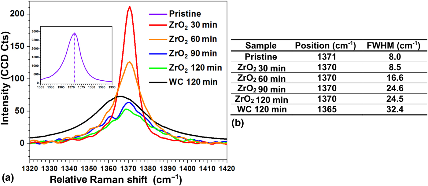
For all dh-BN powders considered in Fig. 1, the Raman and mid-infrared fingerprint of h-BN was affected by the introduction of defects. As both spectroscopies constitute direct measures of the molecular vibrations of chemical bonds in the material, the energy of the B-N lattice vibration may hold some information on the density and types of defects present in the flakes. Raman spectra of the h-BN and dh-BN powders collected for the different durations of milling are presented in Fig. 2. The positions and full width at half maximum (FWHM) of the BN mode at 1371 cm−1 were extracted using a Lorentz fit. A summary of the values is provided in Fig. 2(b). The band at 1371 cm−1 corresponds to the E2 g phonon vibrational mode originating from the in-plane vibration of B and N atoms in opposite directions.[Reference Li and Chen29] After the first 30 min of ball milling, a large decrease of the band intensity and broadening become apparent. Extended ball milling process intensified this effect. This can be attributed to the disorder introduced along the basal plane as the flakes get smaller. A slight red shift of the band from 1371 cm−1 for the pristine h-BN powder to 1370 cm−1 was observed within the first 30 min of ball milling. No further peak shift was measured despite the confinement of flake size as the milling duration increased from 30 to 120 min. Similar strain-induced red shifts of the analogous Raman G mode in graphene have previously been observed.[Reference Graf, Molitor, Ensslin, Stampfer, Jungen, Hierold and Wirtz26, Reference Hashimoto, Suenaga, Gloter, Urita and Iijima31] Based on these, we attribute the shift in dh-BN to the strain induced upon initial ball milling. FWHM exhibited a stronger dependence on milling duration as it increased gradually from 8.0 to 8.5 cm−1 after 30 min of treatment, and reached 24.5 cm−1 for 90 min of milling but remained constant for longer treatments. This significant increase in FWHM can be ascribed to a complex contribution from the increase in the strain or defect level inside the material and from the variation in surface scattering partly due to the distribution of dh-BN clusters with different dimensions and orientations, compared with the pristine h-BN flakes.[Reference Peng, Crean, Dearden, Huang, Wen, Bordas and De13] When milling the h-BN flakes with WC milling balls for 120 min, the E2 g band shifted of about 6 cm−1 from 1371 cm−1 (pristine h-BN in purple in Fig. 2) to 1365 cm−1 (black in Fig. 2). In addition, a large broadening of the band to ~32 cm−1 was measured. The increase in the impact force and kinetic energy resulting from the higher mass of WC compared with ZrO2 can explain this effect. These higher forces were found to result in more in-plane disorder.
(a) Raman spectra exhibiting the E2 g band ~1370 cm−1 of pristine h-BN (purple in inset) and dh-BN after ball milling with ZrO2 for 30 min (red), 60 min (orange), 90 min (blue), 120 min (green), and with WC for 120 min (black). (b) Table summarizing the position and FWHM of the E2 g band in ball-milled h-BN obtained with different durations of treatments with ZrO2 and WC milling balls.

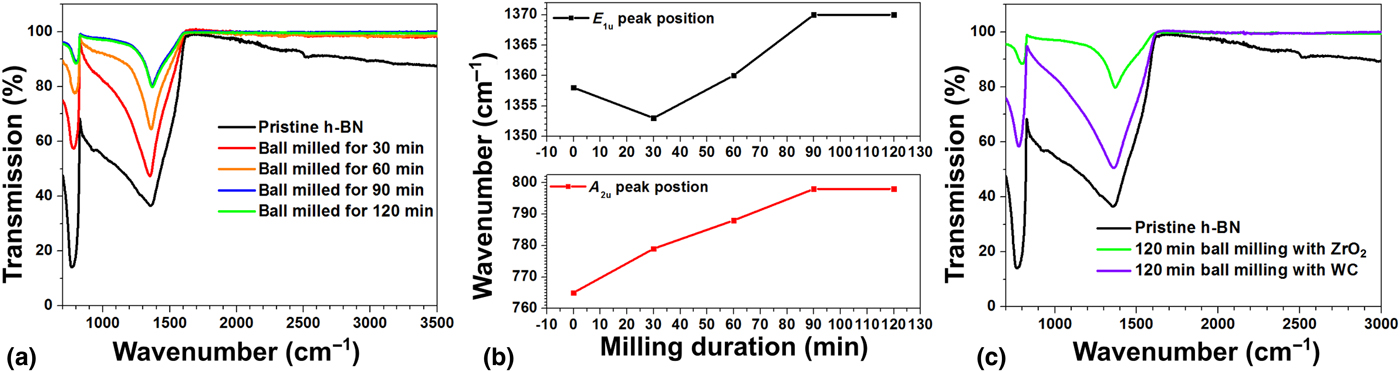
Next, we compared the IR fingerprint of the h-BN powders obtained with FTIR spectroscopy (Fig. 3). h-BN exhibits two primary bands around 763 and 1358 cm−1. The vibrational mode near 763 cm−1 corresponds to A2u out-of-plane bending vibration of B-N-B, while the peak near 1358 cm−1 represents the E1u in-plane stretching vibration of the B-N bond.[Reference Fakrach, Rahmani, Chadli, Sbai, Bentaleb, Bantignies and Sauvajol33, Reference Aradi, Naidoo, Billing, Wamwangi, Motochi and Derry34] In addition, we note that for pristine h-BN powder, small bands around 2342 and 2542 cm−1 are present. They have previously been attributed to small molecules and impurities adsorbed on the pristine h-BN in air.[Reference Nithya and Pandurangan35, Reference Baraton, Merle, Quintard and Lorenzelli36] The first 30 min of ball milling seemed to remove contamination as the bands above 2000 cm−1 disappeared and the E1u in-plane stretching band became narrower. Another weak band about 925 cm−1, previously reported[Reference Li, Xiao, Xu, Lin, Huang, Xue, Jin, Zou and Tang37] as a B-O or B-N-O stretching mode in pristine BN,[Reference Li, Xiao, Xu, Lin, Huang, Xue, Jin, Zou and Tang37, Reference Tang, Bando, Huang, Zhi and Golberg38] disappeared after 30 min of ball milling. This indicates that some oxidation of h-BN or oxygen adsorption may take place in storage, but can be reversed by ball milling. This is in agreement with the induction period, i.e., the time required for h-BN to be processed in the ball milling reactor before any catalytic activity can take place, which has been observed in our measurements.
(a) FTIR spectra of h-BN and dh-BN obtained for milling times of 30 min, 60 min, 90 min, and 120 min with ZrO2. (b) Evolution of E1u (top, black) and A2u (bottom, red) IR bands as a function of milling duration. (c) FTIR spectra of h-BN and dh-BN obtained after ball milling for 120 min with ZrO2 (green) and WC (purple) milling balls.

After increasing ball milling treatment durations, the respective intensities of the two h-BN characteristic bands were found to decrease. The quantity of powder and the force applied on the ATR crystal were maintained constant for all measurements. Hence, the reduction in the size of the BN flakes, leading to smaller crystalline regions to absorb the light, is likely responsible for the smaller band intensity. Another interesting point is that the ratio of Absorption(A2u)/Absorption(E1u) was above 1(1.36) for pristine h-BN but continuously decreased from 0.81 at 30 min ball milling to 0.55 as 90 and 120 min (Table S2 in the supplementary material). This may suggest that the out-of-plane bending vibrations decreased as the exfoliation of the flakes progressed. However, the ratio observed for the powder obtained with WC balls was higher than for the powders obtained with the ZrO2 balls. This remains unexplained at this time and should be investigated further. Next, we considered the position of the two bands. As shown in Fig. 3(b), the two bands shifted to higher wavenumber with increasing milling duration. For the A2u peak, the position moved from 765 cm−1 for pristine h-BN to 779 cm−1 after 30 min milling before reaching a maximum at 798 cm−1 after 90 min milling. Similarly, the position of the E1u band shifted from 1358 to 1370 cm−1 as the milling time increased from 0 to 90 min. No additional changes were found between 90 and 120 min. This is likely due to the change in strain the flakes as a result of exfoliation. Lastly, we compared the effect of milling with ZrO2 or WC balls on the IR fingerprint of dh-BN powder obtained after 120 min [Fig. 3(c)]. We found that the position of the A2u and E1u bands did not significantly change given the 4 cm−1 resolution of the FTIR system. The results suggest that after 120 min, milling may have achieved its highest potential for defect creation in the volume of h-BN used when milling with ZrO2 media. This is in agreement with a previous study by Torii et al.[Reference Torii, Jimura, Hayashi, Kikuchi and Takagaki32] in which the surface area available was found to decrease after a threshold of treatment duration, as an aggregation of the small flakes became predominant.
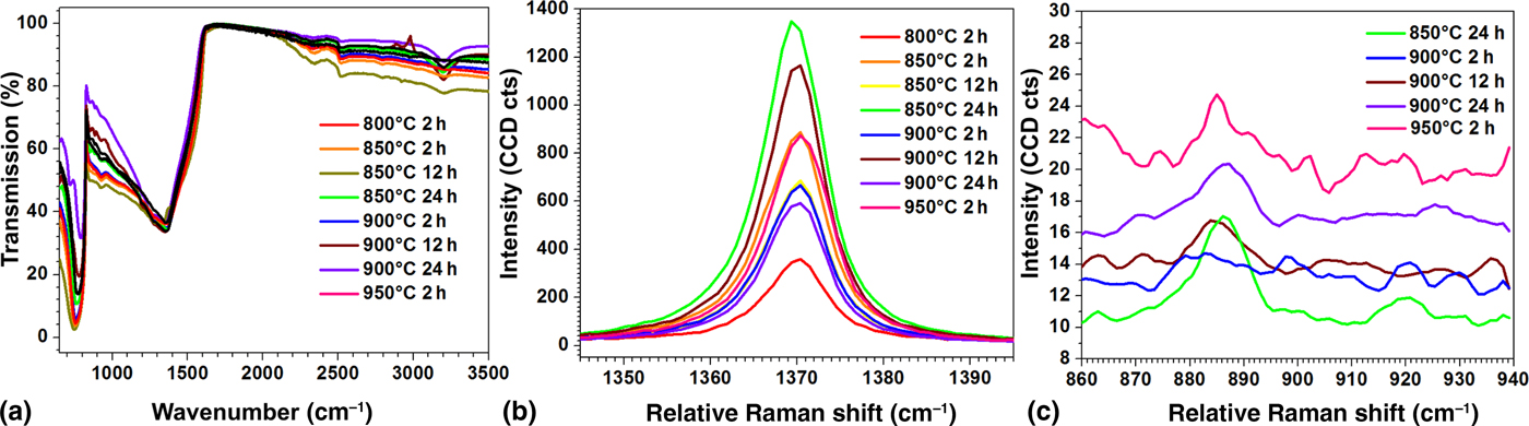
The effect of heat treatment on h-BN was investigated next. As shown in Fig. 4, no Raman shift or FWHM broadening was observed following the treatment in the optimization phase, carried out in the air (Fig. 4 and Table S3 in the supplementary material). This is in agreement with a study by Li et al.[Reference Li, Cervenka, Watanabe, Taniguchi and Chen39] that found the Raman E2g band unchanged by heat treatment of bulk h-BN. Based on our SEM images, we could estimate the flakes in the powder to be more than 50 layers thick. However, for powders heat treated with 850 °C for 24 h, 900 °C for 12 and 24 h, and 950 °C for 2 h, a Raman band centered near 886 cm−1 was observed, corresponding to the breathing mode of oxygens within a boroxol ring according to previous reports.[Reference Liao, Tu, Han, Hu, Connell, Chen and Lin40, Reference Yang, Sha, Wang, Zou, Niu, Cui and Yang41] These observations and various DFT reports in the literature[Reference Nash, Restrepo, Parra, Giesler, Penabade, Aminpour, Le, Li, Farha and Harper3, Reference Sajid, Reimers and Ford14] allow us to infer that the creation of defects, most likely VN, led to local h-BN oxidization into B2O3 as a result of the treatment under ambient conditions. After reproducing the measurements in the conditions of defect creation (950 °C for 2 h) but under the argon filled atmosphere, the Raman signature of the heat treated powder was obtained and indicated no sign of oxidation in the 800–900 cm−1 (Fig. S2). The FTIR signatures of the treated h-BN flakes exhibited the two bands at around 770 and 1360 cm−1 respectively, indicating that the crystal structure remains after heat treatment. This is also confirmed by the SEM image (Fig. S1). A strong (B-) O—H stretching IR peak near 3200 cm−1[Reference Liao, Tu, Han, Hu, Connell, Chen and Lin40] appeared for heat treated BN powder beyond 12 h (850 °C for 12 h, 850 °C for 24 h, 900 °C for 12 h, and 900 °C for 24 h). The powder treated at 900 and 950 °C for 2 h exhibited no detectable IR signals from (B-) O—H about 3200 cm−1. However small bands at ~925 cm−1 and between 2000 and 3000 cm−1 previously associated with impurities in the powder,[Reference Li, Xiao, Xu, Lin, Huang, Xue, Jin, Zou and Tang37, Reference Tang, Bando, Huang, Zhi and Golberg38] which could be removed with short ball milling, remained on the flake for all heat treated powders. This could impede the reactivity of the flakes given our prior observations of a required induction period to remove impurities in ball milling before reactions can take place in the ball milling reactor.
Effect of heat treatment on h-BN powders (a) FTIR spectra of BN powders heat treated at different conditions: 800 °C for 2 h (red), 850 °C for 2 h (orange), 850 °C for 12 h (green), 850 ° for 24 h (light green), 900 ° for 2 h (blue), 900 ° for 12 h (brown), 900 ° for 24 h (purple), 950 ° for 2 h (pink), (b) Corresponding Raman spectra representing the E2 g mode Raman peak of the heat treated BN powders (c) Raman band corresponding to the B-O bond for oxidized BN powder around 885 cm−1.

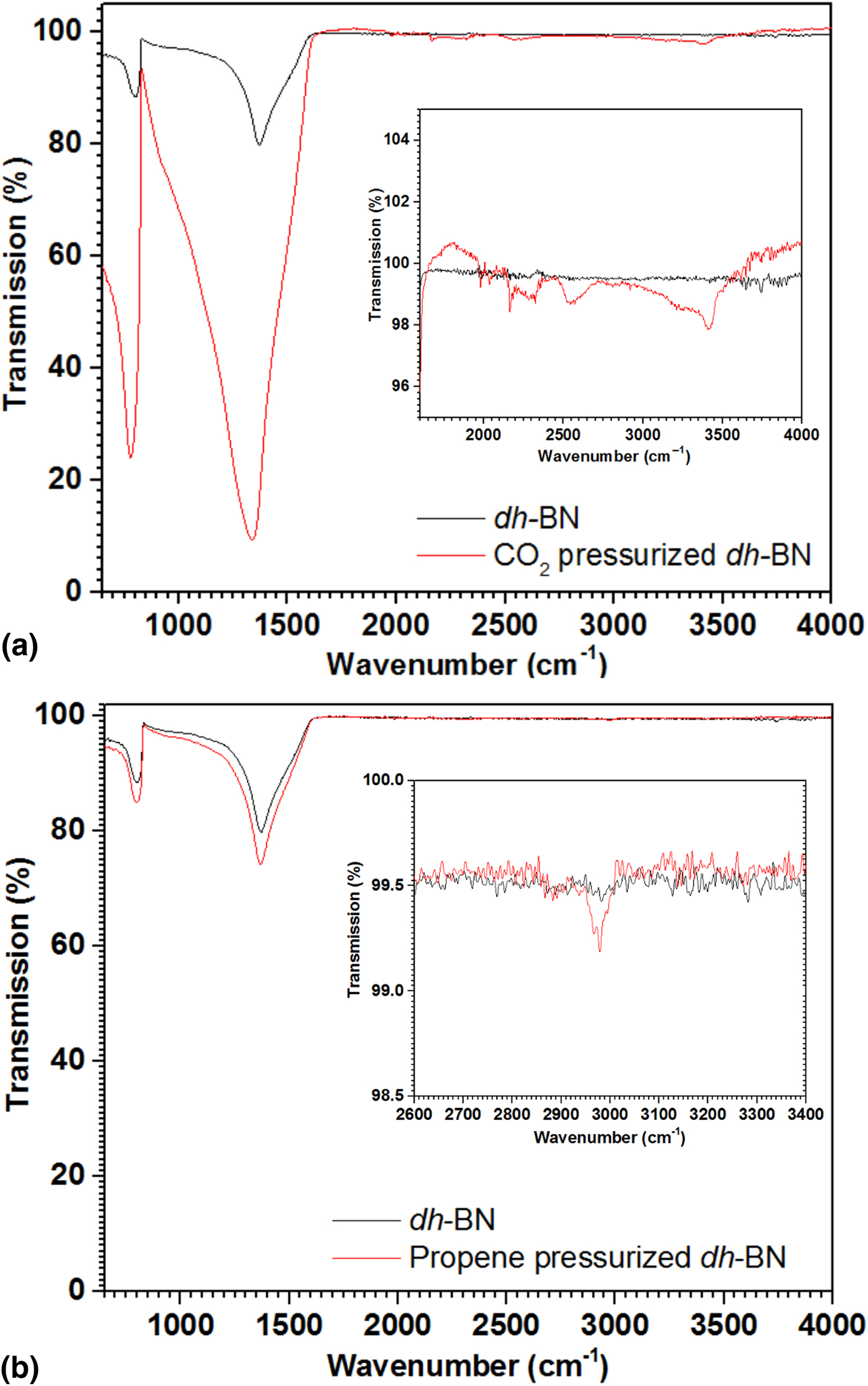
These results indicate that both treatments affect the vibrational modes of the h-BN lattice with signs of defect formation. This is only meaningful if dh-BN exhibit catalytic properties for targeted reactions. To test this, we pressurized a representative powder of each treatment, the powder ball-milled with ZrO2 for 2 h and the powder heat-treated at 950 °C for 2 h, with molecules of interest. We evaluated the reaction with olefin by pressurizing the cell at 40 PSI with propene for 48 h. For the reaction with CO2, we pressurized the cell at 40 PSI with CO2 for 48 h. The largest mass uptake, an about 6.8% increase, was observed for the ball-milled powder exposed to propene. The corresponding exposure with heat treated powder only saw a 0.2% uptake. After exposure to CO2, a smaller mass uptake of 3% was found for the ball-milled powder while a 0.4% uptake was measured for the heat treated powder. The nature of the uptake on the ball-milled flakes was confirmed with FTIR spectroscopy (Fig. 5). The presence of two bands at ~2550 cm−1 (B-H stretching) and ~3400 cm−1 (region for O-H stretch in alcohol R2CH2OH or in carboxylic acid R2COOH) in the CO2-pressurized dh-BN powder [inset in Fig. 5(a)] suggests that the new bands corresponds to the products of a reaction on the flakes. In addition, the C-H asymmetric stretching mode ~2980 cm−1,[Reference Baraton, Merle, Quintard and Lorenzelli36] in agreement with the presence of propene or propane, was observed in the propene-pressurized dh-BN powder [inset in Fig. 5(b)]. Neither band was present in the dh-BN spectrum acquired prior to placing the powder in the pressure cell. No signal could be detected on the heat treated powders, likely due to the small concentration of molecules.
FTIR spectra of ball-milled BN powders before (black) and after pressurization for 48 h under 40 PSI (red) in CO2 (a) and in propene (b). Insets provide details of the IR bands in the 1500–4000 cm−1 region.

Conclusion
Ball milling and heat treatment lead to the formation of different defects in h-BN flakes. Particularly the microstructure and surface area of the flakes can be controlled by varying the duration of the treatment and the composition of milling media. For heat treatment, varying the temperature and duration of treatment was also found to affect the properties of the flakes. Raman and FTIR spectroscopy can be used to monitor defect creation, strain induction or oxidation of the sites created depending on the conditions of the treatments. For instance, signals from impurities (~925 cm−1) could be differentiated from oxidation taking place in the flakes resulting in the stronger band at 3200 cm−1. In addition to deepening the fundamental understanding of processing–structure–property relationship of h-BN, our study has practical implications for the induction of catalytic sites in h-BN for reaction activation. Initial demonstration of the reactivity of dh-BN shows that the reaction is greatly treatment dependent. The detailed of catalytic activity, including the mechanism and the kinetics of the processes taking place after defect induction, will be the subject of future studies.
Supplementary material
The supplementary material for this article can be found at https://doi.org/10.1557/mrc.2018.113.
Acknowledgments
The authors would like to thank Saint-Gobain Ceramic Materials for the generous donation of hexagonal boron nitride powders used in this study. We acknowledge the NSF support for CHE-1465105 entitled “SusChEM: Defect-laden 2D Catalysts for Carbon Sequestration and Safer Hydrogenation”.