I. INTRODUCTION
Iron-deficiency is the most prevalent cause of anemia, and has been estimated to affect over two billion people, owing to a variety of causes including malnutrition, chronic blood loss and disease (Crohn's disease, chronic kidney disease, inflammatory bowel disease, etc.) among others (Camaschella, Reference Camaschella2015). Oral iron supplementation is the typical therapy, and ferrous gluconate, Fe(C6H11O7)2·xH2O, is a common iron supplement with a long history of usage (Reznikoff and Goebel, Reference Reznikoff and Goebel1937). Ferrous gluconate has seen recent consideration for iron-deficiency prevention via fortification of food (Pizarro et al., Reference Pizarro, Boccio, Salgueiro, Olivares, Carmuega, Weill, Marque, Frereux and Noirt2013) and milk for infants (Villalpando et al., Reference Villalpando, Shamah, Rivera, Lara and Monterrubio2006). Ferrous gluconate used in conjunction with ascorbic acid has also been studied for replenishing iron stores for regular blood donors (Radtke et al., Reference Radtke, Tegtmeier, Rӧcker, Salama and Kiesewetter2005).
Despite longstanding and widespread usage, and a number of Mössbauer spectroscopic studies (Kulgawczuk et al., Reference Kulgawczuk, Ruebenbauer and Sepiol1989; Coe et al., Reference Coe, Bowen and Bereman1995), the only pattern for iron gluconate currently in the powder diffraction file (ICDD, 2015) is an old, un-indexed pattern with visually estimated intensities (PDF entry 00-005-0257). This work provides a high quality, indexed pattern for phase identification.
II. EXPERIMENTAL
A ferrous gluconate tablet manufactured by Apotex Inc. (product number 00545031, lot MP3827) was cut in half, and powder was scraped from the tablet centre to avoid the surface coating. The powder was loaded, with minimal grinding, into a 0.3 mm ID Kapton capillary that was sealed at both ends with a Loctite adhesive.
Powder X-ray diffraction (PXRD) patterns were collected using a Canadian Macromolecular Crystallography Facility beamline (CMCF-BM or 08B1-1, Fodje et al., Reference Fodje, Grochulski, Janzen, Labiuk, Gorin and Berg2014) at the Canadian Light Source (CLS). 08B1-1 is a bending magnet beamline with a Si (111) double crystal monochromator. Two-dimensional (2D) data were obtained using a Rayonix MX300HE detector with an active area of 300 × 300 mm2. The patterns were collected at an energy of 18 keV (λ = 0.688 80 Å) and capillary-detector distance of 250 mm.
The 2D PXRD patterns were calibrated and integrated using the GSASII software package (Toby and Von Dreele, Reference Toby and Von Dreele2013). The sample-detector distance, detector centering and tilt were calibrated using a lanthanum hexaboride (LaB6) standard reference material (NIST SRM 660a LaB6) and the calibration parameters were applied to all patterns. After calibration, the 2D patterns were integrated to obtain standard 1D powder diffraction patterns. A pattern collected from an empty Kapton capillary (using the same conditions) was subtracted from the sample data during integration.
Ab initio indexing was performed using the programs DICVOL (Boultif and Louer, Reference Boultif and Louer2004) and McMaille (Le Bail, Reference Le Bail2004), while EXPO2014 (Altomare et al., Reference Altomare, Cuocci, Giacovazzo, Moliterni, Rizzi, Corriero and Falcicchio2013) was used to examine possible space groups.
Potential solutions were examined with Le Bail refinement (Le Bail et al., Reference Le Bail, Duroy and Fourquet1988) using the FullProf program (Rodriguez-Carvajal, Reference Rodriguez-Carvajal2001).
III. RESULTS AND DISCUSSION
Initial indexing attempts on the first 20 reflections in the PXRD pattern performed using DICVOL, for monoclinic and higher symmetries, suggested three to five possible cells. The highest figures of merit (M 20 = 22.1, F 20 = 133.2) were obtained for a monoclinic cell (a = 19.9518 Å, b = 5.5127 Å, c = 18.4689 Å, β = 111.386°, V = 1891.5 Å3). Subsequent indexing with McMaille yielded a larger number of monoclinic cells, but the same cell was identified with a McMaille figure of merit (Mc 20 = 195.98) that was four times larger than the next closest candidate cell.
Assuming 17 Å3 per non-hydrogen atom would yield 111 atoms in the unit cell, while four formula units of Fe(C6H11O7)2 yields 108 non-hydrogen atoms, suggesting the cell is consistent with Z = 4. While the hydration level is not definitively known, the label suggests that each tablet yields 35 mg of elemental iron contained in 300 mg of ferrous gluconate. Based on mass balance considerations, this would suggest the compound is a dihydrate, Fe(C6H11O7)2 · 2 H2O (expected yield of 34.7 mg of elemental iron). The anhydrous compound would yield 37.6 mg of elemental iron for 300 mg of ferrous gluconate.
Space group tests using EXPO2014 suggested space group I2 (#5) as the most likely candidate, with a general multiplicity (4) consistent with Z. Space group P21 (#4) was also examined closely, because every metal gluconate entry found in the Cambridge Structural Database (Groom et al., Reference Groom, Bruno, Lightfoot and Ward2016) belonged to either space group P21 or P212121. But P21 yields numerous low angle Bragg reflection positions that are unobserved in the experimental pattern, and would require two formula units in the asymmetric cell (Z′ = 2, based on a general multiplicity of 2). Therefore, I2 is believed to be the correct space group.
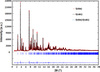
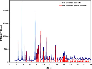
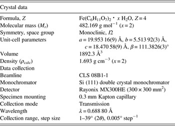
Table I lists the crystal data and data collection parameters, while Figure 1 illustrates the final Le Bail refinement obtained using space group I2, resulting in a reduced χ 2 of 1.01. Visually, the pattern is reasonably consistent with an unindexed pattern obtained by Coe et al. (Reference Coe, Bowen and Bereman1995), after accounting for the different wavelengths. The final reflection list extracted from the Le Bail refinement is plotted in Figure 2 with the raw data. The reflection list was prepared by summing reflections closer than 0.02°2θ as multiple reflections and assigning a weighted average reflection position, then including all reflections with relative integrated intensities of 0.5% or greater up to 23°2θ. The full Bragg reflection list and raw data are contained in a crystallographic information file (CIF) in the online supplementary material.
(Color online) The final Le Bail refinement obtained for ferrous gluconate using a monoclinic cell (a = 19.953 16(9) Å, b = 5.513 92(3) Å, c = 18.47058(9) Å, β = 111.3826(3)°) with space group I2 (reduced χ 2 = 1.01).

(Color online) The Bragg reflection list for ferrous gluconate obtained with Le Bail refinement. The reflection list is contained in a CIF file in the online supplementary material.

The crystal data and data collection parameters obtained for ferrous gluconate.

SUPPLEMENTARY MATERIAL
The supplementary material for this article can be found at https://doi.org/10.1017/S0885715616000671
ACKNOWLEDGEMENTS
Research described in this paper was performed using beamline 08B1-1 at the Canadian Light Source, which is supported by the Canadian Foundation for Innovation, the Natural Sciences and Engineering Research Council of Canada, the National Research Council Canada, the Canadian Institutes of Health Research, the Government of Saskatchewan, Western Economic Diversification Canada, and the University of Saskatchewan.