





Introduction
Psychosis is increasingly recognised as a multisystem disorder in which the hypothalamic–pituitary–adrenal (HPA) axis plays an important role (Pillinger, D’Ambrosio, McCutcheon, & Howes, Reference Pillinger, D’Ambrosio, McCutcheon and Howes2019). Cortisol, the principal stress hormone of the HPA axis, reflects HPA-axis activity and mediates physiological responses to stress (Chrousos, Reference Chrousos2000; McEwen & Stellar, Reference McEwen and Stellar1993). HPA-axis dysregulation is well documented in psychosis, including elevated cortisol levels, a blunted cortisol awakening response (CAR), and altered stress reactivity (Berger et al., Reference Berger, Kraeuter, Romanik, Malouf, Amminger and Sarnyai2016; Borges, Gayer-Anderson, & Mondelli, Reference Borges, Gayer-Anderson and Mondelli2013; Girshkin, Matheson, Shepherd, & Green, Reference Girshkin, Matheson, Shepherd and Green2014; Karanikas, Antoniadis, & Garyfallos, Reference Karanikas, Antoniadis and Garyfallos2014; Zorn et al., Reference Zorn, Schür, Boks, Kahn, Joëls and Vinkers2017). These findings are consistent with the neural diathesis–stress model, which proposes that genetic vulnerability and stress-related HPA-axis abnormalities increase risk for psychosis through effects on dopaminergic and glutamatergic pathways (Cullen et al., Reference Cullen, Rai, Vaghani, Mondelli and McGuire2020; Pruessner, Cullen, Aas, & Walker, Reference Pruessner, Cullen, Aas and Walker2017; Walker & Diforio, Reference Walker and Diforio1997; Walker, Mittal, & Tessner, Reference Walker, Mittal and Tessner2008). Further support comes from evidence linking adverse childhood experiences to increased risk of psychosis (Varese et al., Reference Varese, Smeets, Drukker, Lieverse, Lataster, Viechtbauer, Read, van Os and Bentall2012), potentially through HPA-axis sensitisation and heightened stress responsivity (Aas et al., Reference Aas, Dazzan, Mondelli, Melle, Murray and Pariante2014).
Robust evidence indicates that cognitive impairment is a core feature of schizophrenia-spectrum and other psychotic disorders (Bora, Binnur Akdede, & Alptekin, Reference Bora, Binnur Akdede and Alptekin2017; Bora, Yücel, & Pantelis, Reference Bora, Yücel and Pantelis2009, Reference Bora, Yücel and Pantelis2010; Lewandowski, Sperry, Cohen, & Öngür, Reference Lewandowski, Sperry, Cohen and Öngür2014; McCutcheon, Keefe, & McGuire, Reference McCutcheon, Keefe and McGuire2023; Sheffield, Karcher, & Barch, Reference Sheffield, Karcher and Barch2018). Deficits span multiple domains, including attention, processing speed, memory, and executive functioning, and are typically associated with large effect sizes (Heinrichs & Zakzanis, Reference Heinrichs and Zakzanis1998; Mesholam-Gately et al., Reference Mesholam-Gately, Giuliano, Goff, Faraone and Seidman2009; Schaefer, Giangrande, Weinberger, & Dickinson, Reference Schaefer, Giangrande, Weinberger and Dickinson2013). Present from the first episode of psychosis and in drug-naïve patients (Fatouros-Bergman et al., Reference Fatouros-Bergman, Cervenka, Flyckt, Edman and Farde2014; Mesholam-Gately et al., Reference Mesholam-Gately, Giuliano, Goff, Faraone and Seidman2009), these impairments contribute substantially to functional disability (Green, Horan, & Lee, Reference Green, Horan and Lee2019; McCutcheon et al., Reference McCutcheon, Keefe and McGuire2023; Reichenberg & Harvey, Reference Reichenberg and Harvey2007).
Several lines of evidence suggest an interplay between HPA-axis dysfunction and cognitive impairment in psychosis. Prolonged exposure to elevated cortisol may exert neurotoxic effects on the hippocampus and prefrontal cortex, brain regions critical for memory and executive functioning and implicated in psychosis (Giovanoli et al., Reference Giovanoli, Engler, Engler, Richetto, Voget, Willi, Winter, Riva, Mortensen, Feldon, Schedlowski and Meyer2013; Knight et al., Reference Knight, McCutcheon, Dwir, Grace, O’Daly, McGuire and Modinos2022; Lupien et al., Reference Lupien, de Leon, de Santi, Convit, Tarshish, Nair, Thakur, McEwen, Hauger and Meaney1998; Lupien, Juster, Raymond, & Marin, Reference Lupien, Juster, Raymond and Marin2018; McEwen, Reference McEwen1999; Mondelli et al., Reference Mondelli, Pariante, Navari, Aas, D’Albenzio, Di Forti, Handley, Hepgul, Marques, Taylor, Papadopoulos, Aitchison, Murray and Dazzan2010). Owing to their high density of glucocorticoid receptors, these regions are particularly vulnerable to chronic stress, which may lead to dendritic retraction, synaptic loss, and reduced neurogenesis (Arnsten, Reference Arnsten2009). Such alterations provide a plausible mechanism linking cortisol dysregulation to cognitive deficits in psychosis.
Two narrative reviews have highlighted a potential link between HPA-axis dysregulation and cognitive impairment in psychosis. Labad (Reference Labad2019) proposed that cognitive deficits may be partly driven by the neurotoxic effects of chronic cortisol elevation, while Aas et al. (Reference Aas, Dazzan, Mondelli, Melle, Murray and Pariante2014) reviewed evidence suggesting that cognition in first-episode psychosis may be modulated by cortisol. Together, these reviews support the hypothesis that cortisol dysregulation may contribute to cognitive dysfunction in psychotic disorders.
To our knowledge, no previous study has quantitatively synthesised associations between cortisol and cognition in psychosis. To address this gap, we investigated the relationship between cortisol levels and cognitive performance in psychotic disorders using a combined narrative review and meta-analytic approach. By examining both global cognitive functioning and specific cognitive domains, we aimed to determine whether cortisol–cognition associations differ across distinct aspects of cognition.
Methods
Literature search strategy
The current systematic review and meta-analysis was prospectively registered in PROSPERO (registration number CRD42024564051) and followed the Preferred Reporting Items for Systematic Reviews and Meta-Analyses (PRISMA) guidelines (Page et al., Reference Page, McKenzie, Bossuyt, Boutron, Hoffmann, Mulrow, Shamseer, Tetzlaff, Akl, Brennan, Chou, Glanville, Grimshaw, Hróbjartsson, Lalu, Li, Loder, Mayo-Wilson, McDonald and Moher2021; see Figure 1). The literature searches were conducted using three databases: Web of Science (Core Collection), PsycINFO, and MEDLINE. The publication period ranged from the inception of the database to 15th June 2026. Two researchers (HZC, JXMY) independently screened the titles and abstracts to identify potentially eligible studies, with a third researcher (EK) adjudicating if differences in opinion arose. The search strategy combined search terms related to cortisol, cognition, and psychosis (see Supplementary Appendix A).
PRISMA flow diagram of the study selection process.

Figure 1. Long description
The flowchart is organized into three vertical phases labeled on the left: Identification, Screening, and Included.
1. Identification Phase:
- Top-left box: Records identified from databases: Web of Science Core Collection (n = 397), PsycInfo (n = 476), and Medline (n = 392).
- An arrow points right to a box: Records removed before screening: Duplicate records removed (n = 356).
- A downward arrow from the identification box leads to the Screening phase.
2. Screening Phase:
- First central box: Records screened (n = 909).
- An arrow points right to a box: Records excluded (n = 877).
- A downward arrow leads to the next central box: Reports assessed for eligibility (n = 32).
- An arrow points right to a box listing specific exclusion reasons: Neural processes other than ‘cold’ cognition (n = 5), Non-adult sample (n = 1), Not clinically diagnosed patient (n = 2), Non-research papers (n = 5), and Correlational data not available after request (n = 7).
3. Included Phase:
- A final downward arrow leads to the bottom central box: Studies met search criteria (n = 12) and Studies included in meta-analysis (n = 9).
Study selection
We selected studies satisfying the following inclusion criteria: (1) primary research study published in a peer-reviewed journal; (2) written in English; (3) studied adult human samples; (4) included participants clinically diagnosed with schizophrenia, other primary psychotic disorders, or mood disorders with psychotic symptoms according to the Diagnostic and Statistical Manual of Mental Disorders (DSM) or the International Classification of Diseases (ICD); (5) measured naturally occurring cortisol; (6) conducted cognitive tests; and (7) reported correlations between cognition and cortisol. Exclusion criteria were: (1) participants in the psychotic group had cortisol-related disorders (e.g., Cushing’s disorder); (2) the study reported mental or physical health comorbidities that are associated with cognitive impairment; and (3) the study assessed neural processes other than ‘cold’ cognition (e.g., social cognition, stress reactivity, emotion or stress perception, and neurophysiological responses). Two researchers (HZC, JXMY) independently conducted full-text screenings, and all decisions on eligibility were based on consensus. When targeted data were missing from an otherwise eligible study, attempts were made to acquire the data before excluding the study from further consideration. Sample overlap was checked among the included studies.
A total of 12 studies met our eligibility criteria. In line with previous studies (Myung, Reference Myung2023), we set the minimum number of independent studies required to conduct a meta-analysis to five. Nine studies with non-overlapping participant samples examined basal cortisol, enabling a meta-correlation. CAR was examined in two studies, whereas diurnal cortisol slope, daytime cortisol area under the curve, and chronic hair cortisol each were examined in one study, allowing a narrative synthesis.
Data extraction and quality assessment
Two researchers (HZC, JXMY) extracted the following data: (1) authors and year of publication; (2) sample size and gender composition; (3) mean age of sample; (4) range of diagnoses among participants; (5) cortisol specimen type; (6) cortisol collection time; (7) cognitive batteries, tests and variables; (8) correlation coefficients between cortisol and cognitive variables. The study by Cherian, Schatzberg and Keller (Reference Cherian, Schatzberg and Keller2019) reported data from two separate cohorts, a schizophrenia spectrum disorder group and a psychotic major depression group, which represented two separate studies/samples in our meta-analyses. A third researcher (EK) compared the two datasets and confirmed that the data were extracted correctly. Cognitive variables were categorised into one of the following cognitive domains: attention, working memory & processing speed (AWP); executive function (EF); verbal learning and memory (VM); visual–spatial perception, organisation & learning; language function (VS); and general/composite cognitive function (GC). In line with previous work by our group (Millgate et al., Reference Millgate, Hide, Lawrie, Murray, MacCabe and Kravariti2022), attention, working memory and processing speed were grouped into a single cognitive domain (AWP). This classification was adopted because many neuropsychological measures used in psychosis research tap overlapping attentional, working-memory, and processing-speed processes, making reliable separation of these constructs difficult across studies. Detailed information on the included studies and their characteristics is presented in Table 1.
Characteristics of eligible studies (n = 12)

Table 1. Long description
The table contains 11 columns: Study, Country, Sample size (female percentage), Mean age, Diagnosis, Cortisol type, Cortisol time, Cortisol measure, Cognitive measurement, Effect sizes (n), and Cognitive domains.
Key entries include:
* Misiak et al. (2021): Poland, 85 participants (47.1% female), mean age 37.1, S S D diagnosis, morning blood basal cortisol, R B A N S measurement, 5 effect sizes, domains A W P, V M, V S, L A.
* Cherian et al. (2019)-S S D: U S, 31 participants (45.2% female), mean age 38.48, S S D diagnosis, blood basal cortisol, multiple tests including W A I S-III and Stroop, 8 effect sizes, domains A W P, E F, V M.
* Cherian et al. (2019)-M D: U S, 46 participants (54.3% female), mean age 36.67, P M D diagnosis, blood basal cortisol, 8 effect sizes, domains A W P, E F, V M.
* Allott et al. (2018): Australia, 35 participants (25.7% female), mean age 20.3, F E P diagnosis, morning blood basal cortisol, 5 effect sizes, domains A W P, E F, V M.
* Krkovic et al. (2017): Germany, 35 participants (42% female), mean age 40.5, S S D diagnosis, afternoon saliva basal cortisol, 7 effect sizes, domains A W P, E F, V M, G C.
* Knytl et al. (2019): Czech Republic, 16 participants (50% female), mean age 25.81, F E P diagnosis, morning blood basal cortisol, 6 effect sizes, domains A W P, E F, V M, G C.
* Havelka et al. (2016): Czech Republic, 23 participants (0% female), mean age 23.5, F E P diagnosis, afternoon blood basal cortisol, 8 effect sizes, domains A W P, E F, V M.
* Walder et al. (2000): U S, 18 participants (44.4% female), mean age 36.72, S S D diagnosis, morning saliva basal cortisol, 3 effect sizes, domains E F, V M.
* Inova et al. (2025): Norway, 257 participants (37.74% female), mean age 30.75, S S D diagnosis, morning blood basal cortisol, 4 effect sizes, domains A W P, E F, V M.
* Yazici et al. (2025): Türkiye, 38 participants (0% female), mean age 37.71, S S D diagnosis, morning blood basal cortisol, 1 effect size, domain V S.
* Labad et al. (2016): Spain, 60 participants (35% female), mean age 24.55, S S D diagnosis, saliva C A R and D C S, domains A W P, E F, V M, V S.
* Aas et al. (2011): U K, 30 participants (33.3% female), mean age 30.1, F E P diagnosis, saliva C A R and D C A u C, domains G C, V M, V S, E F, A W P.
* Aas et al. (2019): Norway, 28 participants (50% female), mean age 33.6, S S D diagnosis, hair H C C, domains A W P, E F, V M.
Note: AWP, attention working memory & processing speed; AVLT, auditory verbal learning test; CANTAB, Cambridge neuropsychological test automated battery; CAR, cortisol awakening response; COWA, controlled oral word association test; CVLT, California verbal learning test; DCAuC, daytime cortisol area under the curve; DCS, diurnal cortisol slope; EF, executive function; FEP, first episode psychosis; GC, general/composite cognitive function; HCC, hair cortisol concentration; HVLT-R, Hopkins verbal learning test; LA, language function; MCCB, matrics consensus cognitive battery; PMD, psychotic major depression; RBANS, repeatable battery for the assessment of neuropsychological status; RBMT, rivermead behavioural memory test; SSD, schizophrenia spectrum disorder; TMT, trail making test; VM, verbal learning and memory; VS, visual–spatial perception organisation and learning; WAIS, Wechsler adult intelligence scale; WCST, Wisconsin card sorting test; WMS, Wechsler memory scale.
a Studies included in meta-analysis.
b Only measures with a reported correlation coefficient with a cognitive variable are included in the Table.
Two researchers (HZC, JXMY) independently assessed the overall quality of the studies using the NIH Quality Assessment Tool for Observational Cohort and Cross-Sectional Studies (National Heart, Lung, and Blood Institute, NIH, U.S.). The tool contains 14 items, each scored with 1 (yes) or 0 (no, not applicable, not reported, or cannot determine). In instances of score discrepancies, the researchers engaged in discussion to reach a consensus. An overall quality score was calculated by summing all item scores (≥ 7, good; 4 to 6, fair; < 4, poor; Li et al., Reference Li, Jia, Yan, Hua, Han, Yang, Ma and Ma2023). All studies were rated as good quality (see Supplementary Appendix B).
Statistical analysis
Multilevel meta-analyses
To generate an overall correlation coefficient between basal cortisol and composite cognitive function among patients with psychotic disorders across eligible studies (n = 9), we first performed a multilevel meta-analysis of correlation coefficients across all studies and cognitive variables. Three further cognitive domains – AWP, EF and VM – met our threshold (5 studies) for performing domain-specific meta-analysis. Study findings relating to the remaining cognitive domains were synthesised narratively.
Several studies reported two or more effect sizes within the same cognitive domain. To account for the dependence arising from multiple effect sizes contributed by individual studies, multilevel meta-analysis was conducted as recommended by López-López, Page, Lipsey, and Higgins (Reference López-López, Page, Lipsey and Higgins2018). This approach accounts for effect size dependence by estimating variance at multiple levels (Supplementary Figure 1), thereby improving the accuracy of statistical inference (Cheung, Reference Cheung2015, Reference Cheung2019) and avoiding the limitations of averaging effect sizes within studies, which can fail to account for within-study variance and reduce analytic precision. Accordingly, associations between basal cortisol and cognition were synthesised using three-level meta-analysis, with variance partitioned into within-effect-size, within-study, and between-study components (Supplementary Figure 1). Correlation coefficients were converted to Fisher’s z prior to analysis and back-transformed to Pearson’s r for reporting.
Given the circadian rhythm of cortisol secretion, which may influence cortisol–cognition associations (Gamble, Berry, Frank, & Young, Reference Gamble, Berry, Frank and Young2014), sampling time was coded as morning (relatively high cortisol levels) or afternoon/evening (relatively low cortisol levels) and included as a moderator in all models. Combining afternoon and evening cortisol measurements was supported by previous studies (e.g., Engert et al., Reference Engert, Efanov, Dedovic, Dagher and Pruessner2011; O’Byrne, Yuen, Butt, & Liu, Reference O’Byrne, Yuen, Butt and Liu2021).
Three-level meta-analyses were first conducted on the full dataset (55 cognitive effect sizes across studies and cognitive domains) and subsequently within the AWP, EF, and VM domains. For each analysis, moderated and unmoderated models were compared using ANOVA, and moderated models were reported only when they provided a significantly better fit. For moderator analyses, a fixed-effect model was used when at least one moderator level contained five or fewer effect sizes (Tufanaru, Munn, Stephenson, & Aromataris, Reference Tufanaru, Munn, Stephenson and Aromataris2015); otherwise, a random-effects model was applied. When dependence among effect sizes remained within significant moderator subgroups, or when unmoderated models were analysed, robust variance estimation (RVE) was used to obtain robust standard errors and valid statistical inference (Hedges, Tipton, & Johnson, Reference Hedges, Tipton and Johnson2010; Tipton, Reference Tipton2015; Tipton & Pustejovsky, Reference Tipton and Pustejovsky2015). Heterogeneity was assessed by τ 2 and I 2, with the caveat that I2 estimates may be biased when meta-analyses include a small number of studies (von Hippel, Reference von Hippel2015). Publication bias was assessed using contour-enhanced funnel plots (visual inspection), Egger’s regression test, and Begg’s rank correlation test (Harbord, Egger, & Sterne, Reference Harbord, Egger and Sterne2006).
Sensitivity and further analyses
To assess the robustness of our findings to different analytical decisions, biological sample (blood/salivary cortisol), additional potential moderators, and influential studies (studies whose inclusion has a substantial impact on the results of a meta-analysis), we conducted sensitivity analyses by: (1) replacing the multilevel approach with the traditional approach of averaging dependent effect sizes within studies and cognitive domains; (2) excluding the two studies that measured cortisol using saliva samples; (3) examining age and sex as additional potential moderators; and (4) applying study-level influence diagnostics to identify potential outliers.
Influence diagnostics followed established approaches for meta-analytic models (Viechtbauer & Cheung, Reference Viechtbauer and Cheung2010), including standardised deleted residuals (SDR), Cook’s distance (CD), and DFBETAS. We used conservative screening criteria requiring all three indicators (SDR, CD and DFBETAS) to exceed their respective thresholds: (1) a significant study-level SDR test, (2) CD greater than 4/k, and (3) maximum absolute DFBETAS greater than 1. Studies flagged by these screening diagnostics were not removed mechanically but were used to guide sensitivity analyses.
Finally, we compared basal cortisol levels and cognitive performance between patient and control samples across eligible studies using standard meta-analytic models.
Statistical software
Multilevel meta-analyses were conducted in R 4.4.1 using the metafor package (Viechtbauer, Reference Viechtbauer2010). Random-effects models were fitted using restricted maximum likelihood (REML) estimation. Model comparisons were refitted using maximum likelihood estimation because REML is inappropriate for comparing models with different fixed effects (Zuur et al.,Reference Zuur, Ieno, Walker, Saveliev, Smith, Zuur, Ieno, Walker, Saveliev, Smith, Zuur, Ieno, Walker, Saveliev and Smith2009, p. 122). Robust variance estimation was performed using the robumeta package (Fisher, Tipton, & Zhipeng, Reference Fisher, Tipton and Zhipeng2023), assuming a within-study correlation of 0.8 between dependent effect sizes (package default). Statistical significance was set at p < .05.
Results
In all, 12 studies, reporting on 13 independent samples, met eligibility criteria for our systematic review (n = 12) and meta-analyses (n = 9). The 13 samples comprised 940 participants (39.57% female), including 702 patients with psychotic disorders (36.32% female), with mean ages ranging from 20.3 to 40.5 (mean age across studies = 31.83). The studies were conducted in 9 countries in Europe & Asia (n = 8), North America (n = 3), and Oceania (n = 1). The study characteristics are summarised in Table 1. The subset of studies that were included in the meta-analyses (n = 9) comprised 822 participants (39.78% female; mean age = 32.45), including 584 patients (35.96% female, mean age = 32.72).
Multilevel meta-analyses of correlations between basal cortisol level and cognitive function
Composite cognitive function
There was no significant difference between the model fit of moderated and unmoderated models for composite cognitive function (
$ {\chi}_1^2 $
 = 3.40; p = .07). Therefore, we report on the unmoderated three-level model with robust variance estimation. The pooled correlation of all the effect sizes was not significant (r = 0.003; 95% CI: −0.15 − 0.15; p = .96). The estimated heterogeneity was high (
$ {\tau}^2 $
 = .04;
$ {I}^2 $
 = 65.3%; low
$ {I}^2 $
 < 25% ≤ moderate
$ {I}^2 $
 < 50% ≤ high
$ {I}^2 $
 ; Higgins, Thompson, Deeks, & Altman, Reference Higgins, Thompson, Deeks and Altman2003). The forest plot is presented in Supplementary Figure 2. For the list of cognitive variables used, see Supplementary Appendix C.
Attention, working memory & processing speed
The moderated model for AWP did not provide a significantly better fit than the unmoderated model (
$ {\chi}_1^2 $
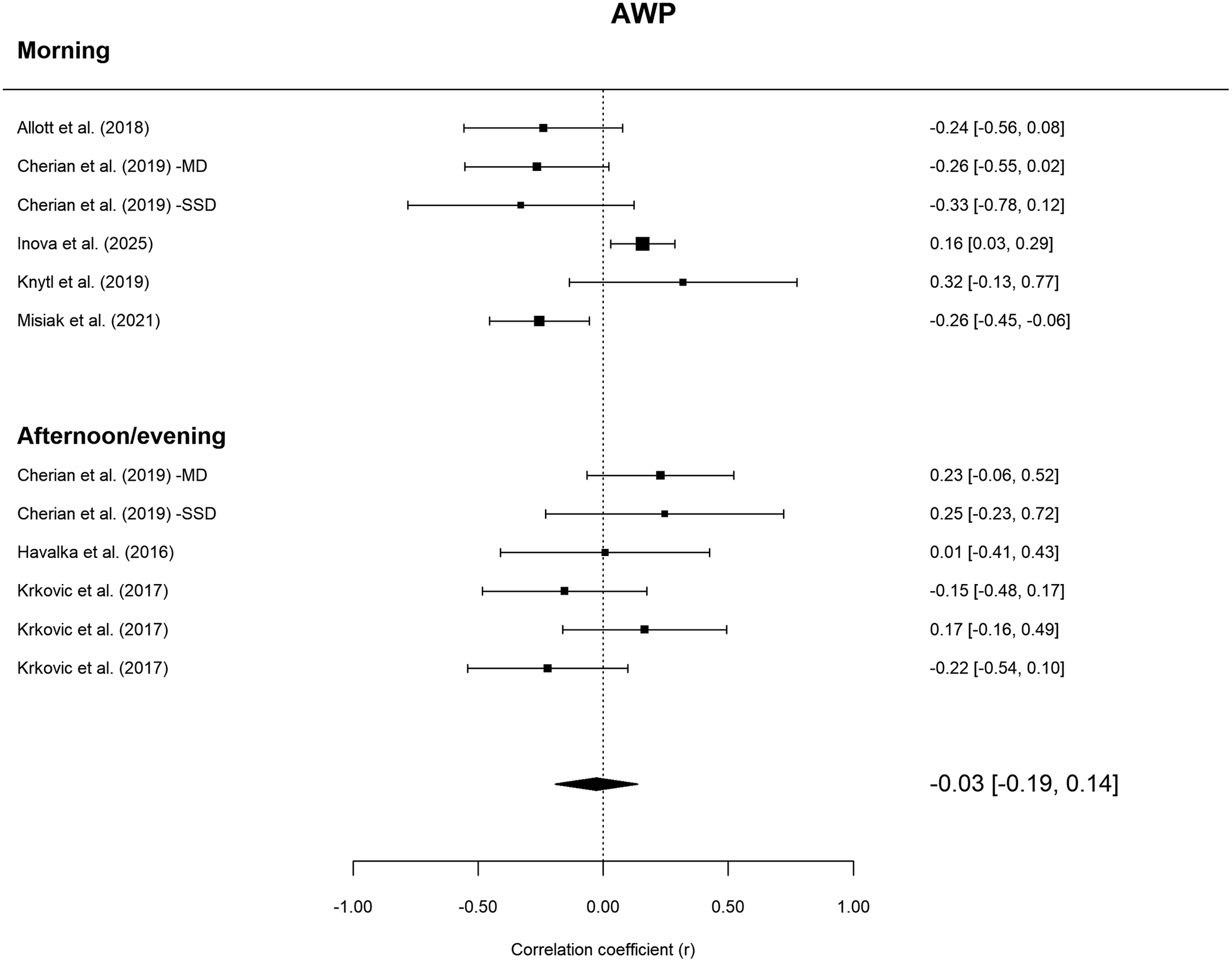
 = 1.02; p = .31). Across all sampling times, basal cortisol was not significantly associated with AWP (r = −0.03; 95% CI: −0.19 to 0.14; p = .71). Total heterogeneity in the all-times AWP model was
$ {\tau}^2 $
 = .03 and
$ {I}^2 $
 = 58.3%. For the forest plot, see Figure 2.
Forest plot of meta-correlations between cortisol and attention/working memory/processing speed via a three-level model. Note that Cherian et al. (Reference Cherian, Schatzberg and Keller2019) included two different cohorts, and they were treated as two independent studies in the analyses.

Figure 2. Long description
The forest plot displays correlation coefficients (r) on an x-axis ranging from -1.00 to 1.00, with a vertical dashed line at 0.00 representing no effect.
Morning Subgroup:
* Allott et al. 2018: -0.24 [-0.56, 0.08]
* Cherian et al. 2019 -M D: -0.26 [-0.55, 0.02]
* Cherian et al. 2019 -S S D: -0.33 [-0.78, 0.12]
* Inova et al. 2025: 0.16 [0.03, 0.29]
* Knytl et al. 2019: 0.32 [-0.13, 0.77]
* Misiak et al. 2021: -0.26 [-0.45, -0.06]
Afternoon/evening Subgroup:
* Cherian et al. 2019 -M D: 0.23 [-0.06, 0.52]
* Cherian et al. 2019 -S S D: 0.25 [-0.23, 0.72]
* Havalka et al. 2016: 0.01 [-0.41, 0.43]
* Krkovic et al. 2017 (first entry): -0.15 [-0.48, 0.17]
* Krkovic et al. 2017 (second entry): 0.17 [-0.16, 0.49]
* Krkovic et al. 2017 (third entry): -0.22 [-0.54, 0.10]
At the bottom, a black diamond represents the pooled effect size, centered at -0.03 with a confidence interval of [-0.19, 0.14], crossing the line of null effect.
Executive function
The moderated three-level model for EF showed a significantly better fit than the unmoderated model (
$ {\chi}_1^2 $
 = 8.76; p = .003). The random-effects model demonstrated a significant difference between the morning and afternoon/evening cortisol sampling frames (
$ {F}_{1,17} $
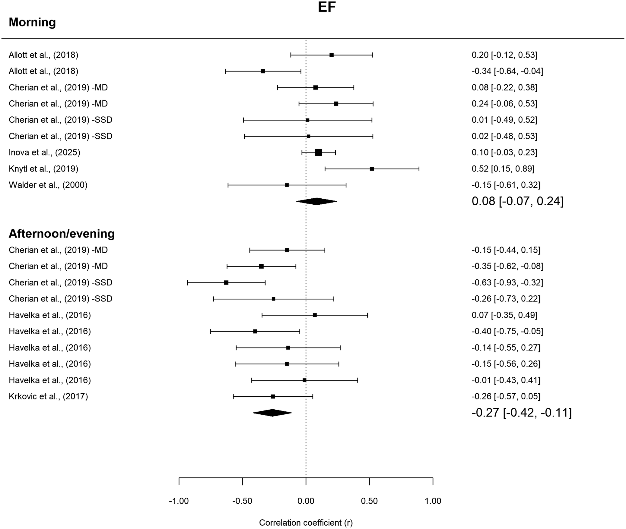
 = 10.98; p = .004). The pooled correlation was not significant for the morning sampling (r = 0.08; 95% CI: −0.07–0.24; p = .30) but was significant for the afternoon/evening one (r = −0.27; 95% CI: −0.42–0.12; p = .01). The forest plot is shown in Figure 3. The within-study (
$ {\tau}_{level2}^2 $
 = .04) and between-study
$ \Big({\tau}_{level3}^2 $
 = .01) heterogeneity explained 8.54% and 20.68% of the total variance, respectively. Applying the robust variance estimation model for the afternoon/evening sampling did not substantially alter the level of significance (r = −0.27; 95% CI: −0.42–0.11; p = .01). The forest plot is shown in Figure 3.
Forest plots of meta-correlations between cortisol and executive function via a three-level model.

Figure 3. Long description
The forest plot is titled E F at the top. The x-axis at the bottom represents the Correlation coefficient r, ranging from -1.00 to 1.00 with a vertical dashed line at 0.00 indicating no effect.
Morning Subgroup:
* Includes 9 entries from studies by Allott et al., Cherian et al. -M D, Cherian et al. -S S D, Inova et al., Knytl et al., and Walder et al.
* Individual correlation coefficients range from -0.34 to 0.52.
* The summary diamond at the bottom of this section is centered at 0.08 with a confidence interval of [-0.07, 0.24], crossing the zero line.
Afternoon/evening Subgroup:
* Includes 10 entries from studies by Cherian et al. -M D, Cherian et al. -S S D, Havelka et al., and Krkovic et al.
* Most data points are positioned to the left of the zero line, with individual correlations ranging from -0.63 to 0.07.
* The summary diamond at the bottom of this section is centered at -0.27 with a confidence interval of [-0.42, -0.11], which does not cross the zero line, indicating a significant negative correlation.
Verbal learning and memory
There was no statistically significant difference in fit between the moderated and unmoderated three-level models (
$ {\chi}_1^2 $
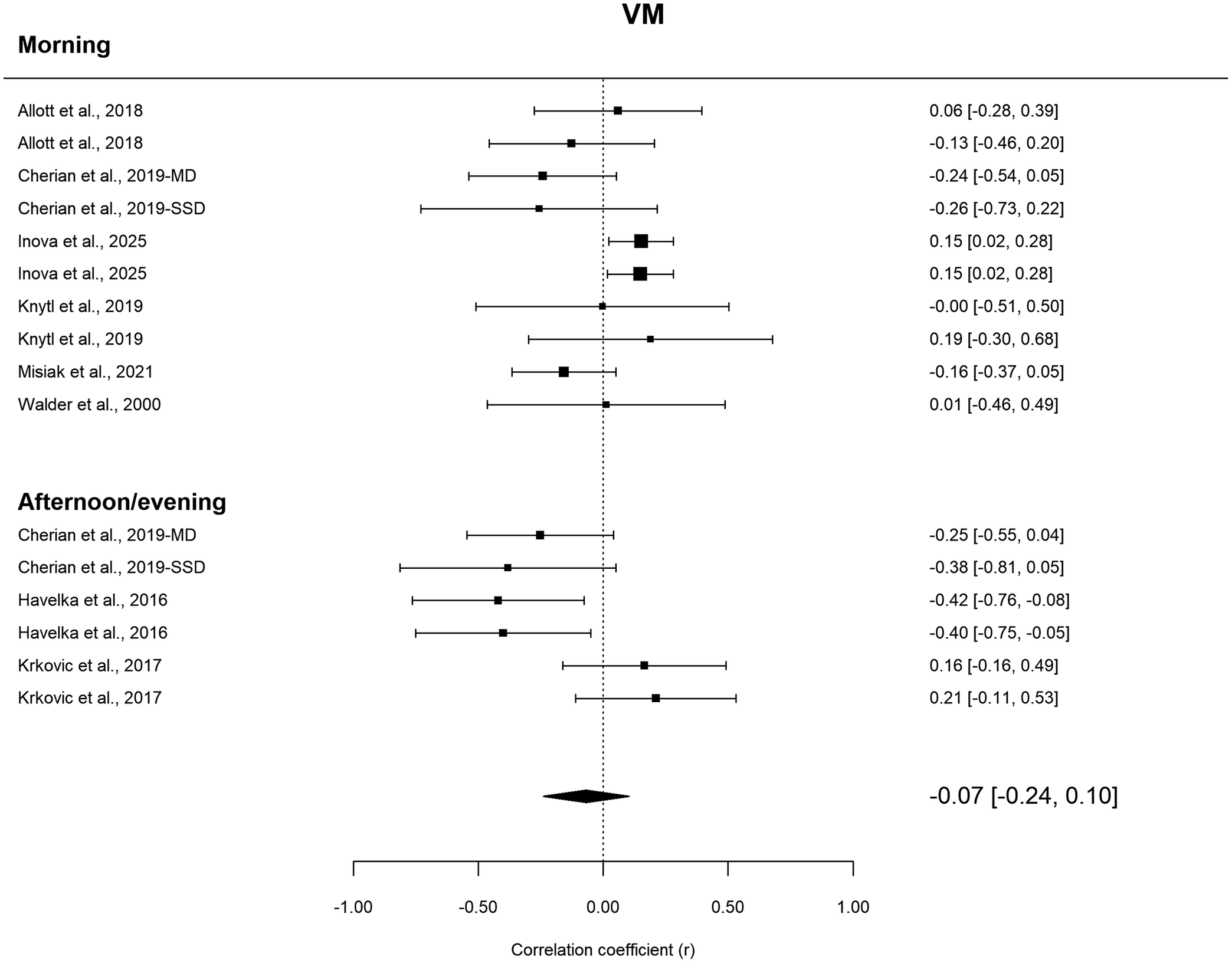
 = 1.95; p = .16). In the robust variance estimated random-effect three-level model with small-sample correction, the pooled correlation was non-significant (r = −0.07; 95% CI: −0.24 − 0.10; p = .39; see Figure 4). Heterogeneity was high (
$ {\tau}^2 $
 = .03;
$ {I}^2 $
 = 59.0%).
Forest plot of three-level meta-correlation between cortisol and verbal learning and memory.

Figure 4. Long description
A forest plot with a vertical dashed line at zero representing the null effect. The x-axis at the bottom measures Correlation coefficient (r) from -1.00 to 1.00.
Morning Subgroup:
* Allott et al., 2018: 0.06 [-0.28, 0.39]
* Allott et al., 2018: -0.13 [-0.46, 0.20]
* Cherian et al., 2019-M D: -0.24 [-0.54, 0.05]
* Cherian et al., 2019-S S D: -0.26 [-0.73, 0.22]
* Inova et al., 2025: 0.15 [0.02, 0.28]
* Inova et al., 2025: 0.15 [0.02, 0.28]
* Knytl et al., 2019: -0.00 [-0.51, 0.50]
* Knytl et al., 2019: 0.19 [-0.30, 0.68]
* Misiak et al., 2021: -0.16 [-0.37, 0.05]
* Walder et al., 2000: 0.01 [-0.46, 0.49]
Afternoon/evening Subgroup:
* Cherian et al., 2019-M D: -0.25 [-0.55, 0.04]
* Cherian et al., 2019-S S D: -0.38 [-0.81, 0.05]
* Havelka et al., 2016: -0.42 [-0.76, -0.08]
* Havelka et al., 2016: -0.40 [-0.75, -0.05]
* Krkovic et al., 2017: 0.16 [-0.16, 0.49]
* Krkovic et al., 2017: 0.21 [-0.11, 0.53]
At the bottom, a black diamond represents the pooled effect size, centered slightly to the left of the zero line with a value of -0.07 [-0.24, 0.10].
Publication bias
An evaluation of publication bias is presented in Supplementary Table 1. All dependent effect sizes were averaged before analysis. Both Egger’s and Begg tests were statistically non-significant, suggesting an absence of publication bias. Funnel plots are shown in Supplementary Figure 3.
Sensitivity analyses
Replacing the multilevel analytic strategy with averaging effect sizes within cognitive domains/studies did not alter the pattern of findings (Supplementary Figures 4–6). Likewise, after removing the two studies that measured saliva cortisol, the pattern of findings remained unaltered for all cognitive domains (Supplementary Table 2). Age and gender were not significant moderators of effect sizes in the domain-specific meta-analyses (Supplementary Table 3).
Applying study-level influence diagnostics to identify studies that may exert undue influence on model estimates or exhibit outlying effect sizes flagged Inova et al. (Reference Inova, Birkenæs, Quintana, Ormerod, Ueland, Ueland, Djurovic, Andreassen, Steen and Aas2025) as an influential and outlier study in relation to the AWP sampling-time moderator model (SDR 
$ {\chi}_1^2 $
 p = .009; CD = 1.19, CDthreshold = 0.50 |; DFBETA|max = 1.57). After excluding Inova et al. (Reference Inova, Birkenæs, Quintana, Ormerod, Ueland, Ueland, Djurovic, Andreassen, Steen and Aas2025), the AWP sampling-time moderator became statistically significant (
$ {F}_{1,9} $
 p = .03) and a significant negative correlation emerged between morning cortisol and AWP (r = −0.21; 95% CI: −0.34–0.08; p = .002), while the afternoon/evening correlation was not significant (r = 0.04; 95% CI: −0.11–0.18; p = .63) (Supplementary Figure 7).
Further analyses: Case–control comparisons
Among the nine meta-correlated studies, seven (excluding Havelka, Prikrylova-Kucerova, Prikryl, & Ceskova, Reference Havelka, Prikrylova-Kucerova, Prikryl and Ceskova2016; and Yazici et al., Reference Yazici, Ogur, Erdogan, Turkmen and Yazici2025) included a control group, and five (excluding Krkovic, Moritz, & Lincoln, Reference Krkovic, Moritz and Lincoln2017; and Walder, Walker, and Lewine, Reference Walder, Walker and Lewine2000) compared cortisol levels between patients and controls. The five studies all measured morning cortisol, but only Cherian et al. (Reference Cherian, Schatzberg and Keller2019) measured afternoon/evening cortisol. A random-effects meta-analysis demonstrated that the difference in morning cortisol between the patient and control groups was not significant (d = 0.11; 95% CI: −0.06–0.28; p = .22). Patients with psychosis demonstrated significantly poorer cognitive performance in all the domains (Supplementary Table 4). Due to the exploratory nature of these analyses, we relaxed our requirement of minimum study number (n = 5) to conduct meta-analyses.
Other cognitive domains and outcomes: A narrative synthesis
The following eligible studies examined cognitive domains that did not meet the threshold for domain-specific meta-analysis. For visual–spatial perception, organisation and learning, Misiak et al. (Reference Misiak, Piotrowski, Chęć and Samochowiec2021) reported a marginal negative correlation with morning cortisol (r = −0.24; p = .06), whereas Yazici et al. (Reference Yazici, Ogur, Erdogan, Turkmen and Yazici2025) reported a significant positive correlation (r = 0.42; p = .008). Misiak et al. (Reference Misiak, Piotrowski, Chęć and Samochowiec2021) found no association with semantic verbal fluency. Several effects could not be assigned to a single cognitive domain, including positive associations with general cognition (Knytl et al., Reference Knytl, Voráčková, Dorazilová, Rodriguez, Cvrčková, Kofroňová, Kuchař, Kratochvílová, Šustová, Čerešňáková and Mohr2019) and negative associations with delayed verbal/visual memory (Misiak et al., Reference Misiak, Piotrowski, Chęć and Samochowiec2021); these were included only in the composite multilevel model.
Three studies examined cortisol measures other than basal cortisol. Aas et al. (Reference Aas, Dazzan, Mondelli, Toulopoulou, Reichenberg, Di Forti, Fisher, Handley, Hepgul, Marques, Miorelli, Taylor, Russo, Wiffen, Papadopoulos, Aitchison, Morgan, Murray and Pariante2011) and Labad et al. (Reference Labad, Gutiérrez-Zotes, Creus, Montalvo, Cabezas, Solé, Ortega, Algora, Sánchez-Gistau and Vilella2016) reported associations between cortisol awakening response (CAR) and poorer cognitive performance, particularly in verbal learning, memory, and processing speed, whereas Aas et al. (Reference Aas, Dazzan, Mondelli, Toulopoulou, Reichenberg, Di Forti, Fisher, Handley, Hepgul, Marques, Miorelli, Taylor, Russo, Wiffen, Papadopoulos, Aitchison, Morgan, Murray and Pariante2011) found no associations with daytime cortisol area-under-the-curve measures. Labad et al. (Reference Labad, Gutiérrez-Zotes, Creus, Montalvo, Cabezas, Solé, Ortega, Algora, Sánchez-Gistau and Vilella2016) also linked a flatter diurnal cortisol slope to poorer spatial working memory in women with early psychosis. Finally, Aas et al. (Reference Aas, Pizzagalli, Laskemoen, Reponen, Ueland, Melle, Agartz, Steen and Andreassen2019) found that higher hair cortisol concentrations were associated with poorer working memory.
Discussion
The present study systematically reviewed and meta-analysed associations between cortisol levels and cognitive functioning in psychosis, examining both global cognition and the domains of attention, working memory and processing speed, executive function, and verbal learning and memory. To our knowledge, this is the first quantitative synthesis of cortisol–cognition associations in psychosis.
Negative correlations between cortisol levels and cognitive function
Our meta-analysis suggests that cortisol–cognition associations in psychosis are both domain- and sampling-time-specific. The strongest and most consistent finding was a negative association between afternoon/evening cortisol levels and executive functioning, which remained significant across multiple analytical approaches despite stringent controls for effect-size dependence. A negative association between morning cortisol and attention, working memory and processing speed also emerged following exclusion of the influential study by Inova et al. (Reference Inova, Birkenæs, Quintana, Ormerod, Ueland, Ueland, Djurovic, Andreassen, Steen and Aas2025), although this effect was not observed in the primary analysis. Together, these findings indicate that cortisol–cognition relationships may be more apparent within specific cognitive domains than at the level of global cognitive functioning.
Previous research in healthy populations has shown that higher daytime cortisol levels are associated with poorer performance across several cognitive domains, including attention, executive functioning, and verbal learning and memory (Evans et al., Reference Evans, Fredhoi, Loveday, Hucklebridge, Aitchison, Forte and Clow2011; Lee et al., Reference Lee, Glass, McAtee, Wand, Bandeen-Roche, Bolla and Schwartz2007). The present findings suggest that a similar relationship may be evident in psychosis, particularly for executive functioning. One potential explanation is that prolonged cortisol exposure affects glucocorticoid-sensitive brain regions involved in cognition, especially the prefrontal cortex (Pruessner et al., Reference Pruessner, Cullen, Aas and Walker2017). Experimental and animal studies indicate that HPA-axis overactivation can disrupt prefrontal cortical pathways through mechanisms such as dendritic atrophy (Radley et al., Reference Radley, Sisti, Hao, Rocher, McCall, Hof, McEwen and Morrison2004) and impaired synaptic plasticity (Cerqueira et al., Reference Cerqueira, Mailliet, Almeida, Jay and Sousa2007, Reference Cerqueira, Almeida and Sousa2008). Cortisol-related hippocampal alterations have also been reported in psychosis (Mondelli et al., Reference Mondelli, Pariante, Navari, Aas, D’Albenzio, Di Forti, Handley, Hepgul, Marques, Taylor, Papadopoulos, Aitchison, Murray and Dazzan2010), although the absence of a significant association between basal cortisol and verbal learning and memory in the present meta-analysis suggests that hippocampal mechanisms provide broader neurobiological context rather than a direct explanation for our findings. More generally, alterations in prefrontal–hippocampal networks may represent one pathway linking HPA-axis dysregulation to cognitive dysfunction in psychosis.
Variations in cognitive measurement and cortisol timing
The cortisol–cognition associations observed in our meta-analysis appeared to be sampling-time-specific. Afternoon/evening cortisol showed the most consistent negative association with executive functioning, whereas associations involving attention, working memory, and processing speed were less stable across sensitivity analyses. One possible explanation is that broad categories such as “morning cortisol” may obscure biologically distinct phases of cortisol secretion. Cortisol levels change rapidly after awakening, and evidence of a blunted cortisol awakening response in psychosis (Aas et al., Reference Aas, Dazzan, Mondelli, Toulopoulou, Reichenberg, Di Forti, Fisher, Handley, Hepgul, Marques, Miorelli, Taylor, Russo, Wiffen, Papadopoulos, Aitchison, Morgan, Murray and Pariante2011) does not necessarily imply uniformly altered cortisol concentrations throughout the morning. Consequently, associations with cognition may differ depending on when samples are collected relative to the post-awakening peak. This issue was also highlighted by Inova et al. (Reference Inova, Birkenæs, Quintana, Ormerod, Ueland, Ueland, Djurovic, Andreassen, Steen and Aas2025), who noted the difficulty of interpreting single morning cortisol measurements without precise timing information. Together, these findings underscore the importance of sampling time when investigating cortisol–cognition relationships in psychosis.
Differences between cognitive domains may also contribute to the observed pattern of findings. Tasks assessing attention, working memory and processing speed are generally less complex than executive function tasks and may be more sensitive to sleep-related fatigue and fluctuations in arousal (Jarraya, Jarraya, Chtourou, & Souissi, Reference Jarraya, Jarraya, Chtourou and Souissi2014; Munnilari et al., Reference Munnilari, Bommasamudram, Easow, Tod, Varamenti, Edwards, Ravindrakumar, Gallagher and Pullinger2024). Given the high prevalence of sleep disturbance in psychosis (Reeve, Sheaves, & Freeman, Reference Reeve, Sheaves and Freeman2015), variability in sleep and morning sampling procedures may therefore be particularly relevant when examining these domains. By contrast, executive function tasks place greater demands on higher-order cognitive control and may be less susceptible to transient fluctuations in arousal, allowing associations with cortisol to emerge more clearly. These findings highlight the importance of precise cortisol sampling protocols, including reporting clock time, time since awakening, and, where feasible, repeated morning measurements.
Cortisol and HPA dysregulation in psychosis: How much is stress involved?
Cortisol is commonly viewed as a biomarker of stress and HPA-axis activity, yet the extent to which psychosocial stress directly contributes to cognitive impairment in psychosis remains unclear. Although stress exposure features prominently in models of psychosis, evidence linking HPA-axis abnormalities to perceived or chronic stress has been inconsistent (Aas et al., Reference Aas, Dazzan, Mondelli, Toulopoulou, Reichenberg, Di Forti, Fisher, Handley, Hepgul, Marques, Miorelli, Taylor, Russo, Wiffen, Papadopoulos, Aitchison, Morgan, Murray and Pariante2011; Cullen et al., Reference Cullen, Rai, Vaghani, Mondelli and McGuire2020). Nevertheless, diathesis–stress models propose that stress-related neurobiological processes may influence cognition through alterations in dopaminergic, inflammatory, and hippocampal–prefrontal pathways (Howes & McCutcheon, Reference Howes and McCutcheon2017; Niwa et al., Reference Niwa, Jaaro-Peled, Tankou, Seshadri, Hikida, Matsumoto, Cascella, Kano, Ozaki, Nabeshima and Sawa2013; Pruessner et al., Reference Pruessner, Cullen, Aas and Walker2017).
The present findings do not permit conclusions about the role of stress per se, as cortisol may reflect biological processes beyond psychosocial stress. Moreover, HPA-axis dysregulation may arise from factors intrinsic to psychosis, including genetic vulnerability, neurodevelopmental alterations, medication effects, and chronic illness burden. Accordingly, the observed cortisol–cognition associations should not be interpreted as evidence that stress directly causes cognitive impairment. Rather, they suggest that HPA-axis dysregulation may represent one component of a broader neurobiological network associated with cognitive dysfunction in psychosis. Future studies combining cortisol measures with direct assessments of stress exposure and stress reactivity may help clarify the contribution of stress-related processes to cognitive impairment in this population.
Limitations and future research
The present systematic review and meta-analysis provides the first quantitative synthesis of associations between basal cortisol and cognitive functioning in psychosis, while also examining the potential moderating role of cortisol sampling time across specific cognitive domains. Several strengths enhance confidence in the findings. First, we used three-level meta-analytic models to account for the dependence arising from multiple effect sizes contributed by individual studies, thereby avoiding the loss of information associated with conventional averaging approaches. Second, the principal findings remained robust across multiple sensitivity analyses, including alternative analytic strategies, exclusion of saliva-based cortisol studies, and adjustment for potentially influential studies. Third, the convergence between the quantitative meta-analytic findings and the broader narrative synthesis strengthens the overall interpretation of the evidence base.
Nevertheless, several limitations should be acknowledged. Although we adhered to recommended methodological guidance regarding the minimum number of studies required for meta-analysis (Cheung, Reference Cheung2019; Myung, Reference Myung2023), the relatively small evidence base limited statistical power and contributed to wide confidence intervals, particularly in moderator and domain-specific analyses. The observed effect sizes were also modest. However, this is not unexpected given the multifactorial nature of psychosis and its substantial clinical and etiological heterogeneity, where any single biological factor is unlikely to account for a large proportion of variance in cognitive functioning. In addition, substantial heterogeneity was observed in several models, reflecting the methodological and clinical diversity of the available literature. The small number of studies also limited the ability to evaluate publication bias and prevented a systematic examination of cortisol measures beyond basal cortisol, such as the cortisol awakening response and diurnal cortisol slope, both of which may be relevant to cognitive functioning in psychosis (Haussler et al., Reference Haussler, Zahid, Day, Ciufolini, Petros, Gifford, Alameda, Quattrone, Dazzan, Pariante, Fisher, Laurens, Di Forti, Wood, Murray, McGuire, Mondelli and Cullen2025).
An additional limitation concerns the grouping of attention, working memory, and processing speed into a single AWP domain. Although these constructs are theoretically distinct and this approach may have obscured more specific associations with cortisol, many neuropsychological measures used in psychosis research draw on overlapping attentional, working-memory, and processing-speed processes and cannot be readily assigned to a single domain. As such, the broader AWP classification may provide a more ecologically valid representation of the cognitive constructs assessed across studies while facilitating meaningful quantitative synthesis.
Finally, we did not formally adjust for multiple comparisons because the domain-specific and sampling-time analyses were not conceptualised as tests of a single omnibus null hypothesis (García-Pérez, Reference García-Pérez2023). Moreover, given the modest evidence base, strict multiplicity correction would have increased the risk of Type II errors and potentially obscured meaningful associations. Nevertheless, given the number of cognitive endpoints and stratified analyses performed, statistically significant findings should be interpreted cautiously and viewed as hypothesis-generating pending replication.
Future research should investigate cortisol–cognition associations across different stages of psychosis, from the premorbid and prodromal phases to chronic illness (Kristensen et al., Reference Kristensen, Mager, Ambrosen, Barber, Lemvigh, Bojesen, Nielsen, Fagerlund, Glenthøj, Syeda, Glenthøj and Ebdrup2024). Longitudinal studies may help clarify the temporal relationship between HPA-axis dysregulation and cognitive impairment, while identifying opportunities for early intervention.
Further work should incorporate more precise assessments of cortisol dynamics, including repeated morning sampling, the cortisol awakening response, and diurnal cortisol slopes. Given the domain- and sampling-time-specific associations observed in the present review, future studies should also examine whether different cognitive domains are differentially sensitive to distinct aspects of HPA-axis functioning. Larger, methodologically rigorous studies will be essential to establish the robustness and clinical relevance of cortisol–cognition associations in psychosis.
Conclusion
Overall, this review provides the first quantitative synthesis of cortisol–cognition associations in psychosis and suggests that these relationships may be both domain- and sampling-time-specific. The most robust finding was a negative association between afternoon/evening cortisol and executive functioning, supporting a potential role for HPA-axis dysregulation in cognitive impairment. Sensitivity analyses also identified a negative association between morning cortisol and attention, working memory and processing speed, although this finding requires replication. Future studies using more precise cortisol assessments will be important for clarifying how cortisol dynamics relate to cognitive functioning across psychotic disorders.
Supplementary material
The supplementary material for this article can be found at http://doi.org/10.1017/S0033291726106266.
Author contribution
Conceptualisation: E.K.; Data curation: E.K., H.Z.C., J.X.M.Y.; Formal analysis: E.K., H.Z.C., J.X.M.Y.; Investigation: E.K., H.Z.C., J.X.M.Y.; Methodology: E.K., H.Z.C., J.X.M.Y., S.M.H.; Software: F.I., H.Z.C., J.X.M.Y.; Supervision: E.K.; Validation: C.M.P., R.M.M., S.M.H.; Visualisation: H.Z.C., J.X.M.Y.; Writing – original draft preparation: E.K., H.Z.C., J.X.M.Y.; Writing – review & editing: C.M.P., E.K., F.I., H.Z.C., J.X.M.Y., R.M.M., S.M.H.
Funding statement
E.K. and R.M.M. are supported by the NIHR Maudsley Biomedical Research Centre at South London and Maudsley National Health Service (NHS) Foundation Trust and King’s College London.
Competing interests
The authors declare no potential conflict of interest.




 Open access

