


Introduction
Next-generation sequencing (NGS)-based preimplantation genetic testing for aneuploidy (PGT-A) is widely used to improve embryo selection in assisted reproductive technology (Practice Committees of the American Society for Reproductive Medicine and the Society for Assisted Reproductive Technology, 2024). However, the transfer of embryos classified as euploid by PGT-A does not always result in successful pregnancy and live birth (Iwasa et al., Reference Iwasa, Kuwahara, Takeshita, Taniguchi, Mikami and Irahara2023; Practice Committees of the American Society for Reproductive Medicine and the Society for Assisted Reproductive Technology, 2024; Dong et al., Reference Dong, Shi, Liu, Liu, Li, Zhao and Xue2025). While multiple factors, including uterine, immunological and embryo-related factors, may contribute to unsuccessful outcomes after euploid embryo transfer, the mechanisms underlying these failures remain incompletely understood (Reshef et al., Reference Reshef, Robles, Hynes, Turocy and Forman2022; Kuroda, Reference Kuroda2024).
One limitation of conventional copy number–based PGT-A is that it may not reliably determine whole-genome ploidy status, including haploidy and triploidy, because normalization-based copy number analysis can fail to distinguish genome-wide ploidy changes from diploidy (Xu et al., Reference Xu, Niu, Peng, Bao, Zhang, Wang, Du, Zhang and Sun2016; Kratka et al., Reference Kratka, Vadapalli, Mendola, Garrisi, Xu, Treff and Marin2023). Single-nucleotide polymorphism (SNP)-based methods can complement conventional copy number–based PGT-A by evaluating allelic patterns across the genome, thereby enabling the detection of whole-genome ploidy abnormalities such as haploidy and triploidy (Walters-Sen et al., Reference Walters-Sen, Neitzel, Bristow, Mitchell, Alouf, Aradhya and Faulkner2022; Kratka et al., Reference Kratka, Vadapalli, Mendola, Garrisi, Xu, Treff and Marin2023).
Previous studies have reported the presence of whole-genome ploidy abnormalities among embryos classified as euploid using SNP-based approaches (Girardi et al., Reference Girardi, Patassini, Miravet Valenciano, Sato, Fagundes Cagnin, Castellón, Cogo, Zambon, Blesa, Jimenez Almazan, Akinwole, Coprerski and Rubio2024; McCallie et al., Reference McCallie, Haywood, Henry, Lee, Schoolcraft and Katz-Jaffe2025). These findings suggest that a subset of embryos considered suitable for transfer may still harbour underlying genomic abnormalities. Nevertheless, additional clinical data are needed to further characterize the occurrence of these abnormalities and their practical relevance in routine PGT-A workflows.
In this study, we retrospectively evaluated embryos classified as euploid or mosaic by NGS-based PGT-A that underwent additional SNP-based ploidy analysis. The aim of this study was to determine the frequency of whole-genome ploidy abnormalities and to describe the distribution of SNP-based ploidy classifications within a clinical cohort.
Materials and methods
Patient and embryo selection
Between May 2022 and January 2025, blastocysts from patients undergoing PGT-A at Sugiyama Clinic were retrospectively analysed. This study was approved by the institutional review board of Sugiyama Clinic, and written informed consent was obtained from all patients. In this study, embryos derived from 2PN and 1PN zygotes were included, whereas embryos derived from multipronuclear (≥3PN) zygotes were excluded. Only embryos classified as euploid or mosaic by initial NGS-based PGT-A were included for subsequent SNP-based ploidy analysis. Mosaic embryos were defined as embryos showing intermediate copy number changes that did not meet the criteria for full aneuploidy. During the study period, the indication for SNP-based analysis changed: both XX and XY embryos were included in 2022, whereas from 2023 onward, SNP analysis was performed only on XX embryos.
Ovarian stimulation and oocyte retrieval
Ovarian stimulation was individualized according to patient age, ovarian reserve and clinical background, using standard clinical protocols at Sugiyama Clinic, including mild ovarian stimulation and controlled ovarian stimulation protocols. Follicular development was monitored by transvaginal ultrasonography and serum hormone measurements. Final oocyte maturation was triggered based on follicular development, and oocyte retrieval was generally performed 35–36 hours later under transvaginal ultrasound guidance.
Insemination and embryo culture
The insemination method was selected as either conventional insemination (C-IVF) or intracytoplasmic sperm injection (ICSI) according to semen parameters and clinical indications. For C-IVF, spermatozoa were prepared using density gradient centrifugation followed by swim-up, and oocytes were inseminated at a final sperm concentration of 1.0 × 10⁵ motile spermatozoa/mL for either 5 h or overnight according to the standard laboratory protocol. For ICSI, spermatozoa were prepared using density gradient centrifugation followed by swim-up or using a microfluidic sperm selection device. Cumulus cells were removed shortly after oocyte retrieval using hyaluronidase, and ICSI was performed at least 2.5 h after confirmation of metaphase II oocytes. ICSI was performed using a piezo-assisted micromanipulation system (PrimeTech, Ibaraki, Japan). Fertilization status was assessed at the fertilization check based on the number of pronuclei. All embryos were cultured in a time-lapse incubator (Geri; Genea Biomedx, Sydney, Australia) under 2% O2 and 6% CO2.
Trophectoderm biopsy
Trophectoderm (TE) biopsy was performed on Day 5 or 6 expanded blastocysts with a TE grade of A or B according to the Gardner criteria (Gardner and Schoolcraft, Reference Gardner and Schoolcraft1999), using a combination of laser-assisted and flicking techniques (Nakagawa et al., Reference Nakagawa, Horikawa, Watanabe, Shirai, Kataoka, Takamizawa, Maruyama, Kuroda and Sugiyama2025). Briefly, laser pulses were applied between adjacent TE cells to induce cellular contraction immediately before biopsy. A small opening was then created in the zona pellucida on the side opposite the inner cell mass, and a biopsy pipette was inserted through the opening. TE cells were aspirated into the biopsy pipette and separated using laser assistance and mechanical flicking. Approximately five TE cells were collected for genetic analysis. For embryos identified as haploid or triploid by SNP analysis, time-lapse recordings were retrospectively reviewed to evaluate the number of polar bodies. This assessment was not performed for all embryos.
NGS-based PGT-A and SNP-based ploidy analysis
Biopsied cells were subjected to whole-genome amplification, followed by NGS-based copy number analysis performed by an external laboratory (Igenomix) using validated protocols. Embryos classified as euploid or mosaic according to laboratory criteria were subsequently analysed using SNP-based methods on the same biopsy samples. SNP-based ploidy status was determined using proprietary bioinformatic algorithms based on Variant Allele Frequency (VAF) distribution (Girardi et al., Reference Girardi, Patassini, Miravet Valenciano, Sato, Fagundes Cagnin, Castellón, Cogo, Zambon, Blesa, Jimenez Almazan, Akinwole, Coprerski and Rubio2024).
The diagnostic process employed a dual-strategy approach: VAF distribution analysis, which evaluates the number and position of peaks within the VAF distribution to identify patterns characteristic of haploid, diploid, or triploid states; and statistical likelihood evaluation, which compares observed genomic data against theoretical ploidy models to determine the most probable state. Final ploidy results were reported as diploid, haploid, or triploid when both algorithms yielded concordant results. Cases with discordant results between the two algorithms or without a definitive classification were categorized as uncertain.
Results
PGT-A outcomes
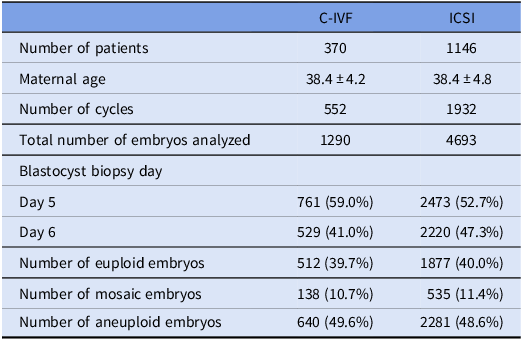
A total of 4693 embryos derived from ICSI and 1290 embryos derived from C-IVF underwent PGT-A. Among ICSI embryos, 1877 were classified as euploid, 535 as mosaic and 2281 as aneuploid. Among C-IVF embryos, 512 were euploid, 138 mosaic and 640 aneuploid (Table 1).
Baseline characteristics and PGT-A outcomes

Table 1. Long description
The table compares baseline characteristics and PGT-A outcomes for embryos derived from C-IVF and ICSI. It has two columns labeled C-IVF and ICSI, and eight rows with the following headers: Number of patients, Maternal age, Number of cycles, Total number of embryos analyzed, Blastocyst biopsy day, Number of euploid embryos, Number of mosaic embryos, and Number of aneuploid embryos. Row 1: Number of patients, 370; 1146. Row 2: Maternal age, 38.4 ± 4.2; 38.4 ± 4.8. Row 3: Number of cycles, 552; 1932. Row 4: Total number of embryos analyzed, 1290; 4693. Row 5: Blastocyst biopsy day, Day 5, 761 (59.0 percent); 2473 (52.7 percent), Day 6, 529 (41.0 percent); 2220 (47.3 percent). Row 6: Number of euploid embryos, 512 (39.7 percent); 1877 (40.0 percent). Row 7: Number of mosaic embryos, 138 (10.7 percent); 535 (11.4 percent). Row 8: Number of aneuploid embryos, 640 (49.6 percent); 2281 (48.6 percent).
Values are presented as number, number (%), or mean ± standard deviation. Percentages for blastocyst biopsy day and PGT-A outcomes were calculated using the total number of embryos analyzed in each fertilization group as the denominator. C-IVF = conventional in vitro fertilization; ICSI = intracytoplasmic sperm injection; PGT-A = preimplantation genetic testing for aneuploidy.
SNP-based ploidy results
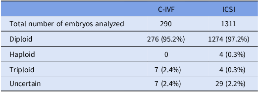
Of the 5983 embryos that underwent NGS-based PGT-A, 1601 embryos classified as euploid or mosaic were subjected to SNP-based ploidy analysis. Of these, 1550 (96.8%) were classified as diploid, 4 (0.3%) as haploid and 11 (0.7%) as triploid. The remaining 36 embryos (2.2%) were categorized as uncertain (Table 2).
SNP-based ploidy results according to fertilization method
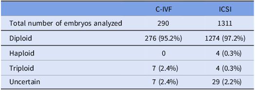
Table 2. Long description
A table comparing embryo ploidy results by fertilization method. The table has two columns labeled C-IVF and ICSI, and four rows labeled Total number of embryos analyzed, Diploid, Haploid, Triploid, and Uncertain. Row 1: Total number of embryos analyzed, C-IVF, 290; ICSI, 1311. Row 2: Diploid, C-IVF, 276 (95.2 percent); ICSI, 1274 (97.2 percent). Row 3: Haploid, C-IVF, 0; ICSI, 4 (0.3 percent). Row 4: Triploid, C-IVF, 7 (2.4 percent); ICSI, 4 (0.3 percent). Row 5: Uncertain, C-IVF, 7 (2.4 percent); ICSI, 29 (2.2 percent).
Values are presented as number (%). Percentages were calculated using the total number of embryos subjected to SNP-based ploidy analysis in each fertilization group as the denominator. C-IVF = conventional in vitro fertilization; ICSI = intracytoplasmic sperm injection; SNP = single-nucleotide polymorphism.
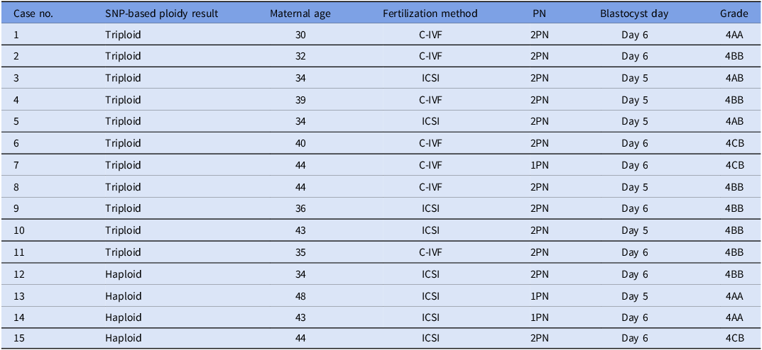
Among the 11 triploid embryos identified among embryos classified as euploid or mosaic by conventional NGS-based PGT-A, 4 were derived from ICSI and 7 from C-IVF. Detailed characteristics of embryos classified as haploid or triploid are shown in Table 3. All haploid and triploid embryos derived from ICSI exhibited second polar body extrusion upon retrospective time-lapse evaluation.
Characteristics of embryos classified as haploid or triploid by SNP-based ploidy analysis

Table 3. Long description
A table comparing characteristics of embryos classified as haploid or triploid by SNP-based ploidy analysis. The table has 15 rows and 7 columns. Column headers are Case no., SNP-based ploidy result, Maternal age, Fertilization method, PN, Blastocyst day, and Grade. Row 1: Case no. 1, SNP-based ploidy result Triploid, Maternal age 30, Fertilization method C-IVF, PN 2PN, Blastocyst day Day 6, Grade 4AA. Row 2: Case no. 2, SNP-based ploidy result Triploid, Maternal age 32, Fertilization method C-IVF, PN 2PN, Blastocyst day Day 6, Grade 4BB. Row 3: Case no. 3, SNP-based ploidy result Triploid, Maternal age 34, Fertilization method ICSI, PN 2PN, Blastocyst day Day 5, Grade 4AB. Row 4: Case no. 4, SNP-based ploidy result Triploid, Maternal age 39, Fertilization method C-IVF, PN 2PN, Blastocyst day Day 5, Grade 4BB. Row 5: Case no. 5, SNP-based ploidy result Triploid, Maternal age 34, Fertilization method ICSI, PN 2PN, Blastocyst day Day 5, Grade 4AB. Row 6: Case no. 6, SNP-based ploidy result Triploid, Maternal age 40, Fertilization method C-IVF, PN 2PN, Blastocyst day Day 6, Grade 4CB. Row 7: Case no. 7, SNP-based ploidy result Triploid, Maternal age 44, Fertilization method C-IVF, PN 1PN, Blastocyst day Day 6, Grade 4CB. Row 8: Case no. 8, SNP-based ploidy result Triploid, Maternal age 44, Fertilization method C-IVF, PN 2PN, Blastocyst day Day 5, Grade 4BB. Row 9: Case no. 9, SNP-based ploidy result Triploid, Maternal age 36, Fertilization method ICSI, PN 2PN, Blastocyst day Day 6, Grade 4BB. Row 10: Case no. 10, SNP-based ploidy result Triploid, Maternal age 43, Fertilization method ICSI, PN 2PN, Blastocyst day Day 5, Grade 4BB. Row 11: Case no. 11, SNP-based ploidy result Triploid, Maternal age 35, Fertilization method C-IVF, PN 2PN, Blastocyst day Day 6, Grade 4BB. Row 12: Case no. 12, SNP-based ploidy result Haploid, Maternal age 34, Fertilization method ICSI, PN 2PN, Blastocyst day Day 6, Grade 4BB. Row 13: Case no. 13, SNP-based ploidy result Haploid, Maternal age 48, Fertilization method ICSI, PN 1PN, Blastocyst day Day 5, Grade 4AA. Row 14: Case no. 14, SNP-based ploidy result Haploid, Maternal age 43, Fertilization method ICSI, PN 1PN, Blastocyst day Day 6, Grade 4AA. Row 15: Case no. 15, SNP-based ploidy result Haploid, Maternal age 44, Fertilization method ICSI, PN 2PN, Blastocyst day Day 6, Grade 4CB.
C-IVF = conventional in vitro fertilization; ICSI = intracytoplasmic sperm injection; PN = pronucleus; SNP = single-nucleotide polymorphism.
Discussion
In this study, whole-genome ploidy abnormalities were detected in 15 of 1601 embryos (0.9%) classified as euploid or mosaic by NGS-based PGT-A. These findings indicate that conventional copy number–based PGT-A does not completely exclude haploidy or triploidy. Because conventional copy number–based PGT-A primarily evaluates relative chromosomal copy number, genome-wide ploidy changes may remain undetected, particularly when no additional aneuploidies or Y chromosome signal are present (Kratka et al., Reference Kratka, Vadapalli, Mendola, Garrisi, Xu, Treff and Marin2023; De Witte et al., Reference De Witte, Baetens, Tilleman, Vanden Meerschaut, Janssens, Van Tongerloo, Szymczak, Stoop, Dheedene, Symoens and Menten2024). Although the frequency observed in this study was quite low, undetected whole-genome ploidy abnormalities may represent one possible embryo-related factor not captured by conventional PGT-A.
Triploidy may arise through dispermic fertilization, fertilization by a diploid spermatozoon, or failure of second polar body extrusion (Rosenbusch, Reference Rosenbusch2008). In the present study, triploid embryos were also observed after ICSI. Because ICSI involves the injection of a single spermatozoon, dispermy is unlikely in these cases. Moreover, retrospective time-lapse review confirmed second polar body extrusion in all triploid embryos derived from ICSI, making failure of second polar body extrusion less likely as an explanation. Therefore, one possible mechanism is fertilization by a diploid spermatozoon. However, because parental origin of the additional chromosome set was not determined, this possibility remains speculative.
Haploidy may be related to abnormal fertilization processes, such as failure of either the paternal or maternal genome to form a visible pronucleus, loss or exclusion of one parental genome, or development through parthenogenetic or androgenetic mechanisms (Hirata et al., Reference Hirata, Goto, Izumi, Taguchi, Hayashi, Fujioka, Ishiko, Nakanishi, Kimura and Murakami2020; McCallie et al., Reference McCallie, Haywood, Henry, Lee, Schoolcraft and Katz-Jaffe2025). In the present study, 2 of the 4 haploid embryos were derived from 1PN zygotes. Previous studies have reported that a substantial proportion of 1PN-derived blastocysts may be haploid, with one report showing a haploidy rate of 36.4% (McCallie et al., Reference McCallie, Haywood, Henry, Lee, Schoolcraft and Katz-Jaffe2025). Therefore, in these 2 embryos, failure of either the male or female pronucleus to form may represent a possible mechanism. However, without parental origin analysis, it was not possible to distinguish between paternal and maternal mechanisms.
The presence of haploid and triploid embryos despite conventional fertilization assessment suggests that morphological evaluation alone may not reliably reflect the underlying genomic constitution (Girardi et al., Reference Girardi, Patassini, Miravet Valenciano, Sato, Fagundes Cagnin, Castellón, Cogo, Zambon, Blesa, Jimenez Almazan, Akinwole, Coprerski and Rubio2024). Furthermore, 12 of the 15 embryos classified as haploid or triploid in this study had good morphological blastocyst quality. If these embryos had been classified as euploid by conventional PGT-A alone, they would likely have been considered candidates for transfer. This finding highlights the limitations of conventional assessment based on fertilization status, blastocyst morphology and copy number–based PGT-A in identifying whole-genome ploidy abnormalities.
In addition to embryos classified as haploid or triploid, 36 embryos were categorized as uncertain by SNP-based analysis. These cases represented results in which a definitive ploidy classification could not be obtained, including discordance between the two internal algorithms. Although uncertain results should not be interpreted as abnormal ploidy, their occurrence highlights a practical limitation of SNP-based ploidy assessment in routine clinical workflows (Cascante et al., Reference Cascante, Besser, Lee, Wang, McCaffrey and Grifo2023; Tšuiko et al., Reference Tšuiko, Vanneste, Melotte, Ding, Debrock, Masset, Peters, Salumets, De Leener, Pirard, Kluyskens, Hostens, van de Vijver, Peeraer, Denayer, Vermeesch and Dimitriadou2021).
From a clinical perspective, although the overall frequency was low, the detection of these abnormalities may be relevant because embryos classified as euploid by conventional PGT-A are generally considered suitable for transfer. SNP-based analysis may therefore provide additional value as a complementary tool to conventional copy number–based PGT-A, particularly in selected clinical settings (Kratka et al., Reference Kratka, Vadapalli, Mendola, Garrisi, Xu, Treff and Marin2023; De Witte et al., Reference De Witte, Baetens, Tilleman, Vanden Meerschaut, Janssens, Van Tongerloo, Szymczak, Stoop, Dheedene, Symoens and Menten2024).
This study has several limitations. First, SNP analysis was performed only in embryos classified as euploid or mosaic, and therefore, the overall prevalence of ploidy abnormalities across all embryos could not be assessed. Second, the indication for SNP analysis changed during the study period; both XX and XY embryos were included until January 2023, whereas SNP analysis was subsequently performed only for XX embryos, which may have introduced selection bias. Third, detailed raw data from the SNP analysis were not available, limiting further characterization of uncertain cases. Fourth, morphokinetic evaluation of polar body extrusion was performed retrospectively and only in selected embryos, which may also introduce selection bias.
In conclusion, a small but measurable proportion of embryos classified as euploid or mosaic by NGS-based PGT-A showed whole-genome ploidy abnormalities detectable by SNP analysis. These findings highlight a residual diagnostic gap in conventional copy number–based PGT-A and suggest a potential role for complementary SNP-based ploidy assessment in selected clinical workflows.
Funding
This research received no specific grant from any funding agency, commercial or not-for-profit sectors.
Competing interests
The author(s) declare none.
Ethical standards
The authors assert that all procedures contributing to this work comply with the ethical standards of the relevant national and institutional committees on human experimentation and with the Helsinki Declaration of 1975, as revised in 2008. This study was approved by the institutional review board of Sugiyama Clinic, and written informed consent was obtained from all patients.
Use of AI tools
The authors used ChatGPT (OpenAI; accessed in July 2026) to assist with English language editing. The tool was not used for data generation, statistical analysis, or image generation. All AI-assisted text was reviewed, edited and approved by the authors, who take full responsibility for the content of the manuscript.