Introduction
The association between gastric cancer, peptic ulcer and gastric inflammation (gastritis) has been recognised since at least the late 19th century (Ref. Reference Faber1). Gastric cancer was one of the most common, if not the most common, cancer in most Western countries throughout the first half of the 20th century (Ref. Reference Graham2). The high prevalence, morbidity and mortality of peptic ulcer disease and gastric cancer resulted in a correspondingly high interest in understanding their pathogenesis as well as the role of the gastritis in their pathogenesis (Ref. Reference Graham and Asaka3). The 20th century saw a marked decline in the incidence gastric cancer and a parallel increase in duodenal ulcer disease (Refs Reference Graham2, Reference Baron and Sonnenberg4). In the early 1980s, Warren and Marshall proved that Helicobacter pylori was a cause of gastritis and suggested that the infection might also be aetiologically responsible for both peptic ulcer disease and gastric cancer (Ref. Reference Marshall and Warren5).
In 1994, based largely on epidemiological studies, H. pylori was defined as a class I human carcinogen (Ref. 6). Although epidemiologic studies showed that approximately half of the world's population was infected with H. pylori, in some areas gastric cancer was rare despite the high prevalence of H. pylori infections (Ref. Reference Yamaoka7). This suggested that H. pylori strains might differ in their propensity to cause cancer and these differences might be responsible for the markedly geographic differences in H. pylori-related diseases (Ref. Reference Graham2). It was also discovered that the clinical manifestations of H. pylori infection could rapidly change (Refs Reference Graham8, Reference Kamada9, Reference Wang, Weber and Graham10) exemplified by the marked decline in the incidence of gastric cancer and increase in duodenal ulcer experienced during the 20th century in Western countries (Ref. Reference Baron and Sonnenberg4).
Research related to H. pylori initially focused on attempting to understand the role of the organism in disease pathogenesis. The importance of the host and environment were most often studied separately. Recently, it has become clear that the interactions between the bacterium, the host and environment are critical in dictating outcome (Refs Reference Graham8, Reference Yamaoka and Graham11).
H. pylori is a necessary agent for gastric cancer
Gastric cancer is an inflammation-associated cancer with H. pylori infection being the most common cause of progressive and persistent inflammation that eventually results in gastric cancer (Ref. Reference Graham8). The infection results in a cascade of histologic changes beginning with mild superficial gastritis and that may eventually result in gastric atrophy during which the normal gastric mucosa is converted into a metaplastic epithelia (Ref. Reference Correa12). This also results in a progressive reduction in the gastric acid barrier that inhibits other bacteria from populating the stomach (Ref. Reference Ruddell13). Cure of H. pylori infection halts the progression of mucosal damage, resolution of the resulting acute and chronic inflammation, and healing of any still reversible damage. It is now recognised that worldwide gastric cancer can be largely be eliminated by H. pylori eradication (Refs Reference Graham8, Reference Lee14, Reference Lee15). Even among those infected and at risk, the risk of developing gastric cancer is reduced following H. pylori eradication. H. pylori eradication prevents further the progression of damage and ‘locks in’ or reduces the level of risk present at the time of eradication, but does not eliminate the risk of subsequent cancer (Ref. Reference Graham and Shiotani16).
H. pylori in gastric cancer pathogenesis
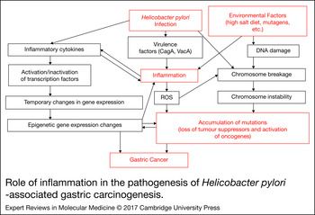
Interactions between H. pylori infection, the host and the environment serve to either potentate or reduce the risks of any particular H. pylori-related clinical outcome (Ref. Reference Hanada and Graham17) (Fig. 1). For example, host genetic polymorphisms associated with an enhanced inflammatory response, such as IL-1β−511 T, IL-1B-31 C alleles and IL-1RN *2/*2, also enhance the risk of gastric cancer (Ref. Reference Xue18). This risk and rate of development of atrophic gastritis and gastric cancer can be further amplified by the infection with a more virulent strain of H. pylori (e.g. CagA-positive strains), which are also associated with an enhanced inflammatory host response (Ref. Reference Figueiredo19). The environment, especially the diet, plays an especially important modifying role in pathogen–host interactions (Ref. Reference Graham2). This critical effect of diet can be seen in Japan, a high gastric cancer country. In Japan, the vast majority of infections are caused by highly virulent H. pylori strains. Between 1965 and 1995, the incidence of gastric cancer in Japan fell approximately 60% in all age groups (Refs Reference Kamada9, Reference Wang, Weber and Graham10). This change occurred despite no change in the virulence of the most common circulating H. pylori strain or in the genetics of the hosts (Refs Reference Kamada9, Reference Wang, Weber and Graham10). The decline was however associated with environmental changes (i.e. in food preservation from salt to refrigeration, in westernising of the diet, and in a reduction in smoking) (Ref. Reference Kamada9).
Role of inflammation in the pathogenesis of Helicobacter pylori-associated gastric carcinogenesis. Adapted from Hanada and Graham (Ref. Reference Hanada and Graham17), with permission.

Role of H. pylori virulence factors
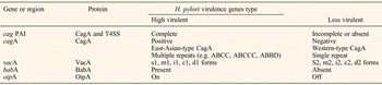
Genomic sequence comparison of clinical H. pylori isolates has shown that approximately 7% of the proteins are strain-specific (Ref. Reference Sgouros and Bergele20). Proteins not present in all strains, or a genotype variety that associate with clinical disease are termed disease-associated virulence factors. More appropriately, they should be called putative virulence factors (Table 1) (Refs Reference Yamaoka7, Reference Lu, Yamaoka and Graham21) in that the association of clinical outcome (e.g. duodenal ulcer) and a putative factor may also occur by chance. Suggested criteria for a valid and biologically important virulence factor include: (1) the presence of the factor should reliably correlate with a disease or a significant increase in mucosal inflammation; (2) the association should exhibit epidemiologic consistency across populations and regions; (3) the effect of the factor should have biologic plausibility (e.g. be involved in a relevant biologic process such as inflammation); and (4) the propertied biologic activity should be reduced or eliminated by gene deletion and restored by complementation (Ref. Reference Lu, Yamaoka and Graham21).
Putative virulence factors possibly related to clinical outcomes

CagA as a risk factor and as an oncoprotein
Most of the currently recognised putative virulence factors are also associated with enhanced mucosal inflammation (Ref. Reference Yamaoka7). The most extensively studied virulence factors are the cag pathogenicity island (cag PAI) and the vacuolating cytotoxin, VacA. The cag PAI encodes a type IV secretion system (T4SS) (Ref. Reference Censini22). The CagA protein is a 120 –145-k immunodominant protein, which is injected into the host cell via the T4SS (Ref. Reference Asahi23). In both humans and experimental animals, CagA expression is associated with a marked host inflammatory response and an increased risk development of mucosal ulcerations and atrophy (Ref. Reference Yamaoka7). The presence of CagA-positive increased the risk for development of gastric cancer (Ref. Reference van Doorn24) approximately 2-fold (odds ratio (OR) = 2.01, 95% confidence interval (CI) = 1.21–3.32) (Ref. Reference Huang25). Gastritis, peptic ulcer and gastric cancer also occur with CagA-negative H. pylori infections, albeit at a lower rate (Ref. Reference Parsonnet26).
Infection with CagA-positive strains is associated with increase in IL-8 production by infected cells (Refs Reference Brandt27, Reference Crabtree28, Reference Yamaoka29). In vitro experiments have also shown that infection of nonpolarised epithelial cells with CagA positive H. pylori produces epithelial cell elongation and scattering, referred to as the ‘hummingbird phenotype’ (Ref. Reference Backert30).
In vivo studies in a number of different experimental animals have confirmed that CagA-positive infections are associated with an increase in gastric mucosal inflammation (Ref. Reference Franco31). For example, Mongolian gerbils infected with wild-type CagA develop more severe gastric inflammation than those infected with CagA-negative strains (Ref. Reference Franco31). Mongolian gerbils infected with CagA-positive H. pylori have been reported to develop gastric dysplasia and even gastric cancer (Refs Reference Watanabe32, Reference Franco33). However, it remains controversial whether the lesions described are actually cancers; the majority of laboratories have been unable to confirm development of cancer (Refs Reference Franco31, Reference Sugimoto34, Reference Sugimoto35) despite use of the same strains of H. pylori and long periods of observation (Refs Reference Sugimoto34, Reference Sugimoto35). In addition, in Mongolian gerbils gastric cancer has been described to develop within 4 weeks of H. pylori inoculation (Refs Reference Franco31, Reference Franco33). These results suggest that the lesions described are unlikely to be cancers but rather likely represent gastritis cystica profunda or some related lesions (Refs Reference Nozaki36, Reference Cao37, Reference Shimizu38). H. pylori-related gastric cancer in mice has also been reported to be cured following H. pylori eradication (Refs Reference Cai39, Reference Tsukamoto40, Reference Freedberg, Abrams and Wang41). Nonetheless, experiments using a combination of a low dose of a carcinogen, such as N-methyl-N-nitrosourea or N-methyl-N′-nitro-N-nitrosoguanidine followed by H. pylori infection have conclusively produced gastric cancers when neither used alone would (Refs Reference Shimizu38, Reference Cao42, Reference Nozaki43). As we discuss later, these experiments may be the most representative of what typically occurs in humans (see gastric cancer and the gastric microbiome other than H. pylori section).
Transgenic mice that express wild-type or phosphorylation-resistant CagA, either in all their cells, or transcendently in the stomach, have been reported to develop gastric epithelial hyperplasia, gastric polyps and adenocarcinoma (Ref. Reference Ohnishi44). Loss of CagA in these transgenic mice prevented development of cancer. These observations resulted in the notion that when CagA is widely expressed it may act as an oncoprotein (Ref. Reference Hayakawa45). However, it remains unclear whether studies with transgenic animals or when CagA is transfected into cells in vitro (in contrast to natural injection via the T4SS) are relevant to the pathogenesis of gastric cancer that occurs following human infection (Ref. Reference Zhang, Lu and Graham46).
Search for a cancer-specific H. pylori gene or virulence factor
CagA subtypes
The suggestion that CagA might be an oncoprotein resulted in investigations as to whether there were differences in CagA obtained from regions that differed in terms of gastric cancer incidence. CagA protein has been subtyped based on the presence of Glu–Pro–Ile–Tyr–Ala (EPIYA) motifs (Ref. Reference Yamaoka7). Variations in the amino acid sequences flanking the EPIYA motif identified four subtypes: EPIYA-A, EPIYA-B, EPIYA-C and EPIYA-D which allowed CagA from separated into Western (i.e. ABC, ABCC, ABCCC) or East-Asian types (ABD) (Ref. Reference Hatakeyama47). East-Asian-type CagA exhibit stronger in vitro binding affinity for the Src homology 2 domain of the Src homology 2-containing protein-tyrosine phosphatase (SHP2) as well as greater ability to induce the hummingbird phenotype compared to Western-type CagA (Ref. Reference Higashi48). Both types are associated with greater mucosal inflammation than are CagA-negative infections (Ref. Reference Hatakeyama47). East-Asian-type CagA H. pylori is also associated with greater mucosal inflammation than infection with the Western-type CagA (Refs Reference Satomi49, Reference Abe50, Reference Matsunari51, Reference Vilaichone52). These finding have led some to postulate that the increased prevalence of gastric cancer in East Asia may be related to the type of CagA circulating (Ref. Reference Yamaoka7).
Studies attempting to show a specific relation of CagA and gastric cancer have generally failed. For example, at the beginning of the 20th century, the highest rates of gastric cancer were present in Europe and those strains did not express the East-Asian type of CagA (Ref. Reference Graham2). Another more current example is that the mountainous regions of Central and South America are very high-risk areas for gastric cancer despite the fact that their CagA structure is Western type (Ref. Reference Yamaoka and Graham11). Mongolia, another high gastric cancer incidence area in Asia, also has non-East-Asian-type CagA as the predominant type (Ref. Reference Matsuhisa53).
Among those with Western-type CagA it was noted that those with cancer were more likely to have a higher prevalence of multiple EPIYA-C than those infected with strains with a single repeat (Refs Reference Xia54, Reference Yamaoka55, Reference Yamaoka56, Reference Argent57, Reference Azuma58) leading some to propose that multiple EPIYA-C repeats enhanced carcinogenicity. There has been considerable experimental work in relation to the presence of multiple repeats influence to the degree and duration of phosphorylation-dependent CagA activity (Refs Reference Naito59, Reference Nagase60). The putative cancer EPIYA-C association is current believed to be spurious as strains with multiple repeats are less able to survive in acid environments (Ref. Reference Yamaoka56) consistent with multiple repeats arising in response to the development of gastric atrophy rather than being the cause (Ref. Reference Yamaoka56). That notion is also supported by the observation that in the populations where frequent multiple repeats are present in strains from cancer patients, they were likely acquired as they are rarely present in children in the same population (Ref. Reference Yamaoka, Reddy and Graham61).
VacA
VacA was originally recognised as a vacuolating cytotoxin based on its ability to cause vacuolation of epithelial cells in vitro (Refs Reference Atherton62, Reference Cover63). In contrast to CagA, VacA is present in almost all H. pylori consistent with it playing an important role in bacterial colonisation or survival. Different vacA genotypes are associated with different abilities of the strains to cause cytotoxicity in vitro (Refs Reference Atherton62, Reference McClain64), although the clinical relevance of cytotoxicity in vitro remains unclear. VacA has been reported to have multiple functions, including a role in the initiation of infection, in modulating lymphocytes, altering membrane permeability and altering autophagy to allow H. pylori to survive within mammalian epithelial cells (Refs Reference Cover and Blanke65, Reference Radin66). Although the potential roles of the cytotoxin in disease pathogenesis have been studied extensively in vitro, in vivo correlations are rare and often inconsistent (Refs Reference Memon67, Reference Saxena68, Reference Chomvarin69).
Different vacA genotypes have been associated with the risk of clinical outcomes such as peptic ulcer or gastric cancer. The gene was divided into two types based on differences in the signal and middle regions (i.e. ‘s’ and ‘m’ types, respectively). The vacA s1m1 genotype is associated with the most cytotoxic strains (Refs Reference Atherton62, Reference Cover63) and with gastric cancer (Ref. Reference Yamaoka7). A systematic review affirmed the association between vacA s1m1 genotypes and peptic ulcer or gastric carcinoma in Latin America, the Middle East, and some Africa countries (Ref. Reference Sugimoto and Yamaoka70). An observational longitudinal study for Spain also reported that vacA s1 and m1 strains were more likely to develop preneoplastic gastric lesions (OR = 2.90 and 3.38, respectively) (Ref. Reference González71). In a Portuguese population, s1m1 was associated with increased risk of gastric carcinoma (OR = 17 and 6.7, respectively) (Ref. Reference Figueiredo19). The vacA genotype was subsequently divided into five subtypes [signal (s), intermediate (i), middle (m), deletion (d) and c-type] and almost every imaginable subtype of vacA has been evaluated in relation to risk of disease, including i- (Ref. Reference Rhead72), d- (Ref. Reference Ogiwara73) and c-region genotypes (Ref. Reference Bakhti74). The associations with disease frequently differ. For example, i1-type strains were strongly associated with gastric adenocarcinoma in an Iranian population (Ref. Reference Rhead72), but with peptic ulcer disease and gastric cancer in Western countries (Refs Reference Memon67, Reference Hussein75, Reference Basso76, Reference Yordanov77).
Despite the interest and the large number of observational studies, the predictive value of vacA subtypes has generally failed to achieve the requirements for valid virulence factors. For example, the reports of the predictive value of vacA s-, m- and i-region genotypes in Western countries were not confirmed in East and Southeast Asia (Refs Reference Ogiwara73, Reference Ogiwara, Graham and Yamaoka78, Reference Yamaoka79). The same failure was found for value of differences in the 3′end region of vacA, denoted by c1 and c2 (c1 thought to be more harmful than the c2), which were reported to be strong predictors for gastric cancer in men (Ref. Reference Bakhti74). The failure of predictions to achieve consistency across populations most likely reflects different evolution of vacA in different populations such that the association is primary with the population with disease associations occurring by chance.
Outer membrane proteins (OMPs)
A number of putative virulence factors enhance the ability of H. pylori to interact with the epithelial surfaces especially the bacterial OMPs (Ref. Reference Backert, Clyne and Tegtmeyer80). One such virulence factor, the outer inflammatory protein (OipA), has been associated with enhanced inflammation and involved in IL-8 production in the gastric mucosa through interactions with transcription factors nuclear factor-kappaB (NF-κB), AP-1 and ISRE-like element (Ref. Reference Yamaoka and Graham11). The ability of OipA to enhance NF-κB activity is independent of the cag PAI (Ref. Reference Yamaoka81). A multiple logistic regression analysis examined the expression status or presence of the cag PAI, VacA, OipA and BabA in various clinical outcomes (Ref. Reference Yamaoka81) and found that only oipA ‘on’ status was an independent determinant predictor of duodenal ulcer disease (OR = 5.0; 95% CI = 2.1–11.9). A nonoverlapping cohort of 200 patients used immunoblot analysis to study expression of four outer-membrane proteins (OipA, BabA, BabB and SabA) (Ref. Reference Yamaoka82). Multiple logistic regression showed only OipA-positive status was an independent determinant predictor of gastric cancer (OR = 4.8; 95% CI = 1.4–16.8) and duodenal ulcer (OR = 4.0; 95% CI = 1.6–10.2) (Ref. Reference Yamaoka82). These results were confirmed by a meta-analysis of 11 studies showing that oipA ‘on’ status showed significant associations with increased risk of peptic ulcer disease (OR = 3.97, 95% CI = 2.89–5.45) and gastric cancer (OR = 2.43, 95% CI = 1.45–4.07) (Ref. Reference Liu83).
Expression of the blood group antigen-binding adhesin (BabA), which binds to monofucosylated (ABO) and difucosylated (Lewisb) is thought to be a determinant of the density of H. pylori colonisation (Refs Reference Sheu84, Reference Moonens85). In humans, BabA-positive strains have been associated with a 2-fold increase in gastric atrophy compared with infections with BabA-negative strains (Ref. Reference Rad86). This observation is consistent with studies showing that BabA-positive strains colonise more densely and induce more IL-8 secretion in the mucosa than BabA-negative strains (Ref. Reference Rad86). As we stress below, it is important to note that OipA, CagA and BabA are typically co-expressed such that the associations found with any putative factor are likely related to the combined action either of different factors or solely to a different factor, or to an as yet unidentified, but linked factor.
Summary of putative virulence factors and disease causation
Associations do not causation make (Ref. Reference Hill87). The study of associations using individual putative H. pylori virulence factors in isolation has generally proved unrewarding. Possibly the most important reason is that the most virulent H. pylori organisms co-express many of the putative virulence factors, whereas less virulent strains often fail to produce any (Ref. Reference Yamaoka7). For example, the Spanish study (Ref. Reference González71) mentioned above that assessed vacA status also assessed CagA status and showed that CagA, vacA s1m1, positive strains had a 4.80 (95% CI = 1.71–13.5) fold higher disease risk than those infected with CagA-negative, vacA s2 m2 strains. The typical virulent strain associated with the presence of marked mucosal inflammation may synergistically interact is CagA-positive, VacA s1, OipA ‘on’ and BabA positive. Studies using panels of coexpressed virulence factors are generally lacking. It also remains unclear whether such studies will provide important insights other than that obtained by simply identifying enhanced mucosal inflammation as the critical variable. As we have illustrated the numerous studies that failed to take into account the presence of multiple putative virulence factors have rarely provided generalisable insights.
Importantly, none of the putative virulence factors identified to date have proved to be disease-specific. A finding that is consistent with the notion that any host or bacterial factors that reliably enhance the inflammatory response to the infection might be expected to increase the risk of a clinical outcome (Ref. Reference Yamaoka and Graham11).
Bacterial factors associated with genetic instability
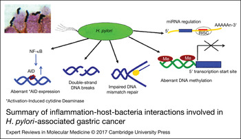
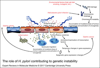
As noted previously, the difference in the ability of H. pylori strains to elicit mucosal inflammation is likely the best predictor of the risk of a clinically significant outcome, such as gastric cancer (Ref. Reference Yamaoka7). The degree and rapidity of gastric mucosal damage are also strongly influenced by both the environment (high-salt diet, smoking, mutagens, etc.) as well as the response of the host to inflammation often reflected in polymorphisms of host genes (Ref. Reference Kikuchi88). All H. pylori strains can potentially cause gastric cancer (i.e. no avirulent strains have been discovered to date). The risk is greater (approximately doubled) with more virulent strains (e.g. CagA-positive) (Ref. Reference Zhang, Lu and Graham46). Genomic instability is a hallmark of cancer. Despite the fact that H. pylori induces chronic often life-long inflammation and the production of reactive oxygen and nitrogen species (ROS and NOS) are important, H. pylori themselves are not innocent bystanders as H. pylori has been shown to be able to induce double-strained DNA breaks both in vitro and in vivo (Ref. Reference Hanada89). The infection is also associated with impaired DNA mismatch repair, aberrant DNA methylation, and abnormal regulation of microRNA regulation (Refs Reference Graham8, Reference Hanada and Graham17, Reference Shiotani, Cen and Graham90) (Fig. 2). Additionally, wherever H. pylori are present in the gastric mucosa, it will stimulate activation-induced cytidine deaminase (AID) enzyme (Ref. Reference Okazaki91) associated with H. pylori induced stimulation of NF-κB that presence in chronic gastritis mucosa with H. pylori infection and around half of gastric cancer (Refs Reference Shimizu92, Reference Hollstein93). The persistent stimulation of AID results an accumulation of nucleotide alterations in the tumour protein p53, a tumour suppressor gene which has been considered as a molecular signature of its involvement in gastric cancer (Ref. Reference Matsumoto94) resulted by direct or indirect involvement of H. pylori virulence factors (Refs Reference Hanada89, Reference Toller95). It is supported by abundant ROS and NOS (Refs Reference Hanada and Graham17, Reference Xu and Price96). Although H. pylori-induced double stranded DNA breaks can be repaired, the prolonged active infection is thought to saturate the cellular repair mechanisms that then contribute to genetic instability and frequent chromosomal aberrations (Ref. Reference Toller95). Inadequate resolution of inflammation caused by H. pylori infection activates transcription factors and signal transducers-activator of transcription-3 (Ref. Reference Mantovani97) to produce epigenetic changes, including alterations in DNA methylation, histone remodelling and the quantity control of messenger RNAs by microRNAs (miRNAs) (Refs Reference Graham8, Reference Shiotani, Cen and Graham90, Reference Nardone98) (Fig. 2). For example, the level of methylation in gastric mucosa of patients with gastric cancers is up to 30-fold greater than in healthy mucosa (Refs Reference Maekita99) and higher in patients with multiple than single gastric cancers. Histone acetylation enhances transcription (Ref. Reference Fischle, Wang and Allis100). Additionally, miRNAs, the post-transcriptional gene frequently altered in gastric cancer that influenced onto cell proliferation and inhibition of apoptosis (Refs Reference Barbarotto, Schmittgen and Calin101, Reference Zhang102, Reference Petrocca103) suggesting a pivotal role in gastric cancer pathogenesis and progression (Ref. Reference Zhang102). Cure of an H. pylori infection reduces or eliminates these H. pylori-associated molecular abnormalities (Refs Reference Graham and Shiotani16, Reference Nagata104). Nonetheless, the risk of cancer, while reduced, often does not return to normal (Refs Reference Shiotani105, Reference Shiotani106). For example, after follow up to 9.5 years of 1131 patients, the risk of developing gastric cancer in the patients cured of H. pylori infection (0.23% at 1 year) was significantly lower than those who had persistent infection (0.70%, P = 0.04) (Ref. Reference Take107). Importantly, the risk of developing gastric cancer is related to the background gastric mucosal atrophy remaining after H. pylori eradication therapy (Refs Reference Shiotani, Haruma and Graham108, Reference Graham109, Reference Shiotani, Matsueda and Graham110). The fact that cancer risk falls significantly even in those with the highest cancer risk (i.e. after endoscopic removal of an early gastric cancer) is consistent with the notion that the organisms themselves play a role in carcinogenesis (Ref. Reference Yoon111).
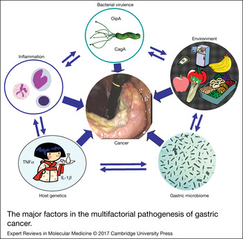
The major factors in the multifactorial pathogenesis of gastric cancer.

Gastric cancer and the gastric microbiome other than H. pylori
Gastric cancer is typically associated with reduced acid secretion leading to hypochlorhydria or achlorhydria. Research on gastric atrophy culminated in the mid-20th when it was recognised that atrophic gastritis was the soil for gastric cancer (Refs Reference Graham and Asaka3, Reference Comfort112). Duodenal ulcer disease and gastric cancer patients are typically infected with the most virulent strains but those with duodenal ulcer develop gastric cancer only if atrophic gastritis occurs (Ref. Reference Graham2). The risk for gastric cancer parallels the increase in gastric atrophy and development of a hypo- or anacidic intra-gastric environment. The absence of acid eliminates the anti-bacterial barrier which promotes the growth of non-H. pylori organisms who would have normally been killed by the acid pH (Ref. Reference Ruddell13). Decades before the discovery of H. pylori it was recognised that this abnormal population of organisms could metabolise dietary constituents such as nitrate and produce carcinogens locally (Refs Reference Forsythe113, Reference Osato114). Of interest, achlorhydria associated with autoimmune gastritis is also associated with an abnormal population of bacteria, local production of carcinogens, but adenocarcinoma of the stomach is rare unless there are also H. pylori present (Ref. Reference Rugge115). These natural experiments suggest that the exponential increase in gastric cancer risk associated with increasing H. pylori-associated gastric atrophy is actually a recapitulation of the experiments of adding subclinical amounts of a carcinogen to an inflammatory condition to produce a marked increase in the development of cancer (Ref. Reference Gaddy116) (Fig. 3). In autoimmune gastritis, the inflammation is primarily lymphocytic and after the gastric corpus is destroyed, inflammation declines. In contrast, in H. pylori the inflammation has a very active neutrophilic component and the H. pylori themselves play a role in producing genetic instability (Refs Reference Hanada and Graham17, Reference Hanada89). H. pylori eradication reduces or eliminates the inflammation-associated inhibition of acid production and also may allow some parietal cells to recover and return some acid secretion to the otherwise achlorhydric stomach, which would then change the microbiota and possibly reduce the production of both inflammation and carcinogens (Refs Reference Graham8, Reference Graham, Shiotani and El-Zimaity117).
Summary of inflammation-host-bacteria interactions involved in H. pylori-associated gastric cancer. Adapted from Graham (Ref. Reference Graham8), with permission.

Research in progress and outstanding research questions
It remains unclear whether the primary role of H. pylori in gastroduodenal diseases is as the source of an acute on chronic infection with chance, diet, host genetics playing the most important roles in defining the eventual outcome (Refs Reference Hanada and Graham17, Reference Chang118) (Fig. 4). Studies searching for disease associations with putative H. pylori virulence factor in isolation of other known factors have been especially unrewarding as have most of the available animal models. Data that H. pylori itself is an oncogenic bacteria are circumstantial at best as the outcomes of the infection (i.e. duodenal ulcer with high acid secretion and very low risk of cancer, and atrophic gastritis with achlorhydria and high cancer risk both result from infection with strains with the same putative virulence factors). The data seem to support the hypothesis that overgrowth of other non-H. pylori organisms in the achlorhydric stomach may be the actual source of carcinogens that interact with the damaged inflamed mucosa to cause cancer. Whole-genome sequencing analysis of H. pylori has provided comprehensive information about the 1600 genes content of the different strains sequenced. It is possible that many new genes associated with carcinogenesis and chronic inflammation may yet be found. Following endoscopic removal of an early gastric cancer, patients remain at high risk for a metachronous cancer despite H. pylori eradication. This group provides an ideal human laboratory to identify factors promoting and retarding gastric carcinogenesis.
The role of H. pylori contributing to genetic instability. Adapted from Shiotani et al. (Ref. Reference Shiotani, Cen and Graham90), with permission.

Acknowledgements
Dr Graham is supported in part by the Office of Research and Development Medical Research Service Department of Veterans Affairs, Public Health Service grant DK56338 which funds the Texas Medical Center Digestive Diseases Center. The contents are solely the responsibility of the authors and do not necessarily represent the official views of the VA or NIH. Professor Yamaoka is supported in part by grants from the National Institutes of Health (DK62813) and the Grants-in-Aid for Scientific Research from the Ministry of Education, Culture, Sports, Science, and Technology (MEXT) of Japan (24406015, 24659200, 25293104, 26640114 and 15H02657). Professor Yamaoka and Dr Miftahussurur are also supported by the Japan Society for the Promotion of Science (JSPS) Institutional Program for Young Researcher Overseas Visits, the Strategic Funds for the Promotion of Science and Technology from Japan Science and Technology Agency.
Conflict-of-Interest
Dr Graham is a paid consultant and has received research funding from RedHill Biopharma regarding novel H. pylori therapies and is a consultant to BioGaia regarding the use of probiotics for H. pylori infections. Professor Yamaoka and Dr Miftahussurur have nothing to declare.