Conjugation of functional groups to DNA nanostructures
DNA is chemically a rather inert molecule and, in Nature, the function of DNA is to store and transfer information. Its chemical inertness provides DNA with the advantages of high stability and high fidelity in forming predesigned DNA structures by DNA hybridization. DNA nanostructures built by self-assembly of DNA strands, therefore, have limited chemical functionality, and DNA is mainly used as a molecular scaffold. Exceptions include artificially selected DNA aptamers that provide selective recognition of other (bio)molecules Reference Song, Lee and Ban1 and artificial DNA structures, the so-called DNAzymes that can catalyze chemical reactions. Reference Willner, Shlyahovsky, Zayats and Willner2
To make functional DNA nanostructures, integration of other materials and molecules is most often required and, with a few exceptions, this requires chemical functionalization of DNA strands. Fortunately, a variety of chemical modifications are available from commercial DNA suppliers, allowing for the introduction of desired artificial functional groups into the DNA sequence.
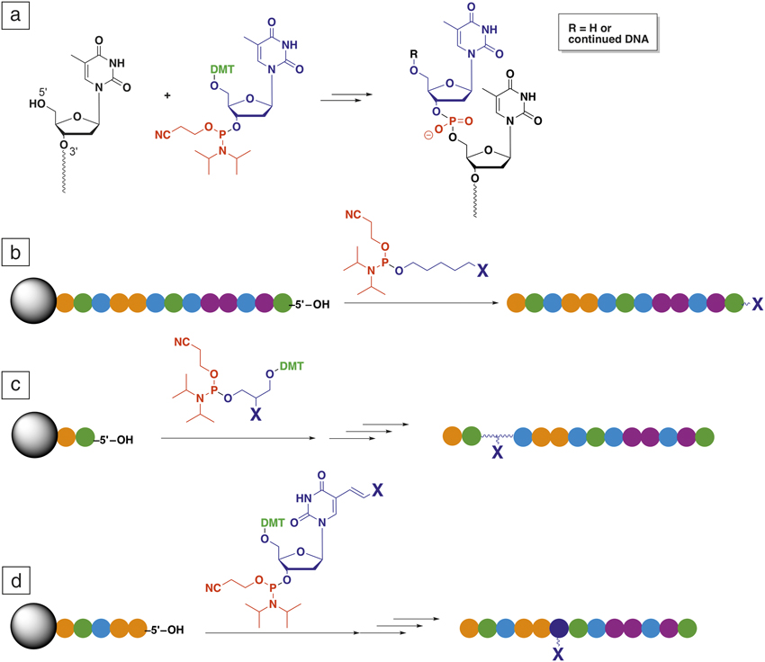
Custom oligonucleotides with any desired sequence are prepared by automated synthesis using phosphoramidite nucleoside building blocks, which are made up of an activated phosphate bridge precursor linked to the protected deoxyribose ring and the nucleobase ( Figure 1a). Reference Lönnberg3 A vast variety of non-nucleosidic phosphoramidites or phosphoramidite nucleoside analogues are available for the introduction of chemical modifications. Some linkers are only used for the introduction of modifications at the 5′-end of oligonucleotides (Figure 1b), while others can be used to introduce nonnucleosidic modifiers at the 3′, 5′ (positions indicated in Figure 1a), or internally in the sequence (Figure 1c). The nucleosidic modifiers contain the modification on the nucleobase (Figure 1d). These modifications can be placed at any position in a DNA strand and conserve the hybridization ability. DNA strands containing such bases may also be compatible with enzymes (e.g., in polymerase chain reaction [PCR] reactions). Reference Lapa, Chudinov and Timofeev4 When such modified DNA sequences are integrated in large DNA nanostructures, the functional groups can be placed at almost any specific position in the structure.
Introduction of modifications in DNA as part of the automated solid-phase DNA synthesis. (a) Illustration of one step in a standard phosphoramidite-based oligonucleotide synthesis, where the 5′ oxygen and the red phosphorus atom are linked in four chemical steps required to add one nucleotide to a DNA strand. (The 3′ and 5′ positions of nucleic acids are indicated in the left structure.) (b) Introduction of 5′-terminal modifiers to a DNA strand (chain of colored dots). (c) Introduction of a 3′-, internal, or 5′ modifiers. (d) Introduction of a 3′-, internal, or 5′-modification by nucleosidic phosphoramidite with the functionality linked to the nucleobase. Note: X, functionality of interest; gray sphere, controlled pore glass bead; colored spheres, nucleotides; DMT, dimethoxytrityl.

The integration of modified DNA strands into DNA nanostructures (e.g., to DNA origami) can essentially be done in two different ways. In the first method, the modified strand(s) is also a staple strand (i.e., an integral part of the origami structure itself) that hybridizes to the long scaffold strand. In the second method, the modified strands are not staple strands. Instead, some of the staple strands are extended with short single-stranded domains that are complementary to the modified strand(s), so they will hybridize and form the double-stranded DNA helix. The first method offers simplicity, since only one hybridization step is required, and it offers higher positional control, since the modification is integrated in the structure rather than extending from the structure. The two major drawbacks are that the modifications must be compatible with the heat-annealing process that is typically required for formation of the origami. This normally implies heating to 95°C, which results in denaturation of most proteins. Furthermore, if more copies of the same modification are to be integrated, they must each be conjugated to a unique DNA staple strand, which increases the cost. Both of these problems are solved by the second method, where the modifications are introduced at room temperature by hybridization of the modified strands to single-stranded DNA domains extending from the prefolded DNA origami structure. As these extensions may be similar, although attached to different staple strands, one modified strand can be introduced to several different locations.
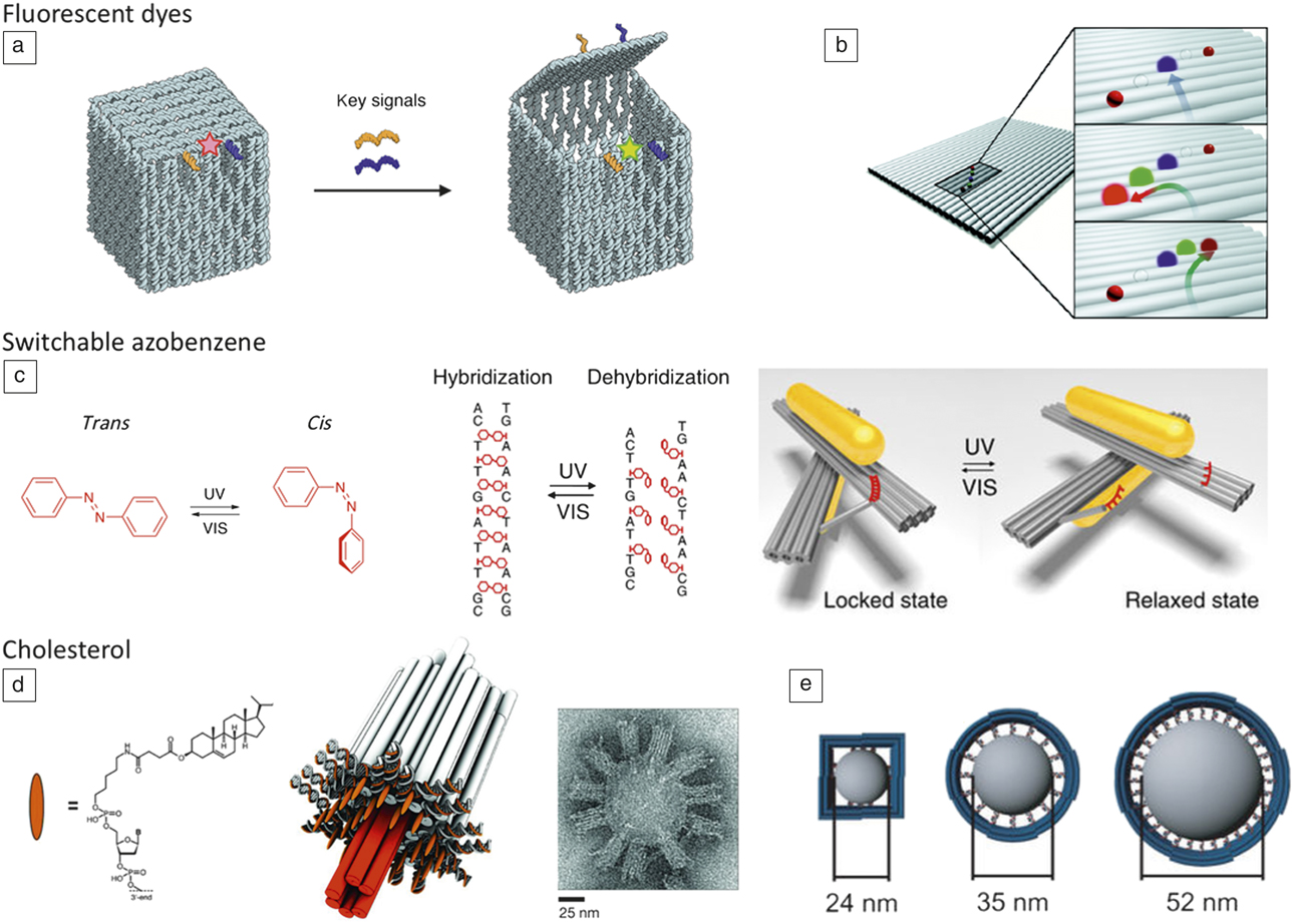
Among the most commonly applied modifications of DNA strands in DNA nanotechnology are dyes. Fluorescent dyes are used to identify DNA structures and for fluorescence microscopy of DNA structures in cells. Reference Beater, Raab and Tinnefeld5 By introducing two or more dyes in specific positions at the DNA structure, the interaction between the dyes, such as fluorescence resonance energy transfer (FRET) and quenching pairs, can be used to monitor dynamic events and distances in DNA structures. This is demonstrated in Figure 2a, where the opening of the lid in a DNA origami box is monitored by FRET. Reference Andersen, Dong, Nielsen, Jahn, Subramani, Mamdouh, Golas, Sander, Stark, Oliveira, Pedersen, Birkedal, Besenbacher, Gothelf and Kjems6 DNA nanostructures offer a unique opportunity to place two or more dyes with well-defined relative distances between the dyes, which allows for the study of their interactions (Figure 2b). Reference Stein, Steinhauer and Tinnefeld7 Biotin (vitamin B7) is another common DNA modification, due to the usefulness of the extremely strong and fast noncovalent binding between the small molecule biotin and the 53 kDa protein streptavidin (or avidin), which contains four binding sites for biotin. Reference DeChancie and Houk8 This binding is used extensively in biotechnology and also in DNA nanotechnology for binding of nanostructures to solid supports and to other molecules via the DNA-biotin-streptavidin-biotin-X linkage (X is the functionality of interest). Reference Clark, Singer, Korns and Smith9
Integration of function in DNA nanostructures by small organic molecules such as florescent dyes. (a) Monitoring the opening of a DNA box by a fluorescence resonance energy transfer (FRET) couple. The keys are DNA sequences that outcompete the orange and blue double-stranded DNA locks in the box and release the lid. The colored stars indicate the change in FRET signal upon opening of the box. Reference Andersen, Dong, Nielsen, Jahn, Subramani, Mamdouh, Golas, Sander, Stark, Oliveira, Pedersen, Birkedal, Besenbacher, Gothelf and Kjems6 (b) A two-way three-station FRET gate on DNA origami. The arrows in the figure indicate transfer of energy between the dyes. Reprinted with permission from Reference Reference Stein, Steinhauer and Tinnefeld7. © 2011 American Chemical Society. (c) Photoswitching of azobenzene and application of the azobenzene in DNA for photocontrolled switching of a chiroptic DNA origami-based device. The panel on the left shows the switching of azobenzene between the trans and cis structure, while the middle structure shows the incorporation of azobenzenes (red) between the bases in double-stranded DNA and how hybridization is controlled by light. In the right panel, the switching between a fixed and a relaxed organization of two gold nanorods by the azobenzene mechanism is shown. Reprinted with permission from Reference Reference Kuzyk, Yang, Duan, Stoll, Govorov, Sugiyama, Endo and Liu12. © 2016 Macmillan Publishers Ltd. (d) Cholesterol-guided insertion of a DNA nanopore in a phospholipid membrane. A transmission electron microscope image of multiple DNA nanopores inserted into the lipid bilayer of a vesicle. Reprinted with permission from Reference Reference Langecker, Arnaut, Martin, List, Renner, Mayer, Dietz and Simmel14. © 2012 AAAS. (e) Shaping micelles caught in a DNA cavity by interactions with hydrophobic molecules. The sizes of the square or circular DNA origami structures, comprising the outer perimeters, define the size of the micelles. Reprinted with permission from Reference Reference Zhang, Yang, Pincet, Llaguno and Lin19. © 2017 Macmillan Publishers Ltd. Note: UV, ultraviolet; VIS, visible; A, adenine; T, thymine; C, cytosine; G, guanine.

In 2001, Asanuma introduced the azobenzene phosphoramidite, which allowed photochemical control over DNA hybridization. Reference Asanuma, Takaradam, Yoshida, Tamaru, Liang and Komiyama10,Reference Asanuma, Liang, Yoshida and Komiyama11 Azobenzene can be switched reversibly between the trans- and cis isomers by UV light at 330 and 400 nm wavelength, respectively. While the trans isomer has a planar geometry that favors intercalation between the bases in double-stranded DNA, the cis isomer yields a geometry where the two benzene rings are twisted out of planarity, which obstructs intercalation and thereby stabilization of the duplex. Incorporation of three or more of the azobenzenes between the bases in two opposing DNA strands provides photochemical control of the hybridization and denaturation of the duplex. This functionality has been extensively applied in DNA nanotechnology for controlling dynamic processes. The advantage is that hybridization can be controlled without additional DNA strands and without changing the physical/chemical conditions such as temperature, salt concentration, and pH. One example is shown in Figure 2c for photochemical switching of a plasmonic device built from gold nanorods integrated in a switchable DNA structure. Reference Kuzyk, Yang, Duan, Stoll, Govorov, Sugiyama, Endo and Liu12 The system is switched between a chiral locked state and an achiral relaxed stated by photochemically controlled hybridization and dehybridization of an azobenzene containing DNA strand (red strand in Figure 2c)
DNA and DNA nanostructures are negatively charged, highly hydrophilic molecules. Recently, hydrophobic modifiers have increasingly been used to change their surface properties. Hydrophobic modifiers such as cholesterol phosphoramidites and others have been integrated in the sequences at selected positions for binding DNA nanostructures to phospholipid membranes, Reference Suzuki, Endo, Yang and Sugiyama13 for integrating DNA nanostructures in membranes as artificial pores (Figure 2d), Reference Langecker, Arnaut, Martin, List, Renner, Mayer, Dietz and Simmel14,Reference Göpfrich, Li, Ricci, Bhamidimarri, Yoo, Gyenes, Ohmann, Winterhalter, Aksimentiev and Keyser15 for growing membranes on origami structures, Reference Zhou, Zhang, Dong, Wu, Wang, Xin and Liu16 for creating artificial monodisperse micelles, Reference Edwardson, Carneiro, McLaughlin, Serpell and Sleiman17 and for integrating and shaping lipid vesicles (Figure 2e). Reference Perrault and Shih18,Reference Zhang, Yang, Pincet, Llaguno and Lin19
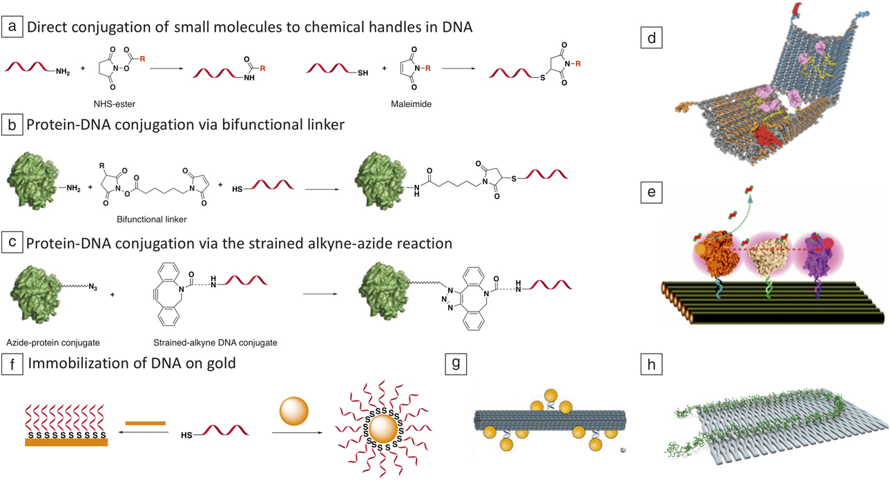
When the desired modification is not commercially available, be it a molecule, protein, or inorganic nanomaterial, the oligonucleotide must be linked to the modification by custom chemistry. For this purpose, a handle on the DNA strand is required. The most common chemical handles are amine modifiers and thiol modifiers that are available from most DNA suppliers ( Figure 3a). The amine modification can be introduced at any position, and the modified DNA strands are normally supplied as the free deprotected amine. Thiol modifications are mainly available for 3′- or 5′-modifications and are most often supplied as disulfides that must be cleaved by reduction before use to produce thiols. The amines react well with acyl groups such as activated esters to form amides, and thiols react well with soft electrophiles such as the maleimide shown in Figure 3a. These reactions are commonly used for direct conjugation or in conjunction with bifunctional linkers that combine amine and thiol functionalized molecules (Figure 3a–b).
Conjugation of small molecules, proteins, and nanoparticles to DNA, and their integration in DNA origami. (a) Conjugation of a small molecule NHS ester to an amine-modified DNA sequence, or a small molecule maleimide to a thiol-modified DNA sequence. (b) Protein-DNA conjugation via a heterobifunctional linker (R = H or SO3 –). (c) Protein-DNA conjugation of DNA premodified with an azide and a protein premodified with a strained alkyne. (d) A DNA nanorobot in the open state where antigen binding fragment (Fab)-DNA conjugates are integrated to induce apoptosis. Reprinted with permission from Reference Reference Douglas, Bachelet and Church24. © 2012 AAAS. (e) Enzymatic cascade reaction between enzyme–DNA conjugates immobilized in DNA origami. Reprinted with permission from Reference Reference Fu, Liu, Liu, Woodbury and Yan25. © 2011 American Chemical Society. (f) Formation of self-assembled monolayers and DNA modified gold nanoparticles from thiol-DNA. (g) A homochiral plasmonic device made out of a spiral of Au nanoparticles arranged around a DNA origami rod. Reprinted with permission from Reference Reference Kuzyk, Schreiber, Fan, Pardatscher, Roller, Högele, Simmel, Govorov and Liedl31. © 2012 Macmillan Publishers Ltd. (h) Routing of a conjugated polymer (red thin line) connecting to DNA strands (green) appending from the backbone, on DNA origami (gray). Reference Knudsen, Liu, Kodal, Madsen, Li, Song, Woehrstein, Wickham, Strauss, Schueder, Vinther, Krissanaprasit, Gudnason, Smith, Ogaki, Zelikin, Besenbacher, Birkedal, Yin, Shih, Jungmann, Dong and Gothelf33

The integration of proteins in DNA nanostructures is particularly important, since proteins offer unique function and molecular recognition capability. Proteins are integrated in DNA nanostructures by conjugating a DNA strand to the protein, and the conjugate is subsequently hybridized to staple strands extending from the origami structure. Functionalization of proteins is frequently made by reaction of a bifunctional linker with an amine-modified DNA strand and subsequent reaction with an amine or a thiol on the protein surface (Figure 3b). Reference Niemeyer20 In recent years, a palette of fast and bioorthogonal bioconjugation reactions (i.e., reactions that proceed under physiological conditions and do not react with natural biomolecules) have also been applied for protein-DNA conjugation. Reference Zheng, Zheng, Zhang, Li and Zhang21 The complementary functional groups are first linked to the protein and DNA separately by small molecule modifiers, and then used for the protein–DNA conjugation reaction. Such chemistries include Cu-free azide-strained alkyne reactions (Figure 3c), aldehyde–O-hydroxylamine condensations, and the tetrazine-alkene reactions.
The conjugation methods previously mentioned often give rise to heterogeneous mixtures of conjugates, since most proteins have multiple amines (lysines) at the surface. To solve this problem, many specialized and site-selective methods are available for conjugation of proteins to DNA, including protein tags Reference Sacca and Niemeyer22 and DNA-templated protein conjugation to native proteins. Reference Trads, Tørring and Gothelf23
Examples of proteins integrated in DNA nanostructures are shown in Figure 3d–e. In the first example, a DNA origami nanorobot was designed containing aptamers that upon recognition of cancer-specific proteins, opens the barrel-shaped structure. Reference Douglas, Bachelet and Church24 The open form, shown in Figure 3d, exposes the antigen binding fragment (Fab) domain of an antibody that, in turn, induces cell death (apoptosis). The Fab domains are integrated in the DNA structure via covalently linked DNA strands. In the second example, redox enzymes were organized on a DNA origami surface to investigate distance and mediator-dependent enzymatic cascade reactions (Figure 3e). Reference Fu, Liu, Liu, Woodbury and Yan25,Reference Rajendran, Nakata, Nakano and Morii26 An alternative site-specific method for integration of DNA strands in DNA nanostructures is by conjugation of a DNA strand to a ligand or cofactor for the protein and subsequent binding of the protein. This approach may, however, impede the function of the protein.
Thiols are also used to immobilize DNA sequences on gold surfaces, including gold nanoparticles, as well as other noble metals and quantum dots (Figure 3f). Reference Samanta and Sarkar27 This is one of the most common methods to graft DNA strands and DNA nanostructures to gold surfaces (e.g., on electrodes). Reference Pei, Lu, Wen, Song, Liu, Yan and Fan28 For covalent immobilization of DNA origami, recent work demonstrates a method for immobilization of DNA origami in patterned arrays on SiO2 surfaces and subsequent covalent coupling to the surfaces via amine-modified staple strands. Reference Gopinath and Rothemund29
There are numerous examples of the integration of small, <20 nm DNA functionalized gold nanoparticles (AuNPs) in DNA nanostructures. Studies have shown that optimal yields are obtained for immobilization of DNA–AuNPs in DNA origami, when more complementary DNA strands, extending from the desired site at the DNA origami, are available for hybridization to the DNA–AuNP. Reference Sharma, Chhabra, Andersen, Gothelf, Yan and Liu30,Reference Kuzyk, Schreiber, Fan, Pardatscher, Roller, Högele, Simmel, Govorov and Liedl31 In a number of examples, metal nanoparticles and nanorods have been integrated in DNA nanostructures for exploiting the plasmonic properties, Reference Samanta, Banerjee and Liu32 including the arrangement of gold nanoparticles in a helix around a DNA origami rod, as shown in Figure 3g. Reference Kuzyk, Schreiber, Fan, Pardatscher, Roller, Högele, Simmel, Govorov and Liedl31 A variety of other materials have been integrated in DNA nanostructures such as polymers (Figure 3h), Reference Knudsen, Liu, Kodal, Madsen, Li, Song, Woehrstein, Wickham, Strauss, Schueder, Vinther, Krissanaprasit, Gudnason, Smith, Ogaki, Zelikin, Besenbacher, Birkedal, Yin, Shih, Jungmann, Dong and Gothelf33 carbon nanotubes, Reference Maune, Han, Barish, Bockrath, Goddard, Rothemund and Winfree34 and nanodiamonds. Reference Zhang, Neumann, Lindlau, Wu, Pramanik, Naydenov, Jelezko, Schüder, Huber, Huber, Stehr, Högele, Weil and Liedl35
Chemistry at DNA nanostructures
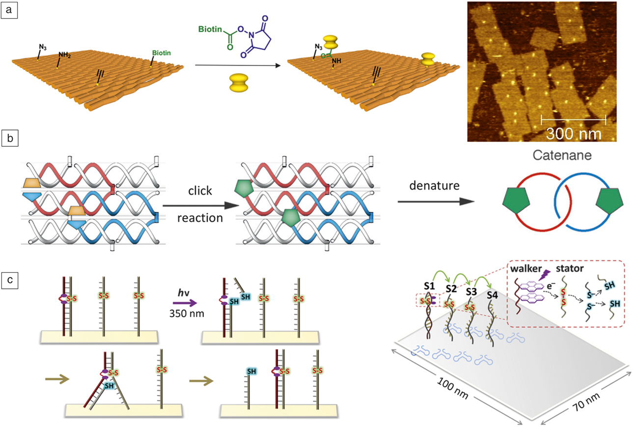
The low chemical reactivity of DNA makes it possible to perform many different chemical reactions in the presence of DNA and at specific modifications in DNA strands. As an example, the exocyclic amines at the bases of DNA have poor reactivity and essentially do not react with NHS esters at room temperature, whereas conventional amine modifiers readily react. Reference Voigt, Tørring, Rotaru, Jacobsen, Ravnsbæk, Subramani, Mamdouh, Kjems, Mokhir, Besenbacher and Gothelf36 DNA origami has been used to study single-molecule reactions by atomic force microscopy (AFM), where the origami functions as an addressable solid support and streptavidin is used as a contrast for imaging by AFM ( Figure 4a). Reference Voigt, Tørring, Rotaru, Jacobsen, Ravnsbæk, Subramani, Mamdouh, Kjems, Mokhir, Besenbacher and Gothelf36
Chemistry in DNA nanostructures. (a) (Left) Single-molecule chemical reaction of an amine on DNA origami with an activated NHS-ester tethered to biotin. (Right) The reaction is imaged by atomic force microscopy after incubation with streptavidin that appears as bright dots. Reference Voigt, Tørring, Rotaru, Jacobsen, Ravnsbæk, Subramani, Mamdouh, Kjems, Mokhir, Besenbacher and Gothelf36 (b) Cyclization of DNA strand by reaction between azide (blue) and alkyne (orange) modified DNA strands in a single-stranded tile structure to form interlocked strands (catenane). When the DNA structure is denatured, the interlocked DNA strands stay linked together. Reprinted with permission from Reference Reference Cassinelli, Oberleitner, Sobotta, Nickels, Grossi, Kempter, Frischmuth, Liedl and Manetto41. © 2015 Wiley. (c) (Left) A light-driven DNA-walker (brown DNA strand containing pyrenes shown as purple dots) is shown, where electron transfer from photochemically excited pyrenes on the walker strand cleaves a disulfide (-S-S-) to release part of the stationary strand. In turn, this makes it thermodynamically favorable for the walker strand to walk to the full length next station. This process is repeated to continue walking. In the right panel, the track of the walker on DNA origami is shown, and the chemical mechanism of the DNA walker is shown in the expansion. S1–S4 are the four stations (physically represented by single-stranded DNA containing the disulfide) that the walker walks along in a stepwise manner. Reprinted with permission from Reference Reference Yang, Goetzfried, Hidaka, You, Tan, Sugiyama and Endo47. © 2015 American Chemical Society. Note: hv, light.

DNA can also template, catalyze, or fuel the formation of metal structures. Global metallization of nonfunctionalized DNA nanostructures has been widely used to form metallic structures that take their shape from the DNA origami template. Reference Shen, Tapio, Linko, Kostiainen and Toppari37,Reference Gates, Dearden and Woolley38
DNA-templated chemistry in DNA nanostructures
Chemical reactions between functional groups attached to DNA strands can be controlled by DNA. Such programmability is obtained by controlling the proximity of the functional groups by DNA hybridization and thus, their ability to react. Reference Gorska and Winssinger39 DNA-templated chemical reactions were first investigated for nonenzymatic ligation of DNA strands. Reference Li and Liu40 Liu and, later, others have expanded the repertoire of chemical reactions and design of DNA geometries significantly. Reference Gorska and Winssinger39,Reference Li and Liu40 In DNA nanotechnology, DNA-templated reactions have been applied to the cyclization of sequences in a single-stranded tile DNA structure by the Cu-catalyzed azide-alkyne cycloaddition reaction, to stabilize a DNA nanostructure and to form geometrically interlinked molecular rings (catenanes) (Figure 4b). Reference Cassinelli, Oberleitner, Sobotta, Nickels, Grossi, Kempter, Frischmuth, Liedl and Manetto41
The most important application of DNA-templated synthesis has been the formation of combinatorial libraries of chemical compounds for drug discovery. Each compound in a library is encoded with a DNA tag that enables easy identification of active binders by PCR and sequencing. Such libraries have been built both from linear DNA designs and in DNA junctions. This is now a commercial technology that has successfully led to the discovery of new drug leads. Reference Goodnow, Dumelin and Keefe42
DNA-templated chemistry has also been applied to build oligomers of non-DNA materials in a parallel fashion Reference Gothelf and Brown43 or in a stepwise manner. Reference McKee, Milnes, Bath, Stulz, O’Reilly and Turberfield44 The groups of Liu Reference He and Liu45 and Turberfield and O’Reilly Reference Meng, Muscat, McKee, Milnes, El-Sagheer, Bath, Davis, Brown, O’Reilly and Turberfield46 have developed sophisticated autonomous DNA-controlled chemical systems for synthesis of short oligomers with defined sequence.
Such advanced chemical reaction systems have not been incorporated in more complex DNA systems such as DNA origami. However, there is the potential to make individual origami into local synthesis machines in the future. One example is the photochemically driven DNA walker on DNA origami, which was recently developed by Sugiyama and Endo (Figure 4c). Reference Yang, Goetzfried, Hidaka, You, Tan, Sugiyama and Endo47
The examples shown here of chemical reactions applied in DNA nanotechnology are far from exhaustive, but provide an overview of some of the ways chemical modifications can be applied to functionalize DNA nanostructures. Although only DNA nanostructures have been described, most of the modifications can also be incorporated in RNA and artificial oligonucleotide analogues.
A range of different modifiers are commercially available and most of them require little chemical training to handle and use. The custom modification of DNA strands with small molecules is also relatively straightforward, since many dyes and other molecules are available as activated reagents for bioconjugation. Purification and identification of such homemade conjugates, however, require access to high-performance liquid chromatography equipment and mass spectrometry analysis. DNA protein conjugation is more demanding, but a variety of kits and reagents are available for such reactions, and it is a field under rapid development. Basically, any nanomaterial that tolerates water, and to which DNA can be conjugated, can be integrated into DNA nanostructures, and the opportunities for exploring the properties of other materials in DNA nanostructures are vast.
Conclusion
DNA nanostructures offer unmatched opportunities for arranging nanomaterials relative to each other to study fundamental chemical, physical, and biological phenomena at the nanoscale. The ability to design structures, introduce modifications, and engineer surface properties also holds great potential for the development of advanced medicines and diagnostic tools, and currently, there is intense activity in this area. Origami structures also offer an addressable platform for the arrangement of optical and electronic components, which has potential for integration with solid-state electronics and optics. This is exemplified by a recent study, where DNA origami structures were immobilized in an array of engineered patterns on silicon nitride, which allows controllable coupling of organic dyes at the origami to photonic crystal cavities. Reference Gopinath, Miyazono, Faraon and Rothemund48 This excellent example of the integration of DNA nanotechnology, organic chemistry, and solid-state physics shows the power of chemical functionalization of DNA nanostructures.

Kurt Gothelf has been full professor and director of the Centre for DNA Nanotechnology at Aarhus University, Denmark, since 2007. He received his PhD degree in organic chemistry and asymmetric catalysis in 1995 from Aarhus University. He completed postdoctoral research at Duke University. He joined Aarhus University in 2002 as an associate professor. His research focuses on surface chemistry and DNA nanotechnology. Currently, Gothelf is focusing on bioconjugation of DNA to proteins for applications in diagnostics, therapeutics, and DNA nanotechnology. Gothelf can be reached by email at kvg@chem.au.dk.