Case report
A full-term male infant, prenatally diagnosed with Trisomy 21, was born to a 38-year-old G2P0-to-1 mother via caesarean section for failure to progress. Fetal echocardiogram at 21 weeks gestation had shown normal anatomy, normal biventricular size and systolic function, and an unobstructed ductus arteriosus. The pregnancy was also complicated by insulin-dependent gestational diabetes. Cardiac examination was normal and physical examination was only significant for features consistent with Trisomy 21. Delivery room resuscitation was limited to stimulation and the patient was admitted to the newborn nursery.
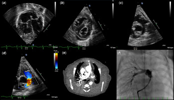
Over the next 12 hours the patient developed signs of respiratory distress with tachypnoea and diffusely course breath sounds. A chest X-ray was unrevealing. He was transferred to the neonatal intensive care unit for initiation of noninvasive positive pressure ventilation. An echocardiogram early on the 2nd day of life demonstrated severe right ventricular hypertrophy and dysfunction, right ventricular hypertension, and a nearly-occlusive thrombus in the left pulmonary artery. No ductus arteriosus was identified. Computed tomography angiography demonstrated left pulmonary artery thrombo-occlusion as well as additional thrombus-burden in the right internal iliac artery (Fig 1). Notably, the patient’s mother was diagnosed with pulmonary embolism post-partum and later revealed a history of pulmonary emboli and deep vein thrombosis in multiple family members.
Given the lack of a patent ductus arteriosus on initial imaging, the presence of both left pulmonary artery and iliac artery thrombosis, and the presence of echo-bright substrate in the pulmonary artery ductal ampulla, the diagnosis of fetal ductal thrombosis was proposed. In this case, fetal ductal thrombosis was associated with thrombotic extension into both the pulmonary and systemic arterial circulations. In this setting, the patient was started on heparin infusion and subsequently switched to low-molecular weight heparin therapy due to difficulty achieving adequate anticoagulation. Serial echocardiography was performed and the study on day 8 of life, after 4 days of anticoagulation therapy, was significant for a completely occlusive left pulmonary artery thrombus with unchanged right ventricular morphology and function. Due to the lack of improvement and clinical evidence of right ventricular failure, after multi-disciplinary discussion, the patient was referred for cardiac catheterisation with thrombectomy.
Pre-intervention imaging. Echocardiography prior to intervention (a–d) demonstrates RV hypertrophy, interventricular septal flattening in systole consistent with elevated RV pressure, and patent main and right PAs with left PA occlusion. CT angiography (e) reveals an unobstructed RV outflow tract and right PA without contrast opacification of the proximal left PA, although the distal vessel is opacified. Invasive pulmonary angiography (f) shows essentially complete occlusion of the left PA with normal flow into the right PA. RV = Right ventricle; PA = pulmonary artery.

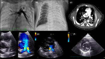
Post-intervention imaging. Following intervention, invasive pulmonary angiography (a) demonstrates unobstructed bilateral branch PAs, as well as a ductal ampulla that now fills densely, but has areas of non-opacification consistent with the presence of intraductal thrombus. Contrast opacification of the aorta is visualized, consistent with the presence of right to left ductal shunt, which was not present pre-intervention. Following the catheterization, chest x-ray (b) shows impressive unilateral left reperfusion injury. CT angiography (c) reveals unobstructed RV outflow tract and bilateral branch pulmonary arteries. Echocardiography immediately post intervention (d) demonstrates patent main and branch PAs. Repeat echocardiography 3 months post intervention (e,f) shows unobstructed PAs and complete resolution of RV hypertrophy with normal interventricular septal curvature. RV = Right ventricle; PA = pulmonary artery.

Under ultrasound guidance, a 5 French sheath was placed in the right femoral vein and an arterial monitoring line was placed in the right femoral artery after which a limited right heart catheterisation was performed. This was significant for nearly systemic right ventricular pressure. Angiography confirmed an unobstructed right ventricular outflow tract and right pulmonary artery with nearly complete occlusion of the left pulmonary artery. A 0.035” angled Glidewire hydrophilic-coated guidewire (Terumo Interventional Systems, Somerset, NJ) was maneuvered past the thrombus in the left pulmonary artery, facilitating distal placement of a 0.018” interventional guidewire. Selective left pulmonary artery angiography was performed, followed by serial balloon angioplasty across the left pulmonary artery thrombus to macerate the thrombus and create a pathway for passage of larger interventional equipment. Next, rheolytic thrombectomy was performed with a 6 French AngioJet catheter (Boston Scientific, Marlborough, MA), with two limited 6 second passes made in an effort to reduce the risk of hemodynamic compromise. Reference Fleming, Khan, Janssen and Doyle1 The patient experienced self-limited bradycardia and 10 beats of 2:1 heart block which resolved spontaneously. Post-intervention angiography demonstrated almost complete resolution of left pulmonary artery thrombus burden but new distal thromboemboli in the left lower and right middle lobes (Fig 2). In addition, the ductus arteriosus was now visible, with intraductal thrombus evident, and some right to left shunt present. The new distal pulmonary arterial thrombus burden was addressed by balloon angioplasty with overall improvement but persistent mild filling defects peripherally. Descending aortography demonstrated no abdominal aortic or tributary thrombus burden, suggesting no overt intraprocedural left to right ductal thromboembolism had occurred. Moreover, this demonstrated resolution of the iliac artery thrombus.
After the procedure, the patient recovered in the cardiac intensive care unit. Chest X-ray demonstrated substantial left lung reperfusion injury, which improved over the course of the following days. Low molecular weight heparin therapy was continued. Follow-up imaging with computed tomography angiography 1 week later revealed no residual pulmonary or systemic arterial thrombus burden and a closed ductus arteriosus. The patient was discharged home 2 weeks post-catheterisation. At 3-month follow-up, echocardiography revealed normal ventricular size and systolic function, normal right ventricular pressure estimate, and unobstructed branch pulmonary arteries without evidence of a ductus arteriosus. The child was thriving clinically and uneventfully transitioned from low molecular weight heparin to aspirin therapy.
Discussion
Fetal ductal thrombosis is a rare form of premature ductal closure which leads to in utero right ventricular strain, elevated right ventricular and pulmonary artery pressure, and complicated postnatal transition. The infrequent occurrence of this event makes diagnosis challenging, especially when premature ductal closure is not diagnosed in utero. Newborns with ductal thrombosis have been reported to present with initial clinical stability followed by the onset of respiratory distress, need for increased ventilatory support, and progressive signs of right ventricular failure. Reference Sequeira, Lemos and Palma2-Reference Gewillig, Brown and De Catte4 Risk factors for in utero thrombus development in this case include maternal diabetes and familial thrombophilia. Reference Mitanchez, Yzydorczyk and Simeoni5
Prior reports have detailed extension of fetal ductal thrombus into the pulmonary arteries, but to our knowledge this is the first reported case of extension and embolisation leading to obstructive lesions in both the pulmonary and systemic circulations. Reference Pagotto, Tani, Raetz, McGough and Minich6
Reported approaches to neonatal systemic and pulmonary vascular thrombo-occlusion include systemic anticoagulation therapy, systemic thrombolytic therapy, surgical thrombectomy, and catheter-based interventions. Reference De Blanche, Schmitz, Johnson, Best and Drummond-Webb7,Reference Ferrari, Vagnarelli and Gargano8 Each approach presents unique risks and benefits. Therapy tailored to the degree of extremis, overall clinical presentation, and centre-experience is necessary. Vascular thrombus leading to rapid clinical decline may preclude therapy such as systemic anticoagulation, which requires days for adequate effect. Systemic thrombolysis in neonates presents substantial risk for intracranial and intraventricular hemorrhage. Transcatheter thrombectomy may offer unique benefit with locally-directed therapy that can be implemented rapidly and provide immediate resolution of thrombus burden, without the invasiveness of surgery.
Rheolytic therapy has been used for treatment of post-operative thrombus in neonates with congenital heart disease, but this is the first reported use in a patient with ductal thrombosis-related pulmonary artery obstruction. Reference Feldman, Feinstein, Lamberti and Perry9,Reference Qureshi, Petit, Crystal, Liou, Khan and Justino10 Our case also highlights the complexities associated with deployment of such a therapy in the neonate, including haemodynamic instability, pulmonary emboli, and ductal thrombus removal which has the potential to facilitate right to left ductal shunt and systemic arterial thromboembolus.
Financial support
This research received no specific grant from any funding agency, commercial, or not-for-profit sectors.
Conflicts of interest
Dr. Goldstein reports consulting relationships with Medtronic, W.L. Gore & Associates, Inozyme Pharma, and is a consultant and scientific advisory board member for PECA Labs. There are no other disclosures to report.