INTRODUCTION
Cryptosporidium is a protozoan enteric parasite of humans and other vertebrates (Fayer et al. Reference Fayer, Speer, Dubey and Fayer1997). Numerous Cryptosporidium species have been described (Smith et al. Reference Smith, Nichols and Grimason2005) most of which are specific to their vertebrate host. The species C. parvum is of medical and economic relevance as it affects both humans and cattle with its primary site of infection being the gastrointestinal tract. It affects the epithelial lining of the ileum, resulting in self-limiting diarrhoea in immunocompetent individuals or in life-threatening diarrhoeal diseases in immunocompromised individuals.
Belonging to the phylum of apicomplexan parasites, Cryptosporidium shares common life-cycle features and morphological characteristics with other members of this phylum (Tetley et al. Reference Tetley, Brown, McDonald and Coombs1998). Initially, Cryptosporidium was categorized as a coccidian parasite (Levine, Reference Levine1988). However, more recent studies show that Cryptosporidium lacks key morphological characteristics of coccidians and is insensitive to anti-coccidial agents (O'Donoghue, Reference O'Donoghue1995; Fayer et al. Reference Fayer, Speer, Dubey and Fayer1997; Carreno et al. Reference Carreno, Martin and Barta1999). Further phylogenomic analysis has since revealed that Cryptosporidium is most closely related to gregarines (Barta and Thompson, Reference Barta and Thompson2006). Cryptosporidium shares many features in common with gregarines, including an extracytoplasmic location and connection to the host cell via a myzocytosis-like feeding mechanism (Barta and Thompson, Reference Barta and Thompson2006). The primary difference between these two groups is that Cryptosporidium induces the host cell to overlay it with the host cell apical membrane (Barta and Thompson, Reference Barta and Thompson2006; Butaeva et al. Reference Butaeva, Paskerova and Entzeroth2006). The parasite appears on the surface of cells, residing in a parasitophorous vacuole (PV) between the cytoplasmic membrane and the apical membrane (Huang et al. Reference Huang, Chen and LaRusso2004).
Critically, the mechanisms of Cryptosporidium pathogenesis are not fully understood, but both parasite stimuli as well as host immune responses are thought to play critical roles (Barta and Thompson, Reference Barta and Thompson2006). The life cycle and the mechanisms of infection by Cryptosporidium have recently been reviewed in detail by Smith et al. (Reference Smith, Nichols and Grimason2005) and Borowski et al. (Reference Borowski, Clode and Thompson2008). Importantly, previous studies by Hijjawi et al. (Reference Hijjawi, Meloni, Morgan and Thompson2001, Reference Hijjawi, Meloni, Ng'anzo, Ryan, Olson, Cox, Monis and Thompson2004) described the life cycle of C. parvum in vitro, using light microscopy while more recent studies by Valigurova et al. (Reference Valigurova, Jirku, Koudela, Gelnar, Modry and Slapeta2008) described the morphology of various life-cycle stages of 2 different Cryptosporidium species from mice and toads in vivo using electron microscopy.
The aim of this study was to expand on this earlier work and to gain a better understanding of the biology and relationship with host cells of the economically and medically important species, C. parvum. In this study the human ileocecal epithelial cell line HCT-8 was used as an in vitro model to monitor the developmental process of C. parvum and to study the effects upon target cells. Cryptosporidium was observed to proliferate in our culture system for 5 days. Hence, infected cells were monitored for this period, with data obtained using scanning (SEM) and transmission (TEM) electron microscopy. From this, a more complete life cycle of C. parvum has been visualized and the behaviour and morphological characteristics of numerous life-cycle stages described for the first time with the aid of SEM.
MATERIALS AND METHODS
Cell culture
The C. parvum cattle isolate used during this study was originally obtained from the Institute of Parasitology, University of Zurich. Oocysts were subsequently passaged through, and purified from, infected ARC/Swiss mice as described by Meloni and Thompson (Reference Meloni and Thompson1996). For routine passaging, HCT-8 cells were cultured in RPMI medium with 2 g L−1 sodium bicarbonate, 0·3g L−1 L-glutamine, 3·574 g L−1 HEPES buffer (15 mmol L−1) and 10% fetal calf serum (FCS) at pH 7·4, 37°C with 5% CO2.
Pre-treatment of oocysts
C. parvum oocysts were bleached with 200 μl of household bleach in 10 ml of water for 30 min at room temperature (RT). Sterilized oocysts were inoculated into excystation medium (0·5% trypsin, pH of 2·5) for 30 min at 37°C. Excysted oocysts were resuspended in maintenance medium consisting of the RPMI medium described above plus 3 g L−1 sodium bicarbonate, 0·2 g L−1 bovine salt, 1 g L−1 glucose, 250 μg L−1 folic acid, 1 mg L−1 4 amino benzoic acid, 500 μg L−1 calcium pentothenate, 8·75 mg L−1 ascorbic acid and 1% FCS.
Cell line infection and cell-free culture
Twenty-four h prior to an infection of cells with C. parvum, HCT-8 cells were plated onto thermonox cover slips in 24-well plates. Each cell was then infected with C. parvum pre-treated oocysts (15 000 per cm2) in 1 ml of maintenance medium and maintained at 37°C with 5% CO2. Infected cultures were sampled and processed for microscopy at 6 h, 7 h, 24 h, 48 h, 72 h, 96 h and 120 h post-inoculation.
Sample preparation for electron microscopy
Cover-slips with adherent cells were fixed in 2·5% glutaraldehyde in 1×PBS. Additionally, to investigate extracellular C. parvum stages present within the supernatant of infected cells, medium from cells was aspirated and added to an equal volume of 5% glutaraldehyde in 2×PBS. Cellular material within this supernatant was subsequently attached to poly-L-lysine coated glass cover-slips for SEM investigation by applying several drops of concentrated supernatant and incubating for 20 min.
All samples were post-fixed in 1% OsO4 in PBS, and then dehydrated in a graded series of ethanols using a Pelco Biowave Microwave Processor. Samples destined for SEM were then critical-point dried, mounted on stubs with carbon tabs, and coated with 3 nm platinum for high-resolution imaging. Samples destined for TEM were infiltrated and embedded in Spurr's Resin. Thermonox cover-slips were removed under liquid nitrogen, and samples re-embedded. Sections, approximately 100 nm thick, were cut on a diamond knife and mounted on copper grids.
Imaging
SEM images were acquired at 3 kV using the in lens secondary electron detector, on a Zeiss 1555VP field emission SEM. TEM sections were viewed unstained at 120 kV using a JEOL 2100 TEM. Images were digitally acquired with a Gatan SC1000 ORIUS digital camera.
RESULTS AND DISCUSSION
All recognized C. parvum life-cycle stages (Table 1) were observed on the surface of epithelial cells or in the supernatant. Small trophozoites (<1 μm) were observed as early as 6 h post-inoculation with well-distinguishable meronts I and free merozoites type I being observed after 24 h. This implies that oocyst excystation and sporozoite invasion must have occurred immediately post-inoculation. Consistent with this, previous studies by Forney et al. (Reference Forney, DeWald, Yang, Speer and Healey1999), observed sporozoites invading host cells as early as 5 min post-inoculation at the point of sporozoite emergence from the oocyst. Subsequent infection increased over the next 2–3 days as a result of ongoing oocyst excystation, and the ability of C. parvum to replicate asexually. After 2 days post-infection, only inoculated oocysts, sporozoites, trophozoites, meronts I and merozoites type I were observed in culture. After 3 days, meronts II as well as merozoites type II were seen, while gametocytes were not observed until 4 days post-inoculation.
Distinguishing characteristics of Cryptosporidium life-cycle stages

This timeline of Cryptosporidium development correlates with previous light microscopic data from in vitro cultures of C. parvum (Hijjawi et al. Reference Hijjawi, Meloni, Morgan and Thompson2001, Reference Hijjawi, Meloni, Ryan, Olson and Thompson2002, Reference Hijjawi, Meloni, Ng'anzo, Ryan, Olson, Cox, Monis and Thompson2004), as well as electron microscopy studies of Cryptosporidium sp. ‘toad’ and C. muris from experimentally infected toads and mice respectively (Valigurova et al. Reference Valigurova, Jirku, Koudela, Gelnar, Modry and Slapeta2008). This consistency with an in vivo system confirms the validity of our in vitro model system of C. parvum infection.
In addition to the widely accepted life-cycle stages, we observed stages that are not commonly reported or known to date (Table 1). Such stages include trophozoites that formed within parent stages without host cell invasion, and extracellular accumulations of trophozoites.
Oocyst excystation and sporozoite host cell invasion
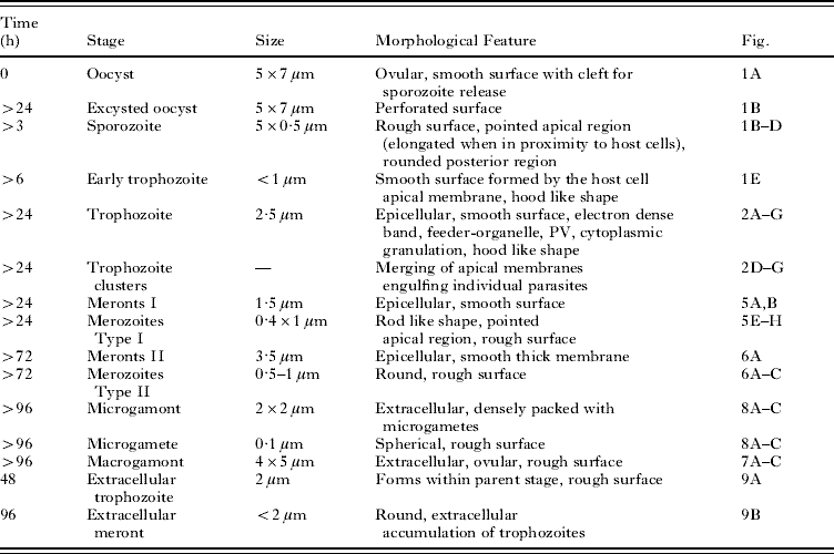
Following inoculation, intact C. parvum oocysts were only ever found in the supernatant and these were ovular, 5 μm×7 μm in size and possessed a smooth surface (Fig. 1A). This correlates with previous findings of Hijjawi et al. (Reference Hijjawi, Meloni, Morgan and Thompson2001). On the surface of these oocysts a cleft was visible and presumably it is along this cleft that the oocyst opens to release sporozoites during the excystation process. Excysted oocysts were observed adhered to cells after 24 h but not before. It is known that oocysts possess surface lectins that are thought to hinder their adherence to host tissue in vivo until the target tissue is reached where they then mediate attachment to host cells (Kuznar and Elimelech, Reference Kuznar and Elimelech2006). In the present study, oocysts were directly applied to target cells but did not attach immediately. Perhaps without passage through the gastrointestinal tract, oocysts do not begin to express the surface lectins needed for host cell adherence until after extended exposure to host cells or simply until a certain progression of time after excystation stimuli are initiated. The outer membrane of excysted oocysts appeared rough (Fig. 1B). This is probably due to membrane perforation during the excystation processes.
Oocyst excystation and sporozoite host cell invasion. (A) Intact oocyst from supernatant, 3 h post-inoculation. (B) Oocyst excystation in vitro, 48 h post inoculation. (C) Free sporozoite isolated from supernatant 3 h post-inoculation. (D) Free sporozoite on host cell, 7 h post-inoculation. Arrows indicate the apical regions of sporozoites. (E) Encapsulated by the host cell apical membrane, invading sporozoites transform into the trophozoite stage epicellularly, at 6 h and 24 h respectively. Scale bars: (A) 4 μm; (B) 2 μm; (C,D,E) 1 μm.

Sporozoites observed in this study measured about 5 μm×0·5 μm and showed well-defined apical ends (Fig. 1B–D). The hatching sporozoite shown in Fig. 1B as well as the sporozoite on the surface of the host cell in Fig. 1D, both showed an enlarged posterior region and a well-defined apical region, which appeared thin and elongated. Sporozoite apical organelle discharge is known to mediate host cell contact and according to previous findings already occurs during excystation (Snelling et al. Reference Snelling, Lin, Moore, Millar, Tosini, Pozio, Dooley and Lowery2007). As the sporozoites shown in Fig. 1B and D appear to be making host cell contact, apical organelle discharge of molecules, which are discharged in the presence of host cells, might account for the occurrence of this thin and elongated apical region. In contrast, sporozoites isolated from supernatant did not display this apical elongation and their shape remained largely regular along their entire length (Fig. 1C). Sporozoites were only observed up to 48 h post-inoculation. As sporozoites are known to invade host cells within 5 min after excystation (Forney et al. Reference Forney, DeWald, Yang, Speer and Healey1999) it can be assumed that after 48 h all oocysts have excysted and that no new sporozoites are produced. Invasion of host cells by sporozoites is shown in Fig. 1E. Invading sporozoites eventually become completely encapsulated by the host cell apical membrane, which initially appeared as a hood-like structure at this early invasion stage (Fig. 1E). A similar process has also been described in the in vivo toad system by Valigurova et al. (Reference Valigurova, Jirku, Koudela, Gelnar, Modry and Slapeta2008). As part of the infection process, sporozoites transform into the trophozoite stage and undergo merogony, which leads to trophozoite growth. This initial trophozoite stage measured <1 μm in diameter (Fig. 1E) and was observed as early as 6 h post-inoculation, showing that host cell infection followed by parasite development occurs rapidly after parasite-host tissue contact.
Trophozoites on the host cell surface 48 h post-inoculation
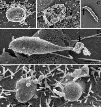
During the invasion process, C. parvum sporozoites always remain epicellular and appear to transform into the trophozoite stage extracytosolic. Thus, stages that result from host cell invasion show a similar cellular location as gregarines, with the only difference being that Cryptosporidium becomes encapsulated by a host cell apical membrane (Fig. 2A–G; Barta and Thompson, Reference Barta and Thompson2006). Trophozoites shown in Fig. 2A–G may have developed from either sporozoites or from merozoites type I after host cell invasion. Trophozoites varied in size depending upon their developmental stage. Early trophozoites observed from 6 h post-inoculation onwards, were ⩽1 μm in size (Fig. 1E), whereas well-developed trophozoites observed after 24 h post-inoculation, were up to 2·5 μm (Fig. 2B). These mature trophozoites appeared attached to the surface of cells, but were separated from the host cell via an electron-dense band (Fig. 2C). The formation of a feeder organelle structure is hypothesized for all C. parvum stages and is shown here in Fig. 2A. Previous morphological studies by Huang et al. (Reference Huang, Chen and LaRusso2004) have already described this extracytosolic location of the parasite and its feeder organelle attachment. Our data show sporozoites becoming engulfed by the host cell apical membrane. This phenomenon was proposed (Perkins et al. Reference Perkins, Riojas, Wu and Le Blancq1999; Elliott and Clark, Reference Elliott and Clark2000; Pollok et al. Reference Pollok, McDonald, Kelly and Farthing2003; Chen et al. Reference Chen, Huang, Splinter, Orth, Billadeau, McNiven and LaRusso2004a, Reference Chen, Splinter, Tietz, Huang, Billadeau and LaRussob, Reference Chen, O'Hara, Huang, Splinter, Nelson and LaRusso2005; Hashim et al. Reference Hashim, Mulcahy, Bourke and Clyne2006) and recently confirmed by Valigurova et al. (Reference Valigurova, Jirku, Koudela, Gelnar, Modry and Slapeta2008). It has been suggested that the parasite induces the host cell to encapsulate itself with the host cell apical membrane (Borowski et al. Reference Borowski, Clode and Thompson2008) to escape the hosts defence mechanisms.
Trophozoites on the host cell surface 48 h post-inoculation. (A) Cross-section through an early trophozoite showing the feeder-organelle (arrow) attachment to the host cell cytosol. (B) Mature trophozoite. (C) Cross-section through a mature trophozoite revealing the electron-dense band (arrow) that separates the parasite from the host cell. (D,E,F,G) Accumulations of trophozoites on the host cell surface. Scale bars: (A) 0·5 μm; (B,C,G) 1 μm; (D,E,F) 2 μm.

Trophozoites were always found to reside within a PV and to be engulfed by the host cell apical membrane, which displayed a smooth surface. In later stages of trophozoite development, the basal membrane developed a hood-like structure and cytoplasmic granulation occurred leading to merozoite development within the trophozoite (Fig. 2A and B). Single trophozoites were regularly seen attached to infected cells (Fig. 2B), but accumulations of 2 or more trophozoites were more frequently observed (Fig. 2D–G). The 4 trophozoites shown in Fig. 2D are likely to have developed from sporozoites that simultaneously excysted from a single oocyst. However, the 6 trophozoites observed in Fig. 2E are more likely to have resulted from merozoite type I host cell invasion, as one meront I is believed to contain 6 or 8 merozoites (Hijjawi et al. Reference Hijjawi, Meloni, Morgan and Thompson2001). A merging of membranes engulfing 2 or more parasites was also often observed (Fig. 2D–G). This leads to the assumption that clusters of 2 or more trophozoites result from invasion of infective zoites (sporozoites or merozoites) in closest proximity. Hijjawi et al. (Reference Hijjawi, Meloni, Ng'anzo, Ryan, Olson, Cox, Monis and Thompson2004) made the observation that zoite stages commonly accumulate in cell-free cultures. Possibly, there is a weak unspecific binding between infective zoites (Fig. 6A–C). Chen et al. (Reference Chen and LaRusso2000) reported that zoite stages express surface lectins that have adhesive properties. It is possible therefore, that these lectins also facilitate the attachment of infective zoites to each other. Taken together, these data suggest that excysted sporozoites and merozoites can be adherent to each other during host cell attachment and invasion of cells (Fig. 6C). This close proximity of invading stages results in the merging of membranes between individuals, forming clusters of rapidly growing trophozoites.
The effect on host cell microvilli
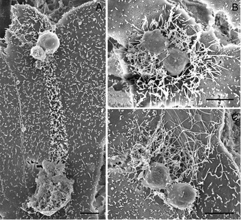
Protozoan parasites possess gliding motility. Gliding motility of sporozoites has been observed in previous studies (Barnes et al. Reference Barnes, Bonnin, Huang, Gousset, Wu, Gut, Doyle, Dubremetz, Ward and Petersen1998; Riggs et al. Reference Riggs, McNeil, Perryman, Stone, Scherman and O'Connor1999; Wetzel et al. Reference Wetzel, Schmidt, Kuhlenschmidt, Dubey and Sibley2005) leading to the suggestion that gliding along the host cell surface is a prerequisite to host cell invasion. C. parvum gliding trails along host cell surfaces were observed in this study (Fig. 3A). These gliding trails were evident as trails of elongated microvilli between an excysted oocyst and newly formed trophozoites; extending up to 15 μm. As C. parvum is known to utilize microvilli material to establish itself in its niche (Bonnin et al. Reference Bonnin, Gut, Dubremetz, Nelson and Camerlynck1995), it can be suggested that gliding sporozoites use microvilli material for their gliding motion and thus cause this abnormality seen in gliding trails. As gliding trails were not observed in all cases of oocyst excystation, it appears that not all sporozoites glide along the host membrane surface until they are ready to invade a cell at a particular area. Some sporozoites are thought to invade directly at their origin of excystation (Fig. 2F), exhibiting gliding movements in a small area only, whereas other sporozoites might even travel through the culture medium (Fig. 1C) before establishing host cell contact and invading cells some distance from excystation.
The parasite's effect on host cell microvilli. (A) Gliding trail composed of elongated microvilli between an excysted oocyst and trophozoites 3 days post-inoculation. (B,C) Abnormal microvilli clusters surround trophozoites 48 h post-inoculation. Scale bars: 2 μm.

Microvillus abnormality has not only been observed in gliding trails, but also around developing trophozoites (Fig. 3B and C). Abnormally abundant microvilli were frequently found to surround trophozoites (Fig. 3B) and in some cases were significantly elongated (Fig. 3C). As the expression of microvilli around trophozoite clusters was found to be higher than in non-infected areas, C. parvum might even induce the production of microvilli to satisfy its need for microvillar components. Microvilli of infected cells were also seen to appear to become incorporated into the membrane engulfing the parasites and to aid in securing the parasite to the host cell surface (Fig. 2B and G). This phenomenon has not been described before, but microvillar material has been identified in the membrane components engulfing the parasite using molecular techniques (Bonnin et al. Reference Bonnin, Gut, Dubremetz, Nelson and Camerlynck1995).
Binary fission and syzygy in the C. parvum life cycle
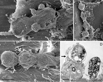
Additional C. parvum stages were also found that were characteristically different from those described above. Figure 4A and B raise the possibility that C. parvum may undergo binary fission, i.e. the splitting of a parent cell into 2 daughter cells, other than during the course of merogony and shizogony. In Fig. 4A, two microgamonts that seem to be in the process of binary fission, are engulfed by a single membrane that may have initially encapsulated the parent trophozoite on the host cell surface. The finding of 2 parasites that appear to be dividing from 1 parent and seem to be the same life-cycle stage (Fig. 4A), demonstrates that C. parvum possibly undergoes asexual reproduction other than in the course of merogony. As explained below, as C. parvum progresses through its life cycle, it appears to become more extracellular. This is evident in stages that appear to have little or no attachment to host cells (Fig. 4A–C). All of these stages are likely to be microgamonts, one of which is clearly visible in Fig. 4A. Microgamonts occur later in the life cycle once sexual reproduction has been initiated. Thus, these stages show a more extracellular location. Our observations raise the question whether C. parvum stages with more epicellular location are still attached to and encapsulated by the host cell apical membrane, or have broken contact with host cells, but still retain the surrounding outer host cell membrane.
Cryptosporidium parvum binary fission and syzygy. (A,B) Binary fission of C. parvum stages 4 days post-inoculation. (C,D) C. parvum syzygy 5 days post-inoculation. Basal discs are indicated by arrows. Scale bars: (A,B,C) 2 μm; (D) 1 μm.

Our data also indicate that C. parvum might employ syzygy (Fig. 4C). Syzygy is defined as the association of gamonts (pre-gametes) end-to-end or in lateral pairing prior to the formation of gametes that may be employed to ensure genetic diversity and has been described in gregarines (Landers, Reference Landers2001; Lacombe et al. Reference Lacombe, Jakowska and Silva2002; Barta and Thompson, Reference Barta and Thompson2006; Toso and Omoto, Reference Toso and Omoto2007). Thus, it is reasonable that syzygy may occur in closely related Cryptosporidium species. Connecting discs between adjacent parasites are visible (Fig. 4C and D) and these appear to be the same as the basal discs already described in Cryptosporidium by Valigurova et al. (Reference Valigurova, Jirku, Koudela, Gelnar, Modry and Slapeta2008). The C. parvum stages involved in syzygy shown in Fig. 4C and D are probably microgamonts. As syzygy was first observed when sexual life-cycle stages occurred in culture, it can be suggested that predominantly gamont stages (here microgamonts) employ syzygy. This correlates with findings on gregarines and further supports the affinity of Cryptosporidium with these apicomplexans (Landers, Reference Landers2001; Toso and Omoto, Reference Toso and Omoto2007).
Meronts and merozoites
Trophozoites that result from sporozoite host cell invasion develop into meronts I. Meronts I with visible internal merozoites type I, were observed as early as 24 h post-inoculation (Fig. 5A and B). Like trophozoites, meronts I were found to be attached to host cells and engulfed by the host cell apical membrane (Fig. 5A and B). Developing meronts measured approximately 1·5 μm in diameter (Fig. 5A and B), whereas mature meronts at the stage of merozoite release were larger, measuring 2·5 μm (Fig. 5C and D). From this, it appears that trophozoites developing into meronts I undergo merogony and begin to form internal merozoites, before they have reached their full size. Merozoites seemed to be aligned in a parallel orientation within intact meronts I (Fig. 5A and B) which is consistent with observations by Hijjawi et al. (Reference Hijjawi, Meloni, Morgan and Thompson2001). Once excysted, the merozoites type I in a meront numbered 6 or 8, which also correlates with previous findings by Hijjawi et al. (Reference Hijjawi, Meloni, Morgan and Thompson2001). Excysting merozoites type I showed a rod-like shape with a pointed apical region. In contrast to excysting oocysts, the membranes of meronts I appear not to become perforated to facilitate infective zoite release. During merozoite release, the membranes engulfing the parasite still appeared smooth (Fig. 5C and D) and seemed to either open up (Fig. 5 D) or rupture (Fig. 5C). When merozoites type I are released from the meront a residual body is left behind (Fig. 5C). Similar observations have been made on the Cryptosporidium sp. ‘toad’ model (Valigurova et al. Reference Valigurova, Jirku, Koudela, Gelnar, Modry and Slapeta2008).
Meronts I and merozoites type I. (A,B) Developing meronts I with internal merozoites type I. (C,D) Mature meronts I at the stage of merozoite I excystation. (E,F,G) Free merozoites type I showing well-defined apical regions (arrow) at 6 h of culture. (H) Merozoite type I host cell invasion 6 h post-inoculation. Scale bars: (A,B,C,E,F) 1 μm; (D) 2 μm; (G,H) 0·5 μm.

Free merozoites type I were seen in cell culture from 24 h post-inoculation onwards (Fig. 5E–H). Merozoites, which previously were probably incorrectly referred to as microgametes (Thompson et al. Reference Thompson, Olson, Zhu, Enomoto, Abrahamsen and Hijjawi2005) type I were 0·4 μm×1 μm, with both a rounded and a pointed end (Fig. 5E–H). The pointed end appeared similar to that of sporozoites, which houses the apical organelles for host cell invasion (Tetley et al. Reference Tetley, Brown, McDonald and Coombs1998). These free merozoites must be adhered to the host cells in some manner (Fig. 5E–5H), otherwise they would have been removed during sample preparation. Surface lectins which have been detected on infective zoite stages (Chen et al. Reference Chen and LaRusso2000) might mediate this attachment to host tissue. Merozoites appear initially to adhere to host cells along their full body length, but become stouter as cellular invasion occurs (Fig. 5H). Thus it can be hypothesized that after initial host cell attachment involving surface lectins, a re-orientation of merozoite organelles occurs, bringing the apical complex into host cell contact to initiate cellular invasion.
Once merozoites type I are present, the parasite employs 2 ways to replicate in its host which both occur concurrently: (i) it progresses via asexual reproduction by the formation of meronts I and (ii) it progresses via sexual reproduction by formation of meronts II which then further develop into micro-and macrogamonts (Borowski et al. Reference Borowski, Clode and Thompson2008).
Meronts II as well as merozoites type II were observed in culture from 72 h post-inoculation. Meronts II were found to be bigger than meronts I measuring 3·5 μm in diameter (Fig. 6). Meronts II appeared to possess a thicker outer membrane than meronts I (Fig. 6A). Merozoites type II were round and measured between 0·5 μm and 1 μm in diameter (Fig. 6A–C). Similar to meronts I, the membrane engulfing the merozoites appeared smooth and not perforated for their release (Fig. 6A) suggesting the host cell origin of this membrane.
Meront II and merozoites type II. (A) Excysting meront II with internal merozoites type II (arrow) 3 days post-inoculation. (B) Merozoite type II host cell invasion. (C) Pairing of a merozoite type II with a merozoite type I 9 h post-inoculation. Scale bars: (A) 1 μm; (B,C) 0·5 μm.

Similar to the adhesion of sporozoites to each other via surface lectins, merozoites type I and type II can also adhere to each other and co-invade cells (Fig. 6B and C).
Microgamonts and macrogamonts
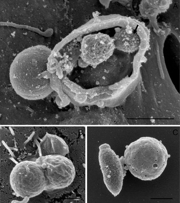
Merozoites type II that are released in culture, invade cells to transform into either micro- or macrogamonts. Four days post-inoculation micro- and macrogamonts were observed for the first time (Figs 7 and 8). Both gamont stages appeared to have less contact with host cells than trophozoites or meronts. This supports our suggestion that the further the parasite progresses in its life cycle, the more extracellular it appears to become. Macrogamonts were ovular and measured about 4 μm×5 μm (Fig. 7A). Feeder organelle attachment of a macrogamont is visible in Fig. 7A. The surface of this macrogamont appeared rough, suggesting that it has already broken host cell contact leaving the host cell-derived membrane that once surrounded it behind. Such empty ‘attachment-zones’ often show a ring-like structure (Fig. 7C), presumably where feeding organelles were attached. These findings correlate with observations by Valigurova et al. (Reference Valigurova, Jirku, Koudela, Gelnar, Modry and Slapeta2008) who described a similar extracellular location of macrogamonts, their detachment from host cells, the granular structure of their feeder organelles, as well as detachment zones in vivo, in their Cryptosporidium sp. ‘toad’ model.
Macrogamonts and their attachment zones. (A) Macrogamont with feeder organelle (arrow) 4 days post-inoculation. (B) Developing macrogamont with feeder organelle (arrow). (C) Cryptosporidium parvum attachment zones 5 days post-inoculation. Scale bars: (A,C) 2 μm; (B) 1 μm.

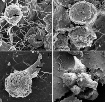
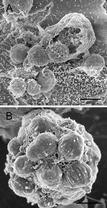
Microgamonts. (A,B) Microgamonts without stalk. The arrows in A indicate a cleft along which the macrogamonts open. (C,D) Microgamonts with stalk (arrow). Scale bars: (A,B) 1 μm; (C,D) 2 μm.

Microgamonts were rounder and smaller than macrogamonts, measuring about 2 μm×2 μm (Fig. 8A–C). They contained a large number of microgametes (Fig. 8A and C), which are released to fertilize macrogamonts. This observation is not consistent with findings by Hijjawi et al. (Reference Hijjawi, Meloni, Morgan and Thompson2001), who identified only 16 microgametes in 1 microgamont with light microscopy. This difference is likely to be due to the difficulty resolving such structures with light microscopy. Microgametes measure about 0·1 μm in diameter and are spherical. Hijjawi et al. (Reference Hijjawi, Meloni, Morgan and Thompson2001) believed that they were non-flagellated which is confirmed by our SEM study. The microgamonts shown in Fig. 8A seem to be surrounded by presumably host cell-derived membrane. Fig. 8C shows a feeder organelle attachment of the same origin as that of the macrogamont in Fig. 7A. Approximately half of all microgamonts seen in culture possessed a stalk-like structure that either appeared as if the stalk had broken (Fig. 8D) or seemed to attach the parasite to the host cell (Fig. 8C). Again, a similar finding has also been reported by Valigurova et al. (Reference Valigurova, Jirku, Koudela, Gelnar, Modry and Slapeta2008). This stalk on microgamonts might result from the parasite being released from the attachment zones described above in a similar manner to macrogamonts. The microgamonts that possess a stalk (Fig. 8C and D) appeared to have less host cell contact compared to those which did not show such a structure (Fig. 8A and B). Additionally, their membrane appeared perforated (Fig. 8C) in contrast to microgamonts without this stalk.
Like trophozoites, microgamonts were also observed to occur in clusters of 2 or more. The accumulation of microgamonts (Fig. 8D) probably resulted from microgamonts that had released from their attachment zone and adhered to each other, whereas the 2 adjacent microgamonts in Fig. 8B are more likely to be a result of merozoite type II host cell invasion in close proximity.
Development of C. parvum in host cell culture without invasion of host cells
The present study is consistent with the intracellular life cycle of C. parvum described by Hijjawi et al. (Reference Hijjawi, Meloni, Morgan and Thompson2001). The stages described above all resulted from host cell invasion, yet, complete cell-free development of C. parvum has also been documented at the light microscope level (Hijjawi et al. Reference Hijjawi, Meloni, Ng'anzo, Ryan, Olson, Cox, Monis and Thompson2004) in vitro. From our own observations, we hypothesize that extracellular life-cycle stages might be part of the C. parvum life cycle, or co-exist resembling rudimentary stages of an ancestral life cycle. Our data show that C. parvum can develop extracellularly in the presence of host cells in an in vitro model, without invasion. We have observed stages of C. parvum that have been described before at the light microscopic level but are not as yet considered as part of the life cycle.
Life-cycle stages of C. parvum that developed extracellularly showed a rough surface like that of previously described free parasite stages (Fig. 5E–H and Fig. 6A–C), and they did not appear to be engulfed by the host cell apical membrane.
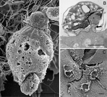
After 48 h inoculation, we observed what we believe to be trophozoite development directly within an oocyst (Fig. 9A). The membrane of the oocyst releasing the trophozoites appeared rough and perforated, a characteristic already observed during the release of sporozoites (Fig. 1B). Similar to these so-called intracellular trophozoites, trophozoites that formed directly within an oocyst measured approximately 2 μm. As these stages were observed as early as 48 h post-inoculation (Fig. 9A) it can be hypothesized that the trophozoites developed within an inoculated oocyst, and were eventually released into culture.
Development of Cryptosporidium parvum in host cell culture without the invasion of host cells. (A) Trophozoite development within an inoculated oocyst after 2 days. (B) Possible extracellular meront after 4 days of culture. Scale bars: (A) 2 μm; (B) 1 μm.

From our observations it can be suggested that each sporozoite and each merozoite is capable of undergoing merogony and transforming into a trophozoite stage, without invasion of host cells (Fig. 9A). This suggestion is supported by our findings of a proposed extracellular meront 4 days post-inoculation (Fig. 9B). The meront measured approximately 8 μm in diameter and was round in shape. The zoite stages observed within the meront, measured up to 2 μm and appeared to be densely packed. It is likely that this extracellular meront has formed through the clumping of infective zoite stages in culture, of which each single one has undergone merogony to form a trophozoite. In support of this, accumulations of merozoites and/or trophozoites, as well as the formations of meronts in cell-free culture, have already been described (Hijjawi et al. Reference Hijjawi, Meloni, Ng'anzo, Ryan, Olson, Cox, Monis and Thompson2004).
Critically, how extracellular trophozoites and meronts progress in their life cycle without cellular invasion is still not understood. Each extracellular trophozoite might progress in a manner typical of an intracellular trophozoite, or each single trophozoite might transform into the next life-cycle stage. The observation that C. parvum also develops extracellularly, despite the presence of host cells, further supports the proposal that Cryptosporidium has a close affinity with gregarines (Carreno et al. Reference Carreno, Martin and Barta1999; Barta and Thompson, Reference Barta and Thompson2006; Valigurova et al. Reference Valigurova, Hofmannova, Koudela and Vavra2007).
CONCLUSIONS
For the first time, our study reveals the morphology of each stage in the currently accepted life cycle of C. parvum. The development of life-cycle stages described here extends previous reports that were based on light microscopic findings (Hijjawi et al. Reference Hijjawi, Meloni, Morgan and Thompson2001, Reference Hijjawi, Meloni, Ryan, Olson and Thompson2002, Reference Hijjawi, Meloni, Ng'anzo, Ryan, Olson, Cox, Monis and Thompson2004). Data from our in vitro model system are further supported by similar morphological observations from in vivo studies on C. muris and Cryptosporidium sp. ‘toad’ (Valigurova et al. Reference Valigurova, Jirku, Koudela, Gelnar, Modry and Slapeta2008). Surprisingly, the species C. parvum observed in our study shows more similarities to the species Cryptosporidium sp. ‘toad’ than to C. muris (Valigurova et al. Reference Valigurova, Jirku, Koudela, Gelnar, Modry and Slapeta2008).
Apart from revealing the morphology of accepted life-cycle stages in C. parvum our study describes previously unreported features of C. parvum life-cycle stages, which include their morphological structure and interaction with host cells. Further, extracellular stages of C. parvum are reported and characterized for the first time in an in vitro model of cultured host cells at an electron microscopic level.
It is not clear whether extracellular stages of C. parvum occur in vivo, and whether they are part of the parasite's life cycle or represent rudimentary stages of an ancestral life cycle. Future studies are needed to identify the existence of these stages in vivo and clarify their nature.