Introduction
Brain tumors are the second most common form of pediatric cancer, making up approximately 18% of all pediatric cancer diagnoses (Howlader et al., Reference Howlader, Noone, Krapcho, Neyman, Aminou, Waldron and Edwards2011) with a prevalence of 4.8 cases per 100,000 children annually (Central Brain Tumor Registry of the United States (CBTRUS), 2008). Approximately 50–60% of pediatric brain tumors arise in the posterior fossa, the region of the brain comprising the cerebellum, brainstem, and fourth ventricle (El-Ghandour, Reference El-Ghandour2011). Although overall survival rates have risen to approximately 72.5%, (CBTRUS, 2008), research suggests survivors are at risk for a wide range of late effects including endocrine deficiencies, cardiac impairment, physical limitations, and emotional distress (e.g., Gurney et al., Reference Gurney, Kadan-Lottick, Packer, Neglia, Sklar, Punyko and Robison2003, Ness & Gurney, Reference Ness and Gurney2007; Zebrack et al., Reference Zebrack, Gurney, Oeffinger, Whitton, Packer, Mertens and Zeltzer2004).
Disruptions in cognitive development are among the most frequently observed late effects in this group of survivors, and include possible long-term disruptions in brain development, cognitive function, and later school and work performance. Numerous independent studies have reported adverse long-term neurocognitive effects in survivors of pediatric brain tumors (e.g., Bonner et al., Reference Bonner, Hardy, Willard, Anthony, Hood and Gururangan2008; Jain, Krull, Brouwers, Chintagumpala, & Woo, Reference Jain, Krull, Brouwers, Chintagumpala and Woo2008; Mulhern & Butler, Reference Mulhern and Butler2004). A recent meta-analysis (Robinson et al., Reference Robinson, Kuttesch, Champion, Andreotti, Hipp, Bettis and Compas2010) indicated that, collapsing across all tumor histologies and locations, survivors are at increased risk of deficits in overall cognitive ability, verbal and nonverbal intelligence, academic achievement (reading, math, spelling), attention, psychomotor skill, visual-spatial skill, verbal memory, and language, with the magnitude of these effects ranging from a Hedges’ g Footnote 1 = −0.45 to −1.43. However, studies have found that survivors of posterior fossa tumors and supratentorial tumors may experience differences in neurocognitive outcome (e.g., Ater et al., Reference Ater, Moore, Francis, Castillo, Slopis and Copeland1996), suggesting that examination of neurocognitive late effect profiles in these groups separately is necessary.
The magnitude of deficits may vary based on several factors, including tumor location (Papazoglou, King, Morris, & Krawiecki, Reference Papazoglou, King, Morris and Krawiecki2008), treatment (Grill et al., Reference Grill, Renaux, Bulteau, Viguier, Levy-Piebois, Sainte-Rose and Kalifa1999), and patient characteristics (Radcliffe, Bunin, Sutton, Goldwein, & Phillips, Reference Radcliffe, Bunin, Sutton, Goldwein and Phillips1994). For example, although radiation therapy has in some cases led to long-term disease control in otherwise surgically inoperable brain tumors (Alexander & Loeffler, Reference Alexander and Loeffler1999), cranial radiation therapy is consistently associated with adverse effects on a variety of domains of neurocognitive functioning, and children treated with this modality have greater declines in these areas than children treated without cranial radiation (Nathan et al., Reference Nathan, Patel, Dilley, Goldsby, Harvey, Jacobsen and Armstrong2007; Reimers et al., Reference Reimers, Ehrenfels, Mortensen, Schmiegelow, Sonderkaer, Carstensen and Muller2003).
Recent studies have examined the relative risk increases at higher cumulative doses of radiation, larger radiation fields, and younger age. One study examined differences in the neurocognitive functioning of survivors of posterior fossa tumors treated with either radiation solely to the posterior fossa, low-dose craniospinal radiation (25 Gy) with a boost to the posterior fossa, or high-dose craniospinal radiation (35 Gy) with a boost to the posterior fossa (Grill et al., Reference Grill, Renaux, Bulteau, Viguier, Levy-Piebois, Sainte-Rose and Kalifa1999). When comparisons were made between the low-dose and high-dose craniospinal radiation groups, the low-dose group performed significantly better than the high-dose group in terms of full scale and verbal IQ, suggesting that lower doses of craniospinal radiation are associated with better neurocognitive outcome (Grill et al., Reference Grill, Renaux, Bulteau, Viguier, Levy-Piebois, Sainte-Rose and Kalifa1999). These findings parallel those from other studies examining the neurocognitive impact of treatment for medulloblastoma that have found a significant negative correlation between post-treatment IQ and whole brain irradiation (Yang et al., Reference Yang, Wong, Cheng, Chang, Hsu, Chen and Chuang1997); and lower full scale, verbal, and performance IQ in children treated with high-dose (35 Gy) versus low-dose (25 Gy) craniospinal radiation (Kieffer-Renauz et al., Reference Kieffer-Renauz, Bulteau, Grill, Kalifa, Viguier and Jambaque2000).
One of the most frequently considered patient characteristics in this area is the age of the patient at diagnosis. Research has suggested that children treated at a younger age are at increased risk for neurocognitive deficits as compared to survivors treated at an older age (Mulhern, Hancock, Fairclough, & Kun, Reference Mulhern, Hancock, Fairclough and Kun1992). Radcliffe and colleagues (1994) found that children younger than 7 years of age at diagnosis showed a progressive decline in full scale IQ, whereas children older than 7 did not show progressive decline. Mulhern and Kun (Reference Mulhern and Kun1985) found that 68% of children less than 6 years of age experienced neurocognitive declines in at least one domain, whereas only 11% of children older than 6 years of age evidenced these declines. Moore, Ater, and Copeland (Reference Moore, Ater and Copeland1992) found significant positive correlations between age at diagnosis and performance on measures of overall intelligence, nonverbal intelligence, and psychomotor skill in a sample of children treated with cranial radiation. Positive but nonsignificant correlations were found between age at diagnosis and verbal intelligence, language, academic achievement, visual-spatial skill, and attention.
Due to the heterogeneity of samples in many studies, identification of moderating factors of deficits in survivors of pediatric brain tumors has proven difficult. Survivors of posterior fossa tumors represent an important population that, for two reasons, lends itself to meta-analytic review. First, studies examining survivors of posterior fossa tumors exclusively are more prevalent and therefore available for synthesis. Second, this population is of particular interest due to the prevalence of brain tumors arising from this region and the large proportion of survivors for whom improved information on potential deficits would be beneficial. Therefore, the focus of the current meta-analysis is to quantify the nature and extent of deficits in children with posterior fossa tumors. Because previous studies have suggested that patient age (Radcliffe et al., Reference Radcliffe, Bunin, Sutton, Goldwein and Phillips1994) and dosage of radiation (Grill et al., Reference Grill, Renaux, Bulteau, Viguier, Levy-Piebois, Sainte-Rose and Kalifa1999) are important to consider, we examined differences in the magnitude of deficits based on these potential moderators. It was hypothesized that survivors would evidence significant deficits in a range of global cognitive, academic, and specific neurocognitive domains. Furthermore, it was expected that deficits would be most pronounced in survivors treated at a younger age and in survivors treated with higher doses of radiation.
METHOD
Literature Search and Selection of Articles
Studies examining the neurocognitive effects of treatment for childhood brain tumors were identified through literature searches of MEDLINE, CINAHL, and PsycInfo using the following key terms: CNS cancer, brain tumor, neoplasm, neuropsychological, neurocognitive, IQ, executive function, memory, attention, pediatric, and child*. Studies’ references were reviewed to identify additional publications for review. Studies were included in data analyses based on several inclusion criteria. First, studies had to be published in English and report original quantitative data on the neurocognitive functioning of survivors of childhood tumors of the posterior fossa. Second, neurocognitive measures had to have adequate psychometric properties (i.e., established reliability and validity), and published normative data. Third, study samples could not be composed of patients with known premorbid medical conditions associated with neurocognitive insult (e.g., neurofibromatosis-type 1). Fourth, studies that included children with other forms of childhood cancer (e.g., leukemia) or survivors of pediatric brain tumors in regions outside the posterior fossa, were excluded unless diagnostic groups were analyzed separately or data were presented by individual participant. Sample and methodological characteristics from studies meeting inclusion criteria were coded using criteria adapted from Lipsey and Wilson (Reference Lipsey and Wilson2000) to ensure consistent and accurate data extraction. Extracted data included (among other things) sample size, demographics, and subgroups, diagnostic and treatment information (where available), outcome domains assessed, and measures used. Full manuscript review and coding were completed by a member of the study team, which included psychology graduate students, a medical student, and undergraduate research assistants. Before participating in coding, study team members completed practice reviews until inter-rater agreement exceeded 90%. During formal reviews, a subset of studies (20%) was double-coded and inter-rater agreement was verified to further assess extraction accuracy. When necessary, consensus was determined through a separate review by the senior graduate student on the study.
Thirty-eight studies contained usable data from which effect sizes could be extrapolated (e.g., means and standard deviations) and were included in analyses. Meta-analytic reviews are susceptible to publication bias (i.e., the “file drawer” effect) because studies with significant effects are more likely to be published, and thus included in the meta-analysis than those with null findings. The general approach to addressing this issue is to calculate a “fail-safe N,” which estimates the number of unpublished or unretrieved studies without significant findings that would need to be found to render the mean effects nonsignificant. Rosenthal's (1991) widely used formula to calculate a fail-safe N was implemented: [(Σz)2/2.706] – k, where Σz is the sum of the combined Z scores squared, 2.706 is the two-tailed critical value for statistical significance at p = .05, and k refers to the number of studies included. Rosenthal (Reference Rosenthal1991) recommends a tolerance level of 5k +10, which yields a highly unlikely number of unpublished studies that would be required to bring the meta-analysis results down to a nonsignificant level. This value was calculated for each effect size.
Moderator analyses were also conducted to determine whether deficits differed based on age at diagnosis or dosage of cranial radiation. To include the largest number of studies possible in moderator analyses, a cutoff of 7 years and older versus younger than 7 years old was established, based on the modal pattern in individual studies. Several studies provided neurocognitive outcome data by individual participants, and also included information about each participant's age at diagnosis; in these instances, cases were split according to a cutoff of 7 years old and means and standard deviations were calculated. For moderator analyses, a minimum of two studies must have provided data at each level of the moderator being considered. Of the 38 studies included in the overall analyses, 7 were subsequently included in our examination of age as a moderator of deficits in survivors, and 5 were included in our examination of the role of radiation dosage as a moderator.
Neurocognitive Domains and Measures
To examine neurocognitive functioning in a variety of domains, we established categories paralleling the approach used in a previous meta-analysis of neurocognitive deficits in survivors of pediatric brain tumors in any region (Robinson et al., Reference Robinson, Kuttesch, Champion, Andreotti, Hipp, Bettis and Compas2010). We assigned the measures used in individual studies to one of these functional domains for analysis. The following neurocognitive domains were examined: (a) Overall Cognitive Functioning (i.e., Full Scale Intelligence) measured by the Wechsler scales of intelligence (i.e., WPPSI, WISC, WAIS), Stanford-Binet, Bayley Scales of Infant Development, McCarthy Scales of Children's Abilities, Raven Colored Progressive Matrices, Leiter International Performance Scales. (b) Academic Achievement (reading, arithmetic, spelling) measured by the Wide Range Achievement Test (WRAT), the Woodcock-Johnson Tests of Achievement, the Peabody Individual Achievement Test (PIAT), the Wechsler Individual Achievement Test. (c) Attention/Concentration, measured by the Trail Making Test A, Connors Continuous Performance Test, WISC Freedom from Distractibility Index, Test of Everyday Attention for Children. (d) Executive Function, measured by the Trail Making Test B, Wechsler Working Memory Index, Stroop Color-Word Task, Wisconsin Card Sorting Test. (e) Information Processing Speed, measured by WISC Coding and/or WAIS Digit-Symbol subtests, Wechsler Processing Speed Index. (f) Psychomotor Skill, measured by Finger Tapping (preferred/dominant hand), Grooved Pegboard (preferred/dominant hand), Purdue Pegboard Test. (g) Verbal Memory, measured by the Wide Range Assessment of Memory and Learning (WRAML) verbal subtest, California Verbal Learning Test (CVLT), Rey Auditory Verbal Learning Test (RAVLT) immediate or delayed recall, Illinois Test of Psycholinguistic Abilities. (h) Visuospatial Skill, measured by the Beery-Buktenica Developmental Test of Visual-Motor Integration (VMI), Rey-Osterrieth Complex Figure Test-Copy. (i) Visuospatial Memory, measured by the WRAML visual subtest, Continuous Visual Memory Test, Rey Complex Figure Test-Delayed Recall. (j) Language, measured by the Peabody Picture Vocabulary Test, Verbal Fluency (F-A-S) Test, Clinical Evaluation of Language Fundamentals (CELF). According to publication and technical manuals, the internal consistency reliability of these measures ranged from 0.61 to 0.98, with the majority of measures reporting internal consistencies between 0.79 and 0.98, and a median internal consistency of 0.91, indicating adequate to excellent reliability. Normative sample sizes ranged from 206 to 8818, with a median sample size of 1700. Additional information regarding the measures included in these analyses is available from the authors upon request.
Methods of Comparison
To compare survivors of pediatric brain tumors to established normative data, Hedges’ g was calculated for each study finding based on a random effects model. Hedges’ g represents the number of standard deviations a posterior fossa tumor group mean on a given neurocognitive test differed from the mean of a normative sample, controlling for potential bias due to small sample sizes. In other words, this value follows a Z-score distribution. Negative effect sizes indicate that the brain tumor sample performed more poorly than the normative sample. According to Cohen (Reference Cohen1988) effect sizes less than 0.2 indicate a negligible effect, those between 0.2 and 0.5 indicate a small effect, those between 0.5 and 0.8 indicate a medium effect, and those greater than 0.8 indicate a large effect. Each effect size was weighted by the inverse of the variance. For our examination of the possible moderating effect of age at diagnosis on the emergence of deficits, analyses were conducted with both categorical age data and continuous age data. When age was entered as a continuous variable, findings are presented as a correlation between age at diagnosis and outcome. The Comprehensive Meta-Analysis computer program (Version 2; Borenstein & Rothstein, Reference Borenstein and Rothstein2005) was used to determine statistical significance and produce 95% confidence intervals for each mean effect size.
Results
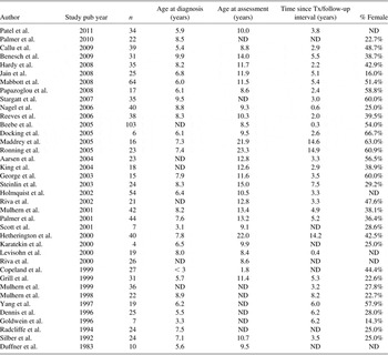
Initial keyword searches of databases identified over 140 empirical articles, reviews, and book chapters. Following screening for inclusion criteria, 53 studies of neurocognitive functioning in survivors of posterior fossa tumors remained, of which 37 presented data usable for meta-analysis. After contacting corresponding authors for additional data (N = 16), one additional study was able to be included in the meta-analysis, for a total 38 studies, published between 1983 and 2011. Data from 1090 children treated for posterior fossa brain tumors from 38 empirical studies published between 1983 and January 2011 were included in the analyses (Table 1).
Empirical studies included in analyses

Note. ND = no data available.
Descriptive Statistics
Descriptive statistics regarding sample characteristics were estimated from the subset of studies that reported necessary information (range, 30–33 of 38 total studies; Table 1). While central tendency (means or medians) and ranges were frequently reported for variables, fewer standard deviations were available to compute an average standard deviation across samples.
The estimated mean age of participants at the time of testing was 11.6 years and they were, on average, 6.6 years old at the time they were diagnosed or began treatment for a pediatric brain tumor. The average time elapsed since treatment was 4.0 years. Thirty-three studies reported the gender compositions of their samples, and 41.3% of participants were female. Only 10 studies reported ethnic or racial composition of their samples. Approximately 80.2% of these participants were White, 11.3% were Black or African-American, 3.0% were Latino or Hispanic, and 5.5% were identified as “other.” Ten studies reported information on socioeconomic status (SES). Various indicators of SES were used across these studies (e.g., Hollingshead index; years of parental education), which prevented quantitative comparison. However, it appears that the majority of participants in these studies lived in middle class households and had parents with some college education.
Main Analyses
Domain effect sizes
To estimate overall effect size, Hedges’ g were weighted and combined (Shadish & Haddock, Reference Shadish and Haddock1994) across 14 broad and specific domains (see above).
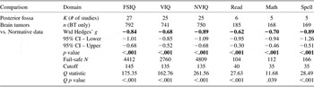
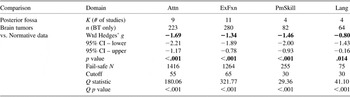
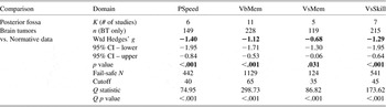
Thirty-eight studies provided the information necessary to examine deficits in survivors of any pediatric brain tumor relative to normative data in the 14 broad and specific neurocognitive domains, and these comparisons yielded 14 significant averaged weighted effect sizes, all of which were in the negative direction (Tables 2–4). The magnitude of these effects ranged from −0.62 to −1.69 with a mean Hedges’ g of −1.03. Specifically, survivors showed deficits of significant medium to large effects in the areas of overall cognitive ability, verbal intelligence, nonverbal intelligence, academic achievement in reading, math, and spelling, attention, executive function, psychomotor skill, language, processing speed, verbal memory, visual memory, and visual-spatial skill.
Deficits in Global Cognitive and Academic domains

Note. FSIQ = Full Scale IQ; VIQ = Verbal IQ; NVIQ = Nonverbal IQ; Read = Academic Achievement in Reading; Math = Academic Achievement in Math; Spell = Academic Achievement in Spelling.
Deficits in specific Neurocognitive domains

Note. Attn = Attention; CI = confidence interval; ExFxn = Executive Function; PmSkill = Psychomotor Skill’ Lang = Language.
Deficits in specific Neurocognitive domains (continued)

Note. CI = confidence interval; PSpeed = Processing Speed; VbMem = Verbal Memory; VsMem = Visual Memory; VsSkill = Visual-Spatial Skill.
Publication bias
Because meta-analytic reviews are susceptible to publication bias, a “fail-safe N,” which estimates the number of unpublished or unretrieved studies without significant findings that would be necessary to render the mean effects nonsignificant, was calculated for each mean effect size. The fail-safe Ns calculated for the 14 significant effect sizes in the current study ranged from 75 to 4809 (Tables 2–4). For each domain, fail-safe Ns exceeded the recommended cutoff, indicating that it is unlikely a publication bias led to the detection of significant findings.
Tests of homogeneity
Homogeneity analyses were conducted with the Q statistic to determine whether individual effect sizes included in averaged means for each domain adequately represent a population mean. Statistically significant results indicate that the variability among the effect sizes is greater than would be expected from sampling error alone and suggests that further analyses should examine potential moderator variables. Significant heterogeneity was found for each of the 14 calculated effect sizes (Tables 2–4). This suggests that examination of moderator variables is warranted.
Moderator Analyses
Domain effect sizes
First, studies that were included in the above main analysis section were reviewed to determine whether they contained data presented in a way that made moderator analyses possible. Studies were included in moderator analyses if they presented data by individual subject or divided by subgroup of our moderator variables. For our examination of age at diagnosis as a moderator, age was divided into two groups: older (≥7 years) and younger (<7 years) age at diagnosis, based on the modal pattern of reporting in individual studies. Seven studies were included in analysis of overall cognitive ability (N = 173), and three studies were included in analysis of verbal and nonverbal intelligence (N = 61). Survivors diagnosed at an older age showed significant deficits in the areas of overall cognitive ability (Hedges’ g = −0.65; p < .001), verbal intelligence (Hedges’ g = −0.45; p = .005), and nonverbal intelligence (Hedges’ g = −0.72; p = .003). Survivors diagnosed at a younger age also showed significant deficits in the areas of overall cognitive ability (Hedges’ g = −1.50; p < .001), verbal intelligence (Hedges’ g = −1.27; p < .001), and nonverbal intelligence (Hedges’ g = −1.85; p < .001). When the magnitude of the effects for older versus younger age at diagnosis was compared, survivors diagnosed younger than 7 years of age showed significantly larger deficits than survivors diagnosed at an older age in the areas of overall cognitive ability (Z = 2.943; p = .003), verbal intelligence (Z = 2.138; p = .033), and nonverbal intelligence (Z = 3.229; p = .001).
Five studies presented outcome data by individual participant (N = 130), so age at diagnosis was re-examined as a continuous variable. In this data format, the association between age at diagnosis and overall cognitive ability is presented as a correlation rather than a mean Hedges’ g. Survivors’ age at diagnosis was significantly correlated with overall cognitive ability (r = .39; p < .001), indicating that survivors who were diagnosed at an older age were more likely to have a higher score on measures of overall cognitive ability after treatment.
We also examined whether survivors who received different amounts of radiation therapy (RT) during treatment differed in the magnitude of deficits. For these analyses, we divided radiation dosage into three groups: No radiation (0 Gy), Low-dose RT (≤25 Gy) and high-dose RT (≥35 Gy). Five studies were included in analysis of overall cognitive ability (N = 116). Significant deficits in overall cognitive ability were found for survivors treated with low-dose RT (Hedges’ g = −1.11; p < .001), and high-dose RT (Hedges’ g = −1.41; p = .005), but not for survivors who did not receive RT (Hedges’ g = −0.56; p = .229). When the magnitude of the effects for varying dosages of radiation was compared, deficits in the area of overall cognitive ability did not significantly differ between survivors treated with no radiation relative to low-dose (Z = 1.138; p = .255) or high-dose (Z = 1.250; p = .211) RT. Additionally, deficits in survivors treated with low dose-versus high-dose RT did not significantly differ (Z = 0.577; p = .564).
Publication bias
The fail-safe Ns calculated for the 6 significant effect sizes calculated in our examination of moderators ranged from 23 to 607. For five of these comparisons, the fail-safe Ns exceeded the recommended cutoff, indicating that it is unlikely a publication bias led to the detection of significant findings. It is possible that publication bias led to the finding of a significant correlation between age at diagnosis and overall cognitive ability after treatment.
Tests of homogeneity
When we examined age at diagnosis as a potential moderator, significant heterogeneity was found for the association between age and overall cognitive ability (Q old = 18.41; p = .010; Q young = 20.19; p = .003), but not for the association between age and verbal intelligence (Q old = 0.89; p = .828; Q young = 5.39; p = .068) and nonverbal intelligence (Q old = 6.55; p = .088; Q young = 2.89; p = .235). When age was entered as a continuous variable, analysis of heterogeneity was nonsignificant for the association between age and overall cognitive ability (Q = 4.42; p = .352). When we examined dosage of RT received as a potential moderator, significant heterogeneity was found for the association between radiation dosage and overall cognitive ability when survivors received no RT (Q 0Gy = 5.28; p = .022) or high-dose RT (Q 35Gy = 20.39; p < .001), but not when survivors received low-dose RT (Q 25Gy = 3.48; p = .481).
DISCUSSION
This review is the first to quantitatively examine the direction and magnitude of neurocognitive sequelae for children diagnosed with tumors in the posterior fossa region. This subgroup of survivors is particularly important to investigate, as over half of all brain tumors arising during childhood occur in the posterior fossa.
We identified significant medium to large negative effects across 14 broad and specific domains of global cognitive, academic, and specific neurocognitive functioning, indicating that relative to published normative data, survivors of posterior fossa brain tumors perform significantly more poorly on measures of overall cognitive ability, verbal intelligence, nonverbal intelligence, academic achievement in reading, math, and spelling, attention, executive function, psychomotor skill, language, processing speed, verbal memory, visual memory, and visual-spatial skill.
These findings suggest that children treated for tumors in the posterior fossa region may experience clinically significant difficulties in broad domains including overall cognitive ability and academic achievement, and also in specific domains such as attention and executive function. Many of these effects were large in magnitude, indicating that children treated for posterior fossa tumors are performing near or over a full standard deviation lower than their peers. Prentice and Miller (Reference Prentice and Miller1992) recommend not only consideration of the magnitude of an effect when determining the clinical value of a between-group difference, but also examination of how readily an outcome measure can be changed. It is possible that even small improvements in the size of these effects may be beneficial in terms of survivors’ functioning.
Our examination of moderator variables indicates that children diagnosed at any age exhibit deficits in overall cognitive ability, verbal intelligence, and nonverbal intelligence, with effects ranging from small (−0.45) to large (−1.85). However, when the magnitude of the effects for these two age groups was compared, survivors diagnosed and treated before age 7 experienced significantly larger deficits. Acknowledging that selection of age 7 as a cutoff was based on the modal pattern of reporting in individual studies rather than critical periods of brain development, age was also examined as a continuous variable. When age was examined as a continuous variable, a positive association was found between age and overall cognitive ability, such that as age at diagnosis increased, so did scores on measures of full scale IQ. Overall, these findings suggest that children demonstrate deficits in cognitive ability regardless of the age at which they were diagnosed, but that children diagnosed at an earlier age should be given special attention, as they could be particularly vulnerable.
When radiation dosage during treatment was examined, large deficits in overall cognitive ability were found regardless of whether survivors received low or high-dose RT, whereas survivors who did not receive RT did not experience statistically significant deficits. When the magnitude of the effects at different levels of our moderator was compared, no significant differences were found. This was largely due to the high degree of variability in findings of individual studies, particularly for survivors treated with no radiation or with high-dose radiation. These findings suggest that additional moderating factors are likely involved. However, the presence of significant large effects in survivors treated with radiation highlights the need to continue research on the treatment of pediatric tumors in the posterior fossa and how treatment efficacy can be maximized while toxicity is minimized.
Despite the statistical and scientific advantages of a quantitative review, some limitations of this study must be considered. Although a large number of studies were identified for overall cognitive ability (N = 25–27), fewer studies reported on academic achievement (N = 5–6) or more specific neurocognitive (N = 4–11) domains. Despite adequate power to identify significant effects, and fail-safe Ns that exceeded the recommended cutoff, it is possible that this smaller pool of individual studies do not represent the overall population of survivors of posterior fossa tumors. Variability in the presentation of data and available demographic (e.g., socioeconomic status), diagnostic (e.g., tumor pathology) and treatment-related (e.g., side effects) information also limited our ability to consider moderators of risk. For example, although the presence of adverse perioperative factors (e.g., shunt infection) has been associated with a higher risk of neurocognitive sequelae (e.g., Kao et al., Reference Kao, Goldwein, Schultz, Radcliffe, Sutton and Lange1994), this information was not provided in most studies, or was provided in aggregate, precluding quantitative exploration. Treatment protocols have advanced in the past several years, as clinical trials have begun to identify subgroups of survivors at heightened risk of late-effects. For example, use of cranial radiation before the age of 3 is now quite limited, as potential neurocognitive deficits in children this young can be particularly severe (Merchant, Pollack, & Loeffler, Reference Merchant, Pollack and Loeffler2010). Modification of treatment protocols depends on reliable and systematic research documenting these effects, and consistency in the measurement and reporting of these factors is essential. Furthermore, more specific categorical documentation of subgroups who experience deficits may allow for discussion of the proportion of survivors who experience neurocognitive late effects, which may be quite useful clinically.
This review is limited in that only studies examining neurocognitive sequelae were included. In addition to neurocognitive deficits, research has documented significant social and emotional difficulties in survivors (e.g., Fuemmeler, Elkin, & Mullins, Reference Fuemmeler, Elkin and Mullins2002), and psychosocial and emotional outcomes warrant consideration and provide an important avenue for future research. Additionally, this meta-analysis was largely limited to the findings of retrospective, cross-sectional studies. Prospective, longitudinal studies following consecutively diagnosed children from diagnosis through several years after termination of treatment are necessary to adequately model the progression of late-effects. It is possible that cross-sectional retrospective studies artificially inflate the magnitude of deficits due to reliance on clinically referred samples. Further examination of this possibility will be essential in accurately describing patterns of neurocognitive late effects in these children. Finally, very few papers included in these analyses provided comparisons relative to healthy control groups. Research including a matched comparison group would be helpful in examining differences between survivors and children with whom they may share more in common environmentally or demographically, as opposed to reliance on comparison to normative data.
Despite these limitations, this review provides the first quantification of the breadth and magnitude of deficits in survivors of pediatric posterior fossa brain tumors. This review extends previous research examining these effects in the broader population of survivors. The more specific examination of deficits in subsets of this population may more practically useful, as diagnoses, treatment modalities, and possibly late-effects profiles differ between tumors of varying regions. Documentation of these deficits provides information beneficial to clinicians and caregivers, and may be helpful when making treatment decisions. This review provides evidence suggesting that demographic (i.e., age at diagnosis) and treatment-related (i.e., radiation) will be important to consider in the development of targeted interventions. However, significant heterogeneity in effects indicates that additional research on differences in the types and extent of late-effects is needed. Clinically, the high base rate of neurocognitive deficits in this group of survivors indicates that regular screening and follow-up are necessary to track and provide accommodation for emerging areas of difficulty.
This review provides a foundation for other future avenues of research. There was a great deal of variability in the magnitude of effects among domains, with broader indices of cognitive and academic functioning showing medium to large effects, and more specific areas of neurocognitive functioning evidencing large effects. Several factors may play a role in this variability, including sensitivity and specificity of measures, robustness of normative samples, and differential degrees of impact of diagnosis and treatment on neuroanatomical substrates. Consideration of contributing factors in future research is warranted. Interestingly, although over half of pediatric brain tumors originate in the posterior regions of the brain, adverse effects are found in functioning that is attributed to anterior brain regions that compromise survivors’ neurocognitive abilities (Mabbott, Penkman, Witol, Strother, & Bouffet, Reference Mabbott, Penkman, Witol, Strother and Bouffet2008). Examining possible mediators will be helpful in understanding how this process occurs and the mechanisms through which survivors are affected (e.g., via white matter pathways, functional connectivity among regions). Examination of protective factors that buffer these late-effects would also be beneficial, potentially providing a framework for intervention. Prospective longitudinal studies are needed to identify the developmental sequence of the emergence of deficits to identify early targets for intervention. Furthermore, neuroimaging research can clarify whether changes in white matter density could explain the association between posterior and anterior regions of the brain and help explain why a tumor in the posterior fossa impairs neurocognitive functioning in anterior brain regions.
This work was made possible by the support of the Justin and Valere Potter Foundation and Patricia and Rodes Hart. The authors have no conflicts of interest to report.