INTRODUCTION
Apseudopsis latreillii (Milne-Edwards, Reference Milne-Edwards1828) is a common, widely-distributed tanaidacean species of the north-east Atlantic and Mediterranean coastal waters (Riggio, Reference Riggio1996). It inhabits sandy and muddy bottoms from the intertidal to 138 m, and has been reported in a variety of habitats, including seagrass meadows, seaweeds, estuaries and sandy beaches (Holdich & Jones, Reference Holdich and Jones1983a; De-la-Ossa-Carretero, Reference De-la-Ossa-Carretero, Del-Pilar-Ruso, Giménez-Casalduero and Sánchez-Lizaso2010; Bamber, Reference Bamber2011). This species is often locally found in large numbers, representing an important component of benthic assemblages (Riggio et al., 1996; Lourido et al., Reference Lourido, Moreira and Troncoso2008; Moreira et al., Reference Moreira, Lourido and Troncoso2008; Esquete et al., Reference Esquete, Moreira and Troncoso2011).
The species was first described by Milne-Edwards (Reference Milne-Edwards1828) as Rhœa latreillii, from a specimen dredged near Port Louis, Brittany, on the Atlantic coast of France (Milne-Edwards, Reference Milne-Edwards1840; Bate & Westwood, Reference Bate and Westwood1868). Bate & Westwood (Reference Bate and Westwood1868) gave a brief description and a figure of an ovigerous female attributed to this species (as Apseudes latreillii) on the basis of features of its rostrum, cheliped and first pereopod (which they termed the second pereopod), based on a single specimen sent to them by A.M. Norman, which they recorded as having been collected in Northumberland (north-east England) although this was a lapsus calami on their part (see Norman & Brady, Reference Norman and Brady1909). The same specimen was in the collection used by Sars (Reference Sars, Norman and Stebbing1886) to redescribe Apseudes latreillii, distinguishing it from Apseudes talpa (Montagu, 1808). This was the first detailed description of the species, and was accompanied with accurate figures. Sars' description (1886) differed from the original in the number of segments of the inner flagellum of the antennule, and in the spination of the first pereopod. In spite of these discrepancies, Sars’ description is considered valid, and has been used to distinguish A. latreillii from other new, morphologically similar species (Băcescu, Reference Băcescu1961; Sieg, Reference Sieg, Gruner and Holthuis1983; Gutu, Reference Gutu2001, Reference Gutu2002). Recently, Gutu (Reference Gutu2006) transferred A. latreillii to the genus Apseudopsis Norman, 1899.
Nevertheless, the diagnostic characters for A. latreillii remain unspecified, and full details of its morphology (i.e. mouthparts, pleopods, hyposphenia and pereopods 2 to 4) have never been given. Furthermore, intraspecific variations have not yet been described.
Although juveniles often represent the majority of the individuals within a population, descriptions of early developmental stages of crustaceans are rare in the literature. Knowledge about intraspecific variation between developmental stages is needed owing to the difficulty of identification of the significant number of juvenile specimens within a sample, particularly when sympatric with other apseudid species. There are cases where an excessive number of species has been described within a genus, based erroneously on intraspecific variations resulting from dimorphic characters or meristic differences between juveniles and adults (Gardiner, Reference Gardiner1975; Bamber, Reference Bamber2010).
In this study, A. latreilli is fully redescribed and the postmarsupial development is investigated. The ultimate objectives were to determine those characters that can be used as diagnostic of the species, as well as those that can be used to recognize the developmental stage of a specimen. Finally, a theoretical scheme of the life history of A. latreilli is proposed based on the data obtained.
MATERIALS AND METHODS
Specimens were collected from O Grove inlet (north-west Iberian Peninsula; 42°41′–42°28′N 9°01′–8°44′W). Most of the intertidal and shallow subtidal sediments of the inlet are covered by meadows of Zostera marina L. and Z. noltii Hornem. In December 1996, benthic samples were taken in those Zostera meadows using a van Veen grab, in order to characterize the macrofaunal assemblages. Five replicate samples were taken at each site, representing a total area of 0.28 m2 for each site. Samples were sieved through a 0.5 mm mesh and fixed in 10% formalin for later sorting and identification of the fauna.
Additional material examined included 31 individuals collected in 2010 in the Isles of Scilly, south-west England (see Bamber, Reference Bamber2011), and two specimens (one male and one female) deposited in the invertebrate collection of the Muséum National d'Histoire Naturelle (Paris, MNHN). The type material could not be found in the collection of any European museum, and consequently we can consider it lost.
Line drawings were created using a camera lucida connected to a microscope. The total lengths (from the tip of the rostrum to the end of the telson) of all individuals yielded by a single grab (766 individuals) were measured in order to obtain a significant sample of the population. Measurements were made using a stereomicroscope (Nikon SMZ-1500) connected directly to a computer with an image analyser (Nikon, NIS-Elements). Incomplete or heavily damaged individuals were excluded.
Fecundity was calculated according to Messing (Reference Messing1983), as the median number of offspring per ovigerous female determined as the number of eggs or larvae in the marsupium in one sample. Those with a damaged marsupium were excluded because part of the brood may have been lost.
Statistical analyses were performed using the IBM SPSS 19 software package.
Morphological terminology follows that of Bamber & Sheader (Reference Bamber and Sheader2005). Serially repetitive body-parts, such as the subdivisions of the antennal flagella and of the uropod rami are segments, while those with independent musculature (such as the parts of the pereopods) are articles. Measurements are made axially, dorsally on the body and antennae, and laterally on other appendages.
Identification of the postmarsupial developmental stages followed Gardiner (Reference Gardiner1975), Messing (Reference Messing1983) and Pereira Leite & Pereira Leite (Reference Pereira Leite and Pereira Leite1997).
RESULTS
The type material could not be found in any European collection; therefore, we accept Bate and Westwood's assumption that their material was indeed the Rhœa latreillii of Milne-Edwards (Bate & Westwood, Reference Bate and Westwood1868). Consequently, Sars' description (1886) should be accepted as corresponding to the same species.
The material deposited in the MNHN by Bate and Westwood and those from Isles of Scilly and O Grove did not show any significant morphological differences, and could be attributed to the A. latreillii sensu Sars (Reference Sars, Norman and Stebbing1886) and that described by Holdich & Jones (Reference Holdich and Jones1983b). Moreover, the specimens examined from southern England and western France can be considered to originate from sites close to the type locality cited by Milne-Edwards (Reference Milne-Edwards1828, Reference Milne-Edwards1840; also Bate & Westwood, Reference Bate and Westwood1868). As a result, in spite of discrepancies with the original description and in absence of type material, we can conclude that all these specimens are, effectively, Apseudopsis latreillii (Milne-Edwards, 1828).
Consequently, a female specimen from the MNHN has been designated as a neotype (MNHN-Ta 87), and a male (MNHN-Ta 88) as an alloneotype.
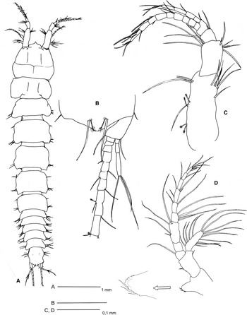
Apseudopsis latreillii: preparatory female. (A) Dorsal view; (B) pleotelson and right uropod; (C) left antennule; (D) right antenna.

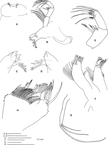
Apseudopsis latreillii: preparatory female. (A) Labrum; (B) left mandible; (C) detail of the left mandible; (D) detail of the right mandible; (E) mandibular palp; (F) maxillule; (G) maxillular palp; (H) maxilla.

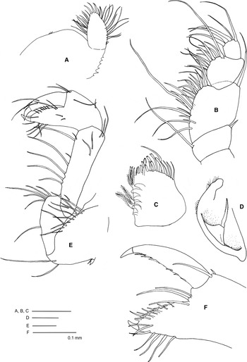
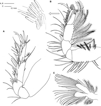
Apseudopsis latreillii: preparatory female. (A) Labium; (B) maxilliped; (C) maxilliped endite; (D) epignath; (E) cheliped; (F) chela.

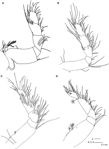
Apseudopsis latreillii: preparatory female. (A) Pereopod 1; (B) pereopod 2; (C) pereopod 3; (D) pereopod 4.

Apseudopsis latreillii: preparatory female. (A) Pereopod 5; (B) pereopod 6; (C) pleopod 1.

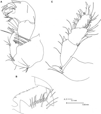
Apseudopsis latreilli: male. (A) Cheliped; (B) chela; (C) pereopod 1.

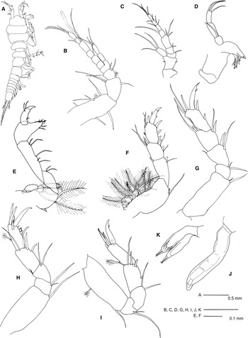
Apseudopsis latreilli: manca II. (A) Dorsolateral view; (B) left antennule; (C) left antenna; (D) left mandible; (E) cheliped; (F) pereopod 1; (G) pereopod 2; (H) pereopod 3; (I) pereopod 5; (J) pereopod 6; (K) pleopod.

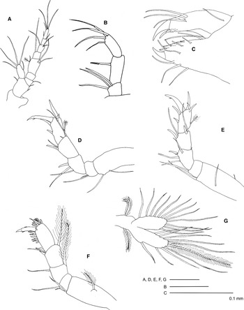
Apseudopsis latreilli: juvenile I. (A) Left antenna; (B) mandibular palp; (C) chela; (D) pereopod 3; (E) pereopod 5; (F) pereopod 6; (G) pleopod.

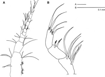
Apseudopsis latreillii: juvenile II. (A) Right antennule; (B) mandibular palp.

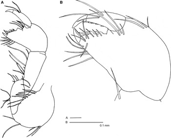
Apseudopsis latreillii: male I. (A) Cheliped; (B) chela.

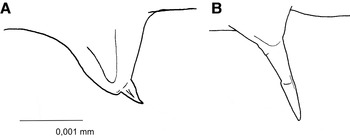
Apseudopsis latreillii: copulatory structures. (A) Penial tubercle of male II; (B) hyposphenium of preparatory female.

Rhœa Latreillii Milne-Edwards, Reference Milne-Edwards1828, pp. 287–301, pl. xiii. A, figures 1–8.
Rhœa Latreillii Milne-Edwards, Reference Milne-Edwards1840, p. 141.
Apseudes latreillii Bate & Westwood, Reference Bate and Westwood1868, p. 153.
Apseudes latreillii Sars, Reference Sars1882, p. 14.
Apseudes latreillii Sars, Reference Sars, Norman and Stebbing1886, pp. 82–84, pl. XVI.
Non Apseudes latreillii, Claus, Reference Claus1888, pp. 319–333, figures 1–19 (= Apseudes acutifrons Sars, Reference Sars1882).
Apseudes latreillei Lister, Reference Lister and Sedgwick1909, p. 477.
Apseudes latreillii Băcescu, Reference Băcescu1961, p. 160, figure 4, pl. 1.
Apseudes latreillii Holdich & Jones, Reference Holdich and Jones1983b, pp. 30–31, figure 7.
Apseudes latreillii Sieg, Reference Sieg, Gruner and Holthuis1983, pp. 61–66.
Apseudes latreillii Riggio, Reference Riggio1996, p. 614–615.
Apseudes latreillei Gutu, Reference Gutu2001, p. 62.
Apseudopsis latreillii Gutu, Reference Gutu2006, p. 61.
MATERIAL EXAMINED
North-west Iberian Peninsula
81♀♀, 14♂♂, 226 juveniles and mancas, 42°29.12′N 08°50.25′W, mud with Zostera marina, 0.8 m, coll. J.S. Troncoso, 11 December 1996; 40♀♀, 10 ♂♂, 141 juveniles and mancas, 42°28.75′N 08°50.75′W, fine sand with shells and Z. marina, 0.3 m, coll. J.S. Troncoso, 4 December 1996; 1152 ♀♀, 165♂♂, 2681 juveniles and mancas, 42°29.75′N 08°50.25′W, mud with Z. marina, 5.9 m, coll. J.S. Troncoso, 4 December 1996; 134 ♀♀, 17 ♂♂, 486 juveniles and mancas, 42°28.25′N 08°50.75′W, fine sand with Z. marina, 0.3 m, coll. J.S. Troncoso, 11 December 1996.
St Martin's, Isles of Scilly, England
16 ♀♀, 5♂♂, 10 juveniles, 49°57.55′N 06°17.76′W, LWST clean sand, coll. R.N. Bamber, 11 September 2010.
Petit Nord, France
Neotype: 1 ovigerous ♀ (MNHN-Ta 87), colls. C. Bate and J.O. Westwood, August 1895. Alloneotype: 1 ♂ (MNHN-Ta 88), coll. E.L. Bouvier.
REDESCRIPTION
Female with oostegites
Total length: 3.2–5.5 mm. Body dorsoventrally flattened, elongated, slightly narrowed posteriorly, 5.7 times as long as wide. Carapace as broad as long; rostrum pointed and slightly downturned, with rounded ‘shoulders’. Ocular lobes present, pointed. Eyes present. All pereonites with rounded corners, and setae on anterolateral and posterolateral margins. First pereonite shorter than the rest, with two posterolateral lobes. Second pereonite trapezoidal. Third to sixth pereonites subquadrangular; fourth and fifth subequal (Figure 1A). Ventral hyposphenia present on pereonites 2 to 6. Pleon one-quarter of total length, longer than wide. Pleonites subequal, narrower posteriorly and with ventral hypophyses. Lateral margins of all pleonites produced posteriorly, bearing lateral setae. Pleotelson as long as broad, bearing a group of 4 setae on two dorsolateral protuberances (Figure 1A), two small setae on posterolateral margins, and one pair of subterminal long setae and one pair of penicillate setae (Figure 1B).
Antennule peduncle 4-articled; first article 3.7 times as long as wide, with two pairs of penicillate setae on outer margin, two medial marginal setae, and two distal groups of setae. Second article half length of first, twice as long as wide, with inner and outer distal tufts of setae. Third article one-third length of second, as long as wide, with single inner and outer distal setae. Fourth article L-shaped, as long as preceding article. Main flagellum of 10 segments, segments 6 and 8 bearing one aesthetasc each. Accessory flagellum 4-segmented (Figure 1C).
Antennal peduncle 5-articled; first article as long as wide, with an inner, serrated lobe bearing 1–3 small setae. Second article 1.3 times as long as preceding, and 1.5 times as long as wide, bearing outer squama with marginal, long setae; inner margin denticulated, bearing one small seta. Third and fourth articles subequal, quadrangular, and 1/3 length of second. Fifth article 1.3 times as long as the two preceding articles together, with one medial, long seta and two long, distal setae. Flagellum of 6 segments, first with four, second and third with one long, outer setae. Terminal segment bearing terminal setae (Figure 1D).
Mouthparts: labrum (Figure 2A) bilobed, setulose. Left mandible (Figure 2B) with strongly dentated pars incisiva and lacinia mobilis, setiferous lobe with 4–5 bifurcate and trifurcate setae; pars molaris crenulated, setulose (Figure 2C). Right mandible (Figure 2D) as left but without lacinia mobilis. Mandibular palp 3-articled; first article 2.2 times as long as broad, bearing numerous setae on inner margin; second article as long as first, bearing 5 setulose setae, decreasing in length distally; third article bearing distal setae and pectinate spines (Figure 2E). Maxillule (Figure 2F) inner endite with short fine setae on inner margin, outer margin with longer fine setae and marginal apophysis, and 5 distal setulose setae; outer endite bearing 11 distal spines and two subdistal setae, inner margin with a group of fine setae, outer margin with longer fine setae; palp (Figure 2G) bearing three long simple setae. Maxilla (Figure 2H) with margins serrated; outer lobe of inner endite with simple setae, and bifurcate, trifurcate and pectinate spines. Inner lobe of fixed endite with a row of simple setae in front of serrated setae. Outer lobe of outer endite with serrate setae and two strong outer setae. Labium (Figure 3A) with outer margin serrated, palp with 3 distal setae and marginal setae and setules. Maxilliped (Figure 3B) basis simple; palp first article with a long inner seta and an outer distal spine; second article inner margin with numerous setae, one as long as whole palp, outer margin with one distal seta; third article with setae along inner margin, one as long as second and third articles together; distal article with long distal setae. Maxilliped endite (Figure 3C) with setulose setae on inner margin and up to six spatulate spines, and simple, bifurcate, trifurcate and pectinate setae on distal margin. Epignath (Figure 3D) bearing a long, distally setulose spine; border sparsely setulose.
Cheliped slender (Figure 3E). Basis 1.6 times as long as broad, with a medial stout spine and a pair of long distal setae on ventral margin, and a row of setae along dorsal margin. Exopodite (not figured) present, 3-articled; first article small, second article cylindrical, naked, distal article with 6 long, plumose setae. Merus elongate, narrower proximally, with one medial ventral seta and a ventral, distal group of setae. Carpus slender, 4.1 times as long as wide, with long setae along ventral margin and shorter ones on laterodistal surface. Chela (Figure 3E, F) fingers about as long as wide. Palm region about as long as wide, with dorsal and lateral short setae; fixed finger with a row of setae distally on ventral margin, shortening approaching claw. Cutting edge convex, with a row of fine setae and pinnate, small spines. Dactylus as long as fixed finger, with a couple of lateral setae near claw; cutting edge with small spines and pinnate spines.
Pereopod 1 (Figure 4A) with oostegite. Pronounced apophysis on coxa. Basis with exopodite, 1.5 times as long as wide, with a ventrodistal spine accompanied by 1–3 long setae. Ischium naked. Merus narrower proximally, half length of basis, with lateral and ventral groups of setae, a ventrodistal stout spine and a dorsodistal spine. Carpus as long as merus, with two ventral and one dorsodistal spine, ventral setae, short laterodistal setae and long dorsodistal setae. Propodus about as long as merus, with three ventral and two dorsal spines, becoming longer distally. Dactylus with one mid-dorsal fine seta, and one mid-ventral denticle. Inguis about 1/3 length of dactylus.
Pereopod 2 (Figure 4B) with oostegite. Basis 3.7 times as long as wide, with long, marginal setae, short setae, and a tuft of ventrodistal setae. Ischium with two ventral setae. Merus, carpus and propodus with long marginal setae. Merus about 1/3 as long as basis, narrower at base, with one long ventrodistal spine. Carpus 1.2 times as long as merus, with one ventrodistal spine and one dorsodistal spine. Propodus about as long as merus, with two ventral spines and one dorsodistal spine, serrated, those on both sides of dactylus longer and slender. Dactylus slender, with one mid-ventral spinule, and one ventrodistal setule. Unguis about 1/4 of total length of dactylus.
Pereopod 3 (Figure 4C) with oostegite. Basis 2.2 times as long as broad, with long proximal setae, smaller dorsal simple and penicillate seta, and a group of long setae on ventrodistal corner. Ischium with a tuft of ventrodistal setae. Merus, carpus and propodus with marginal and lateral long setae. Merus narrower at base, with one long ventrodistal spine, and two ventrolateral shorter spines. Carpus with one long, ventrodistal spine, one long, dorsodistal spine, and two shorter lateral spines. Propodus slender, narrower at base, three times as long as broad and 1.3 times as long as carpus, with two long, ventral spines, two long, slender and one shorter dorsodistal spines, and two stout lateral spines. Dactylus as in pereopod 2.
Pereopod 4 (Figure 4D) with oostegite. Basis fusiform, 2.2 times as long as broad, with dorsal small setae and 3 penicillate setae, and one long and one very short ventrodistal setae. Ischium with 2–3 ventrodistal setae. Merus, carpus and propodus with long ventral and distal setae. Merus with one pair of ventral spines. Carpus 2 times as long as merus, 2.5 times as long as broad, with two rows of 5 ventral spines that become longer approaching propodus. Propodus 2.7 times as long as broad and 1.3 times as long as carpus, with a long penicillate dorsal seta, a group of long terminal spines, and a marginal terminal crown of lanceolate, denticulate spines. Dactylus slender, as long as propodus, inguis about 1/5 of length of dactylus.
Pereopod 5 (Figure 5A) very similar to pereopod 4, but with basis naked, merus with two rows of 4 spines, propodus with two pairs of ventral spines, three terminal, long spines and some lanceolate, serrate spines, not forming a crown.
Pereopod 6 (Figure 5B) basis with dorsal plumose setae and ventral long, simple setae. Merus and carpus with ventral long setae and dorsal very long, plumose setae. Ischium with a tuft of ventrodistal setae. Merus narrower proximally, with one long ventral spine. Carpus with two rows of 3–4 spines, longer when approaching propodus. Propodus ovate, as long as carpus, with a dorsal penicillate seta, and a row of lanceolate, pinnate spines along ventral and frontal border. Dactylus about as long as propodus, with a medial dorsal setule and a distal ventral setule, inguis about one-third of length of dactylus.
Pleopods (Figure 5C) biramous. Articulation not distinct. Pleopod 1 basis with 6 + 5 (six inner, five outer) plumose setae; pleopods 2–3 with 5 + 4 and pleopods 4–5 with 4 + 3 plumose setae. Both rami with numerous marginal plumose setae. Endopod longer than exopod. Inner proximal seta on endopod shorter and robust.
Uropods (Figure 1B) biramous. Basis with two distal setae. Inner ramus with around 30 segments, increasing in length towards end, some with simple or penicillate setae. Outer ramus 3-, occasionally 4-segmented. Distal segment significantly longer than the other two/three together, with long distal setae.
Male
Slightly longer than female: total length 3.6–6.1 mm. Penial tubercle (Figure 11A) on pereonite 6. Hyposphenia absent. Cheliped (Figure 6A) more robust: basis as long as broad, with row of setae on dorsal margin, group of setae and a small spine on ventrodistal margin. Merus about 3 times as long as broad, with ventral, distal and proximal tufts of setae. Carpus narrower at base, 1.2 times as long as basis, with a row of setae on ventral margin and a blunt subterminal apophysis. Chela (Figure 6B) robust, cutting edge of fixed finger straight, with a triangular proximal apophysis. Cutting edge of dactylus with a proximal rounded apophysis; setation and ornamentation as in the female. Pereopod 1 (Figure 6C) merus dorsodistal spine smaller than that of female; ventral spines of carpus and propodus larger. Dorsodistal corner of carpus pronounced.
REMARKS
Apseudopsis latreillii resembles other species of the genus such as A. acutifrons (G.O. Sars, Reference Sars1882), A. anabensis (Gutu, Reference Gutu2002), A. bacescui (Gutu, Reference Gutu2002), A. caribbeanus Gutu, Reference Gutu2006, A. elisae (Băcescu, Reference Băcescu1961), A. hastifrons (Norman & Stebbing, 1886), A. mediterraneus (Băcescu, Reference Băcescu1961) and A. ostroumovi (Băcescu & Cărăşu, 1947). These species share with A. latreillii the presence of a pointed rostrum with rounded shoulders, but they have apophyses or pointed corners on the pereonite margins, and more than four spines on the ventral margin of the propodus of pereopod 1. Apseudopsis apocryphus (Gutu, Reference Gutu2002) and A. minimus (Gutu, Reference Gutu2002) show a similar rostrum and 3 spines on the ventral margin of the propodus of pereopod 1, but do not have a dorsodistal spine on the merus. Apseudopsis bruneinigma (Bamber, 1998), A. isochelatus Gutu, Reference Gutu2006 and A. robustus (G.O. Sars, Reference Sars1882) mostly differ from A. latreillii in the shape of the rostrum.
POSTMARSUPIAL DEVELOPMENT
Up to eight different stages have been identified in the life history of A. latreillii (apart from manca I, which occurs inside the marsupium and is not included in this work). They can be diagnosed as follows:
Manca II: Sixth pereopod with no articulation; pleopods without plumose setae around the border.
Juvenile I: All pereopods developed. Hyposphenia often absent; oostegites absent.
Juvenile II: Hyposphenia present, but no oostegites. Cheliped slender.
Preparatory female: Oostegites present. Cheliped slender.
Copulatory female: Complete marsupium.
Intermediate female: Adult females without oosteguites, nor marsupium nor hyposphenians.
Male I: Rudimentary hyposphenia. Penial tubercle present. ‘Intermediate’ cheliped (see description).
Male II: Penial tubercle present. Cheliped robust.
Morphological characteristics of preparatory females and males II correspond with those of the adults already described. The rest of the developmental stages differ from the preparatory female, and can be described as follows (only those characters that differ from the adult female with oostegites are specified):
Manca II
Total length: 1.2–1.9 mm. Less setose in general than adult. Rostrum broad, slightly pointed. Cephalothorax about 0.22 of total length. Pereonites 1 and 2 significantly broader than rest, about 3 times as broad as long (Figure 7A). Pleotelson as in adult.
Antennular accessory flagellum 2-segmented, inner flagellum 6-segmented, with one aesthetasc on fourth segment (Figure 7B).
Antennal squama bearing three setae; flagellum 4-segmented (Figure 7C).
Mouthparts very similar to adult, except mandibular palp (Figure 7D): proximal article short, as long as broad, bearing one seta; second and third subequal in length, 2.2 times as long as first; second article naked; distal article with three simple setae.
Cheliped (Figure 7E) slender, similar to female; exopod proportionally larger, basis bearing long plumose setae.
Pereopods 1–5 (Figure 7F–I) sparsely setose by comparison to the adult.
Pereopod 1 (Figure 7F) spination as in adult; exopodite proportionally larger; basis bearing long dorsal setae.
Pereopod 2 (Figure 7G) spination similar to adult, but without terminal dorsal spine on the propodus.
Pereopod 3 (Figure 7H) merus lacking spines; two long, slender ventral spines on carpus; propodus with one dorsal penicillate seta, one slender and three lanceolate, serrated ventral spines, two long terminal spines, and one serrated, subterminal spine.
Pereopod 4 (Figure 7I) basis with one ventral penicillate seta; carpus with one ventral spine; propodus with one large penicillate seta, two long, serrated and two simple terminal spines.
Pereopod 5 similar to pereopod 4.
Pereopod 6 (Figure 7J) reduced, with no segmentation or ornamentation.
Pleopods (Figure 7K) all alike, biramous, with two terminal, short, simple setae on each ramus.
Uropod inner ramus of about 10 segments. Outer ramus as in the adult.
Juvenile I
Total length: 1.4–3.0 mm. Rostrum defined. Antennule, cheliped and pereopod 1 as in manca II.
Antenna (Figure 8A) squama bearing 5 marginal setae; flagellum of 4 segments.
Mandibular palp (Figure 8B) first article short, as long as broad, bearing three setae; second article bearing one distally setulose seta; third article as long as second, bearing 4 terminal simple setae.
Cheliped and chela as in manca II (Figure 8C).
Pereopods 1 and 2 as in adult.
Pereopod 3 (Figure 8D) merus with one ventral spine; one spine on carpus; propodus with two ventral spines, two subdistal small spines, and one long distal spine.
Pereopod 4 merus with one ventral spine; carpus with one ventral and one dorsodistal spine; propodus with two ventral and three dorsodistal spines.
Pereopod 5 (Figure 8E) similar to pereopod 4.
Pereopod 6 (Figure 8F) articulated; basis with one plumose seta, and one ventrodistal simple seta; merus with a dorsodistal, long plumose seta and a ventral simple seta; carpus with one dorsodistal plumose seta, one ventrodistal spine and seta; propodus border bearing several lanceolate, serrated spines.
Pleopods (Figure 8G) similar to those of adult, but with one plumose seta on inner and one on outer margin of the basis, and fewer plumose setae on margins of rami.
Uropod inner ramus of 12–16 segments.
Only some of the largest individuals had hyposphenia.
Juvenile II
Total length: 2.1–4.4 mm. Hyposphenia present. Antennular (Figure 9A) inner flagellum 3-segmented; outer flagellum 7–8-segmented, presence of aesthetascs variable, often on fourth and sixth segments.
Mandibular palp (Figure 9B) first article twice as long as broad, bearing a tuft of setae on inner margin; second article as long as first, bearing two distally setulated setae; distal article bearing four simple setae and three pectinate spines.
Cheliped as in adult female.
Pleopod setation variable.
Uropod inner ramus of about 20 segments.
Rest of appendages and mouthparts as in adult.
Male I
Total length: 3.1–5.7 mm. Mostly similar to adult female, bearing vestigial hyposphenia on pereonites 2–5, and penial tubercle on pereonite 6. Antennular accessory flagellum 4-segmented. Cheliped (Figure 10A, B) as in female, but with a triangular apophysis on cutting edge of fixed finger, in proximal position.
REMARKS
A few individuals (i.e. intermediate males and the largest juvenile II) had a different number of segments on the accessory flagellum of each antenna (3 on one, 4 on the other). Similarly, a few of the largest specimens (males and females) had a different number of segments on the outer rami of the uropods (3 and 4). These have been considered abnormalities, owing to the small proportion of cases (<2% of the total).
Apart from a general increase in the number of setae and spines in the pereopods, meristic changes that occur through the development of A. latreilli include: the abrupt appearance of a segmented pereopod 6 and well-developed pleopods; an increase in the number of segments in antennal and antennular flagella and uropod inner rami; and the appearance of a third article on the mandibular palp after the juvenile II. Morphometric changes include the proportional size of the cephalothorax, cheliped, pereopod 1, and the exopodite of the cheliped and pereopod 1. The penial tubercle appears on the pereonite 6 on males I, while in preparatory females a hyposphenium appears as a pointed, conical structure (Figure 11B). Earlier stages present a structure which is not yet differentiated. On the other hand, some characters do not show significant changes, remaining relatively stable from the mancae to the adults; these are the cheliped (which only changes in the males, but is very similar in the manca and the ovigerous female), the pereopod 1, and the uropod outer rami. There are no observable differences on the size of the oostegites among the preparatory females. The hyposphenia appear on the juveniles, and are only absent in the ovigerous females and adult males.
LENGTH–FREQUENCY DISTRIBUTION
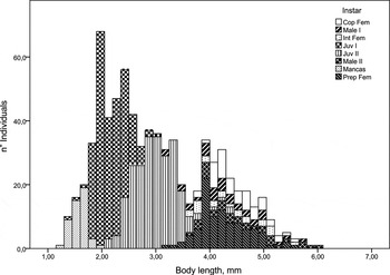
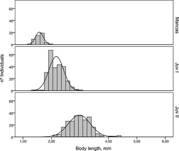
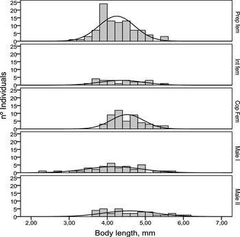
The length–frequency distribution obtained (Figure 12) may, a priori, be divided into two normal distributions, with the juvenile stages on the left and the adult–preparatory stages on the right. There is an overlap in the size of the juvenile stages, including mancas (Figure 13). The adult-stage size-classes overlap completely on the right of the graph (Figures 12 & 14). The total length measured ranged from 1.22 mm to 6.09 mm (Table 1). In general, the largest adults were males (3.66–6.09 mm) and the smallest were preparatory males (minimum size: 3.12 mm) and preparatory females (minimum: 3.19 mm). In general, juvenile stages did not exceed 3.8 mm, although the largest juvenile II reached 4.36 mm.
Statistical values of the size distribution for every developmental stage studied. Values in mm.

Cop Fem, copulatory females; Prp Fem, preparatory females; Int Fem, intermediate female; Tot Fem, total females; Tot Male: total males; Juv I and Juv II, juveniles I and II; Tot Juv, total juveniles.
FECUNDITY AND MALE:FEMALE RATIO
Fecundity ranged from 7 to 23 potential offspring per brood (mean: 13.15; standard deviation: 5.5; mode: 13). Male:female ratio in O Grove ranged from 1:1.7 to 1:8 per sample, and between 1:4 and 1:7 per site.
DISCUSSION
Taxonomic observations and development
Some discrepancies with previous descriptions were detected. For instance, the outer rami of the uropods were described by Sars (Reference Sars, Norman and Stebbing1886), and later on by Riggio (Reference Riggio1996) as 4-segmented; we, however, observed three segments on the vast majority of the individuals, finding four only on a few of the largest specimens. Such discrepancies can be attributed to the difficulty in distinguishing the limit between two consecutive segments. In any case, this character is not specific, and can be disregarded for identifications.
A number of characters remain constant during the postmarsupial development of A. latreilli. Some of these are characteristic of the genus, or at least shared by several species (i.e. morphology of the rostrum, mouthparts, cheliped and pereopods 2–6, the number of segments of the uropod outer ramus (Gutu, Reference Gutu2001, Reference Gutu2006)), and thus not reliable for species identification. On the other hand, there is not a single character that can serve as a definite diagnosis of the species. Therefore, we conclude that A. latreilli can be identified, regardless the developmental stage, from the following combination of morphological characters: a pointed and downturned rostrum, pereonites without apophyses, three ventral spines on the pereopod 1 propodus, and one dorsodistal spine on the merus.
Life history
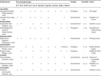
The number of manca and juvenile postmarsupial stages varies among tanaidaceans (Table 2); generally, there are one or two manca stages followed by one or two distinct juveniles, each one originated through a single moult (Messing, Reference Messing1983; Schmidt et al., Reference Schmidt, Siegel and Brandt2002). In contrast, in adults several moults lead to an increase of the body size, without the occurrence of significant meristic changes (Lang, Reference Lang1953). Large size-ranges, like those on Figures 12–14, support this idea (Schmidt et al., Reference Schmidt, Siegel and Brandt2002). In peracarids, growth normally slows down as the individuals become older (Johnson et al., Reference Johnson, Stevens and Watling2001).
Size–frequency distribution of the specimens of Apseudopsis latreillii yielded by a single grab from O Grove Zostera meadows. Cop Fem, copulatory female; Int Fem, intermediate female; Prep fem, preparatory female; Juv I-II, juveniles I and II. Sizes in μm. Interval: 150 µm.

Size–frequency distribution of the immature developmental stages of Apseudopsis latreillii. Juv I-II, juveniles I and II. Sizes in μm. Interval: 150 µm

Size–frequency distribution of the mature developmental stages of Apseudopsis latreillii. Cop Fem, copulatory female; Int Fem, intermediate female; Prep fem, preparatory female. Sizes in μm. Interval: 200 µm.

Developmental stages, proposed sexual development strategy and fecundity of several species of tanaidaceans.

M I, M II and M III, mancas I–III; Juv I and Juv II, Juvenile I and II; Prp Fem, preparatory female; Cop Fem, copulatory female; Int Fem, intermediate female.
Considering that the differentiation of the copulatory structure marks the passage from the juvenile to an adult stage, males I can be considered sexually mature. Consequently, developed hyposphenia (rudimentary on males I) appear to be a characteristic restricted to juveniles and females. Similarly, the 4-articled inner flagellum on the antennule arises as an indication of maturity.
The large range of the body length for copulatory females suggests the presence of more than one copulatory stage separated by moults; similarly, the females with adult characters but no oostegites nor hyposphenia, classified as intermediate, share the same range. This indicates a sequence of copulatory instars followed by intermediate stages on which the female lose the ovisac after the manca release. Lang (Reference Lang1953) proposed this sequence for the Apseudidae, Johnson & Attramadal (Reference Johnson and Attramadal1982) for Tanais dulongii (Audouin, 1826) and Gardiner (Reference Gardiner1975) for the family Neotanaidae Lang, 1956.
The occurrence of two types of male is frequent among apseudomorphans (see Table 2), and have different interpretations. For instance, Pennafirme & Soares-Gomes (Reference Pennafirme and Soares-Gomes2009) proposed protogynic hermaphroditism for Monokalliapseudes schubartii (Mañé-Garzón, 1949), considering that a female-skewed sex-ratio may indicate this strategy. Other authors (Bamber, Reference Bamber2010) consider that in certain tanaidomorphan species males I are simply pre-adults. In A. latreillii males I present rudimentary hyposphenia (absent in the male II but evident on juveniles and females) and a 4-articled inner flagellum of the antennule (an adult characteristic in this species). Therefore, it seems possible that they follow either a juvenile II instar, or a preparatory female. Both origins can be supported by their position on the length–frequency distribution, being larger than most of the juvenile II and a number of the preparatory females. The males I coincide in the size-range with the females. Because there are males I smaller than the smallest males II, it does seem equally possible that males I represent an intermediate state between juveniles and males II. Nevertheless, protogyny cannot be discarded, considering the female-skewed sex-ratio, the occurrence of large males I and the fact that sex reversal may be a facultative strategy (Bückle-Ramírez, Reference Bückle-Ramírez1965; Highsmith, Reference Highsmith1983; Błażewicz-Paszkowycz, Reference Błażewicz-Paszkowycz2001). Experimental work would be needed to solve this question definitely (Highsmith, Reference Highsmith1983).
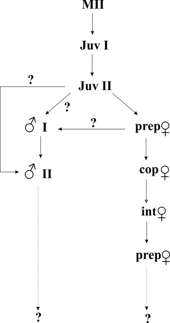
Figure 15 shows a proposed life developmental sequence based on our results. Post-marsupial mancas are common among peracarids (Gardiner, Reference Gardiner1975). While in isopods the sex can be determined from the manca stages, in cumaceans and tanaidaceans the sex is not noticeable in juveniles. Several moults occur in adult females of all peracarids, leading to at least two (usually several) breeding periods in a lifetime. In contrast, peracarid copulatory males are usually considered terminal (Gardiner, Reference Gardiner1975; Johnson et al., Reference Johnson, Stevens and Watling2001). There are cases of terminal males among tanaidaceans (Gardiner, Reference Gardiner1975; Johnson et al., Reference Johnson, Stevens and Watling2001), but several authors indicate more than one moult (Schmidt et al., Reference Schmidt, Siegel and Brandt2002; Pennafirme & Soares-Gomes, Reference Pennafirme and Soares-Gomes2009). Mature males of A. latreilli probably moult several times before dying, during which time they are able to reproduce.
Proposed life cycle scheme for Apseudopsis latreillii. MII, manca II; Juv I, juvenile I; Juv II, juvenile II; prep ♀, preparatory female; cop ♀, copulatory female; int ♀, intermediate female; ♂ I, male I; ♂ II, male II. Black arrows indicate one or more moults. White arrow indicates no moults implicated. Dotted arrows indicate an undetermined number of moults.

Fecundity is comparable to that of other tanaidacean species (Table 2). Although tanaidaceans have the least fecundity per female of the peracarids (Messing, Reference Messing1983), they are abundant and often numerically dominant among crustaceans on sandy and muddy bottoms, reaching high densities (Swennen et al., Reference Swennen, Duiven and Spaans1982; Moreira et al., Reference Moreira, Lourido and Troncoso2008; Esquete et al., 2011, among others).
CONCLUSIONS
– Apseudopsis latreilli can be identified, regardless of the developmental stage, from the following combination of morphological characters: a pointed and downturned rostrum, pereonites without apophyses, three ventral spines on the pereopod 1 propodus, and one dorsodistal spine on the merus.
– Through the life of A. latreilli, the following postmarsupial stages can be recognized: manca II, juvenile I, juvenile II, preparatory females, copulatory females, intermediate females, males I and males II.
– The number of segments of the inner flagellum of the antennule increases through the life history, and can be used as an indication of the developmental stage of the individual. Similarly, the differentiation of the copulatory structure on pereonite 6 is characteristic of the adults. A general increase of the number of setae and spines on the pereopods, as well as number of articles on the inner flagellum of the uropodal endopod occurs through the developmental history of A. latreillii.
– The length–frequency distribution suggests that the adults pass through a variable number of instars–moults before and after the first reproductive period.
ACKNOWLEDGEMENTS
We thank the Adaptations of Marine Animals (University of Vigo, Spain) laboratory staff for their invaluable help with sample collection at the O Grove inlet. Part of the material used for this study was kindly lent by Dr D. Defaye, curator of the Muséum National d'Histoire Naturelle collection (Paris). Constructive comments by two anonymous referees are greatly acknowledged. Measurements of the specimens were performed in the installations of the ECIMAT marine station (University of Vigo). This research received no specific grant from any funding agency, commercial or not-for-profit sectors.