The development and widespread acceptance of mastitis prevention and national dairy herd control programmes (Neave et al. Reference Neave, Dodd, Kingwill and Westgarth1969; National Mastitis Council, 2006) coupled with the introduction of penalty limits for the bulk milk somatic cell count (SCC) and premiums for milk of high quality has led to substantial progress in controlling subclinical mastitis worldwide. In The Netherlands, for instance, the geometric mean bulk milk SCC (BMSCC) decreased from nearly 600 000 cells/ml to 200 000 cells/ml between the introduction of the BMSCC as quality control criterion in 1971 and 2002 (Sampimon et al. Reference Sampimon, Sol and Kock2005). Routine monitoring of BMSCC carried out in all delivering herds by dairies in Finland revealed a decrease of the geometric mean BMSCC from 325 000 cells/ml in 1989 towards 132 000 cells/ml in 2002 (Myllys et al. Reference Myllys, Asplund, Brofeldt, Hivelä-Koski, Honkanen-Buzalski, Junttila, Kulkas, Myllykangas, Niskanen, Saloniemi, Sandholm and Saranpää1998; Pitkälä et al. Reference Pitkälä, Haveri, Pyörälä, Myllys and Honkanen-Buzalski2004). In Flanders, Belgium, the BMSCC decreased from above 300 000 cells/ml in 1991 to 196 000 cells/ml in 1999. Since 2000, however, the geometric mean BMSCC has increased from 202 000 cells/ml to 221 000 cells/ml in 2006 (Annual Report 2006, Milk Control Centre Flanders, Lier, Belgium).
In many countries, the control of subclinical mastitis was associated with a change in the distribution of the involved pathogens. In Finland (Myllys et al. Reference Myllys, Asplund, Brofeldt, Hivelä-Koski, Honkanen-Buzalski, Junttila, Kulkas, Myllykangas, Niskanen, Saloniemi, Sandholm and Saranpää1998, Pitkäla et al. Reference Pitkälä, Haveri, Pyörälä, Myllys and Honkanen-Buzalski2004) and Wisconsin, USA (Makovec & Ruegg, Reference Makovec and Ruegg2003) prevalence of non-aureus staphylococci IMI increased from 39·8% (1988) to 49·6% (2001), and from 12·7% (1994) to 17·5% (2001), respectively, while the proportion of Staphylococcus aureus IMI decreased from 30·9% (1988) to 10·2% (2001) in Finland, and from 17·7% (1994) to 9·7% (2001) in Wisconsin. Results of large-scale surveys on the prevalence of IMI, and the distribution of the involved bacteria in Belgium have not been reported. Regular monitoring of the prevalence and the distribution of mastitis-causing pathogens provides, however, valuable knowledge about the effectiveness of proposed control strategies, and helps in prioritizing specific mastitis control efforts (Barkema et al. Reference Barkema, Schukken, Lam, Beiboer, Benedictus and Brand1999; Pitkälä et al. Reference Pitkälä, Haveri, Pyörälä, Myllys and Honkanen-Buzalski2004). It was also found that bacteriological monitoring of a dairy herd and implementation of specific measures based on the outcome, substantially improved udder health status at the herd level (Hillerton et al. Reference Hillerton, Bramley, Staker and McKinnon1995).
Milk SCC is used worldwide as an indicator of the IMI status of the mammary gland, and so as a guideline in the selection of cows for culturing (Schukken et al. Reference Schukken, Wilson, Welcome, Garrison-Tikofsky and Gonzalez2003). The use of SCC for monitoring udder health and milk quality, and the grounds for SCC thresholds in separating infected from non-infected cows have been extensively presented and discussed (Schepers et al. Reference Schepers, Lam, Schukken, Wilmink and Hanekamp1997; Hillerton, Reference Hillerton1999; Dohoo, Reference Dohoo2001; Schukken et al. Reference Schukken, Wilson, Welcome, Garrison-Tikofsky and Gonzalez2003; Barrett et al. Reference Barrett, Healy, Leonard and Doherty2005). Although currently an international standard for bovine milk SCC does not exist, the cut-off point generally suggested, based on most literature, is 200 000 cells/ml. In Flanders (Belgium), a geometric mean SCC ⩾250 000 cells/ml during the last three monthly test-days is used as threshold for the detection of cows with subclinical mastitis. Whether the proposed threshold is a good predictor for the cows' IMI status, and whether bacteriological analysis of milk samples collected from those cows is representative for the prevalence and distribution of mastitis pathogens at the herd level has not been evaluated.
The aims of this study were two-fold: (1) to determine the prevalence and distribution of mastitis pathogens in quarter milk samples obtained from all cows in herds subjected to a yearly screening in Flanders, Belgium and (2) to determine the prevalence and distribution of mastitis pathogens cultured from cows with a geometric mean composite SCC ⩾250 000 cells/ml during the last three monthly test-days.
Materials and Methods
Data
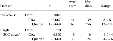
A total of 178 668 quarter milk samples were collected from 1087 cross-sectional herd screenings performed in three consecutive years in Flanders, Belgium. These data were combined into dataset ‘All-cows’ (Table 1). All herds in this dataset had the AA-milk label which is awarded if specified requirements regarding the health of animals, the milking installation and the milk quality are met. Prior to 1 August 2003, an annual cross-sectional bacteriological screening of all lactating cows was required by the Ministry of Agriculture. Samples were taken by the official herd veterinarian. In addition to the official analyses that are conducted for all producers, specific AA-milk regulations require a geometric mean SCC (last 3 months, 12 recordings) <350 000 cells/ml, a geometric mean bacterial count (last 2 months, 4 recordings) <50 000 cells/ml, a geometric mean coliform count <50/ml (last 2 months, 4 recordings), and no antibiotic residues in any delivery.
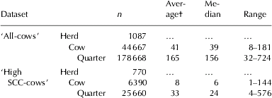
Structure of the dataset ‘All-cows’ representing data from Belgian dairy cows sampled during mandatory cross-sectional dairy herd screenings performed between 2000 and 2002, and of the dataset ‘High SCC-cows’ representing data from Belgian dairy cows with a geometric mean composite milk somatic cell count ⩾250 000 cells/ml between 2000 and 2002
† Average number of cows/quarters per herd
In addition, culture results from 25 560 quarter milk samples were collected from 6390 cows with a geometric mean composite SCC in the last 3 months ⩾250 000 cells/ml (referred to as an ‘high SCC’ throughout this paper) from dairy herds enrolled in the Flemish DHI programme. Data were combined into the dataset ‘High SCC-cows’ (Table 1).
Bacteriological analysis
Bacteriological culture and isolate identification was based on NMC standards (National Mastitis Council, Reference Neave, Dodd, Kingwill and Westgarth1999) and performed by the Animal Health Service Flanders (accreditation BEL-TEST 205-T ISO 17025). In short, from each sample, 0·01 ml of milk was plated on blood-aesculin agar (one plate per cow) and on MacConkey's agar, and incubated aerobically for 36±12 h at 37±1°C. A quarter was considered culture-positive when growth of ⩾1 colonies was detected. Samples yielding three or more different bacterial species were considered to be contaminated. Bacteria were identified by colony morphology and Gram-staining. For Gram-positive cocci, catalase tests were used to differentiate between catalase-positive staphylococci and catalase-negative cocci. Morphology, haemolysis patterns, DNAse-testing and pigment production were used to distinguish Staph. aureus from non-aureus staphylococci, referred to as Staphylococcus spp. throughout this paper (Devriese, Reference Devriese1979). Cocci were subdivided into aesculin-positive cocci and aesculin-negative cocci (Streptococcus agalactiae and Str. dysgalactiae). The CAMP-test was used to differentiate Str. agalactiae from Str. dysgalactiae. Coliforms including Escherichia coli, Klebsiella spp., and Enterobacter spp. were differentiated from each other and from other Gram-negative bacteria based on appearance on MacConkey's agar, KOH-testing, Triple Sugar Iron reactions, indol production and motility.
Somatic cell count
Composite milk samples were collected monthly for all lactating cows from dairy herds enrolled in the DHIA during two successive milkings by personnel from the local DHIA (Flemish Cattle Breeding Association, Oosterzele, Belgium). All milk samples were stored with 0·02% Bronopol (Boots PLC, Nottingham, UK) at 4°C, and were transported to the laboratory of the Milk Control Centre Flanders (Lier, Belgium). SCC were quantified by electronic counting (Fossomatic 5000™, Foss Electric, Hillerød, Denmark).
Results
‘All-cows’
The included herds had an average of 41 lactating cows ranging from 8 to 181. Average BMSCC of all AA-qualified dairy herds in Flanders was 179 000 cells/ml in 2000; 188 000 cells/ml in 2001; and 184 000 cells/ml in 2002 (Milk Control Centre, Lier, Belgium). Average BMSCC of all Flemish herds was 202 000 cells/ml, 211 000 cells/ml and 207 000 cells/ml, in the same years, respectively.
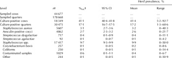
Overall, 17·1% of the 178 668 quarter milk samples were culture-positive (Table 2). The most frequently isolated mastitis pathogens were Staphylococcus spp. (57·2% of the culture-positive samples), followed by Staph. aureus (18·4%) and aesculin-positive cocci (15·9%). Str. dysgalactiae, Corynebacterium bovis and Str. agalactiae were isolated in 717 samples (2·3% of the culture-positive samples), 257 samples (0·8%), and 92 samples (0·3%), respectively. Coliforms were present in 0·8% of the culture-positive samples.
Descriptive statistics of the prevalence and distribution of mastitis pathogens isolated from quarter-milk samples obtained from Belgian dairy cows during cross-sectional dairy herd screenings performed between 2000 and 2002

† Absolute number of cows/quarters
‡ Percentage of total cows/quarters
At the herd-level, a mastitis pathogen was isolated from 41·4% of the cows ranging from 5·3% to 100%, and from 17·2% of the quarters ranging from 1·3% to 84·6% (Table 2). Staphylococcus spp., Staph. aureus and aesculin-positive cocci were cultured on 99·2%, 85·6% and 81·1% of the herds, respectively. For Staphylococcus spp., the mean within-herd quarter prevalence was 9·9%. In 6 herds, >50% of the quarters were infected with Staphylococcus spp. and in one herd Staphylococcus spp. were isolated from as many as 76·9% of the quarters (123 out of 160 quarters) (Table 2). For Staph. aureus and aesculin-positive cocci, the mean within-herd prevalence was 3·2% and 2·6%, respectively, with a range from 0 to 42·8% and 25·0%, respectively. In >75% of the herds from which Staph. aureus was isolated, <5% of the quarters were infected with this major pathogen, and only 66 (10·0%) herds had a prevalence >10% (Table 2). Str. agalactiae was isolated from 41 herds (5·3%). In 28 out of these 41 herds, only one quarter (on average 0·6% of all quarters per herd) was culture-positive for Str. agalactiae.
Proportion of infected cows and quarters was 39·0% and 16·1% in 2000 and 43·0% and 18·0% in 2002, respectively. Prevalence of Staphylococcus spp. and aesculin-positive cocci was 9·1% and 2·5% in 2000 and 10·4% and 3·2% in 2002, respectively. Isolation of Staphylococcus spp. and aesculin-positive cocci was 56·6% and 15·5% of the culture-positive quarters in 2000 and 57·4% and 18·0% in 2002, while the proportion of quarters infected with Staph. aureus and Str. dysgalactiae was 19·1% and 2·8% in 2000 and 16·6% and 1·8% in 2002.
‘High SCC-cows’
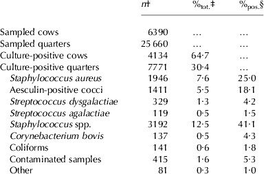
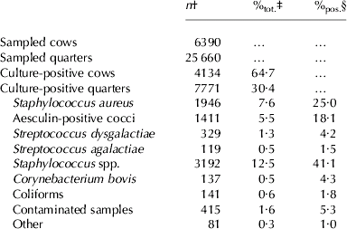
From all cows with a high SCC, 64·7% had at least one culture-positive quarter (Table 3). Similarly to the ‘All-cows’ dataset, Staphylococcus spp. (41·1% of the culture-positive samples), Staph. aureus (25·0%) and aesculin-positive cocci (18·1%) were the three most frequently isolated mastitis pathogens. Str. dysgalactiae, C. bovis and Str. agalactiae were found in 329 (4·3% of the culture-positive samples), 137 (1·8%), and 119 (1·5%) samples, respectively. In this dataset, coliforms were isolated in 1·8% of the culture-positive quarters.
Descriptive statistics of the distribution of mastitis pathogens isolated from quarter-milk samples obtained from Belgian dairy cows with a geometric mean composite somatic cell count ⩾250 000 cells/ml between 2000 and 2002
† Absolute number of cows/quarters
‡ Percentage of total cows/quarters
§ Percentage of culture-positive cows/quarters
Proportion of culture-positive cows and quarters was 62·9% and 29·5% in 2000 and 65·3% and 30·0% in 2002, respectively. Isolation of Staphylococcus spp. and aesculin-positive cocci was 37·5% and 16·6% of the culture-positive quarters in 2000 and 47·9% and 18·8% in 2002, while the proportion of quarters infected with Staph. aureus and Str. dysgalactiae was 27·4% and 5·1% in 2000 and 20·2 and 2·8% in 2002.
Discussion
This is the first time that the prevalence of subclinical mastitis and the distribution of the involved pathogens have been estimated in Flemish dairy cattle. Approximately 40% of the dairy cows had at least one infected quarter, either with a minor or a major pathogen, leaving room for improvement in mastitis control and prevention. As the mean BMSCC of AA-qualified dairy herds was slightly lower than the mean BMSCC of all Flemish dairy herds between 2000 and 2002, and as the data we used were from AA-milk herds, the results were most probably even an underestimation of the actual prevalence of subclinical mastitis during the study period on all herds.
The increase noticed in the average BMSCC in Flanders over the last few years seems to be reflected by the increase in the proportion of infected cows and quarters when comparing 2002 with 2000. However, as this study presents data from only three consecutive years we were cautious in focusing too much on this finding.
Because all lactating cows were sampled, because milk samples were taken by the official herd veterinarian, and because all samples were processed by one central laboratory, the ‘All cows’ dataset was unique and provided a wealth of reliable information allowing us to calculate accurate prevalence data on subclinical mastitis in Flanders, Belgium. Unfortunately, no information about the management and the milking technique on the dairy farms from which samples were submitted was available, so preventing us from relating the findings to management practices.
Similarly to other studies (Myllys et al. Reference Myllys, Asplund, Brofeldt, Hivelä-Koski, Honkanen-Buzalski, Junttila, Kulkas, Myllykangas, Niskanen, Saloniemi, Sandholm and Saranpää1998; Sampimon et al. Reference Sampimon, Sol and Kock2005; Makovec & Ruegg, Reference Makovec and Ruegg2003; Pitkälä et al. Reference Pitkälä, Haveri, Pyörälä, Myllys and Honkanen-Buzalski2004) Staphylococcus spp. were the most commonly cultured bacterial pathogens. Although they are generally considered as minor pathogenic skin flora opportunists (Timms & Schultz, Reference Timms and Schultz1987) the majority of IMI found in quarters from cows with a high SCC were caused by Staphylococcus spp. as well. It has been suggested that antimicrobial treatments, control of contagious mastitis pathogens including Staph. aureus and Str. agalactiae, and introduction of modern milking machines, are responsible for the change in the distribution of mastitis pathogens (Myllys et al. Reference Myllys, Honkanen-Buzalski, Huovinen, Sandholm and Nurmi1994). The large variation of the within-herd prevalence of Staphylococcus spp. IMI may be explained by differences between herds concerning implementation of post-milking teat disinfection and dry cow treatment which seem to be relatively effective in their control (Harmon et al. Reference Harmon, Crist, Hemken and Langlois1986; Hogan et al. Reference Hogan, White and Pankey1987). However, little is known about the epidemiology and pathogenicity of the different species within this group of staphylococci. The data presented in this study definitely emphasize the need for more research in that area.
The prevalence of Staph. aureus IMI was low suggesting that the spread of contagious pathogens during milking time was well controlled. Implementation of standard mastitis prevention programmes such as the ones proposed by Neave et al. (Reference Neave, Dodd, Kingwill and Westgarth1969) led to a sharp decrease of Staph. aureus IMI in many countries, including Belgium. In The Netherlands, the proportion of clinically healthy quarters infected with Staph. aureus decreased from 54·1% in 1950 to 33·8% in 2001 (Sampimon et al. Reference Sampimon, Sol and Kock2005). In Finland, isolation of Staph. aureus decreased from 31·0% in 1988 to 10·2% in 2001 (Myllys et al. Reference Myllys, Asplund, Brofeldt, Hivelä-Koski, Honkanen-Buzalski, Junttila, Kulkas, Myllykangas, Niskanen, Saloniemi, Sandholm and Saranpää1998; Pitkälä et al. Reference Pitkälä, Haveri, Pyörälä, Myllys and Honkanen-Buzalski2004). Nevertheless, Staph. aureus is still the most frequently isolated major pathogen from subclinically infected quarters in Belgium as well as in other regions or countries (Wilson et al. Reference Wilson, Ruben and Das1997; Makovec & Ruegg, Reference Makovec and Ruegg2003; Pitkälä et al. Reference Pitkälä, Haveri, Pyörälä, Myllys and Honkanen-Buzalski2004; Sampimon et al. Reference Sampimon, Sol and Kock2005; Østeras et al. Reference Østeras, Sølverød and Reksen2006; Tenhagen et al. Reference Tenhagen, Köstern, Wallmann and Heuwieser2006). Furthermore, the prevalence of Staph. aureus IMI in quarters from cows with a high SCC indicates indirectly that Staph. aureus still has a large impact on the composite milk SCC.
The prevalence of Str. agalactiae compared favourably with the 8·5% found in the survey of Wilson et al. (Reference Wilson, Ruben and Das1997) and was consistent with the 0·1% found in Finland in 2001 (Pitkälä et al. Reference Pitkälä, Haveri, Pyörälä, Myllys and Honkanen-Buzalski2004). In Denmark, the prevalence of Str. agalactiae varies between 1% and 2% since the mid-1970s (Andersen et al. Reference Andersen, Pedersen, Aarestrup and Chriél2003). In Flanders, Belgium, Str. agalactiae was isolated from only 8 out of 4020 culture-positive clinical mastitis quarter milk samples collected in 2004 and 2005 (Animal Health Service, Annual Report 2004–2005). Generally, this obligatory intramammary pathogen is very contagious, transferring from cow to cow during milking, and therefore infecting a large proportion of cows in any one herd. However, in the majority of the herds included in our study, infection was limited to one or two animals, suggesting an outbreak that has been controlled or the transfer of strains of human origin to the udder (Jensen & Aarestrup, Reference Jensen and Aarestrup1996; Andersen et al. Reference Andersen, Pedersen, Aarestrup and Chriél2003).
Among the environmental pathogens, aesculin-positive cocci were most frequently cultured. Although it is hard to compare results from different studies because of potential differences in sample selection and in culturing techniques, aesculin-positive cocci including Str. uberis, have become more important as cause of subclinical mastitis in many countries (Sampimon et al. Reference Sampimon, Sol and Kock2005; Makovec & Ruegg, Reference Makovec and Ruegg2003; Barrett et al. Reference Barrett, Healy, Leonard and Doherty2005). The primary source of aesculin-positive cocci is the surroundings in which cows live. Therefore, methods developed to control contagious pathogens are not necessarily effective against environmental pathogens. The presence of aesculin-positive cocci is mainly associated with nutrition and general sanitation of lactating cow housing (Barkema et al. Reference Barkema, Schukken, Lam, Beiboer, Benedictus and Brand1999). In contrast to aesculin-positive cocci, Str. dysgalactiae caused <5% of the IMI, corresponding well with results from Finland (Pitkälä et al. Reference Pitkälä, Haveri, Pyörälä, Myllys and Honkanen-Buzalski2004), Ireland (Barrett et al. Reference Barrett, Healy, Leonard and Doherty2005) and Germany (Tenhagen et al. Reference Tenhagen, Köstern, Wallmann and Heuwieser2006). Measures to control mastitis caused by contagious mastitis pathogens may have been helpful in preventing and controlling Str. dysgalactiae, as it was shown that Staph. aureus and Str. dysgalactiae can have similar epidemiological patterns (Barkema et al. Reference Barkema, Schukken, Lam, Beiboer, Benedictus and Brand1999).
The low prevalence of coliforms was expected as they are generally involved in cases of acute clinical mastitis of short duration (Todhunter et al. Reference Todhunter, Smith, Hogan and Schoenberger1991). However, chronic and persistent coliform IMI, mainly caused by Klebsiella spp., have been described (Todhunter et al. Reference Todhunter, Smith, Hogan and Schoenberger1991; Opsomer et al. Reference Opsomer, De Vliegher, Laureyns, Hoflack, Beeckman and de Kruif2001; Dogan et al. Reference Dogan, Klaessig, Rishniw, Almeida, Oliver, Simpson and Schukken2006). Moreover, all samples were collected by veterinarians who may be more aware of proper sample collection compared to farmers.
Although focus should rely on prevention rather than on cure, an early detection of subclinically infected cows is still an important step in the control of mastitis at the herd level (National Mastitis Council, 2006). However, to encourage farmers and veterinarians sampling cows for bacteriological analysis, the probability of isolating a mastitis pathogen from cows selected for culturing should be maximized. At this moment, monitoring of the individual cow composite milk SCC is still the most accepted tool worldwide to detect subclinically infected cows (Hillerton, Reference Hillerton1999; Schukken et al. Reference Schukken, Wilson, Welcome, Garrison-Tikofsky and Gonzalez2003). Still, no international standard for cow milk SCC thresholds is currently defined, even though grounds for SCC thresholds in separating infected from non-infected cows have been extensively presented. In the present study, nearly 65% of the cows with a high SCC had at least one culture-positive quarter. Evaluation and discussion of the value of this threshold in predicting the IMI status of cows compared with other thresholds falls beyond the scope of this paper, however. The parallel distribution of mastitis pathogens in the ‘High SCC-cow’ and ‘All cow’ dataset revealed that bacteriological analysis of milk samples collected from ‘High SCC’ cows in Flanders is representative for the prevalence and distribution of mastitis pathogens at herd level.
Conclusions
More efforts are needed to improve the general udder health in Flemish dairy herds as approximately 40% of the cows had at least one infected quarter. Str. agalactiae is almost eradicated. On the contrary, Staph. aureus is still an important cause of subclinical mastitis as well as of high individual cow milk SCC in dairy cattle in our region. Remarkably, but in concordance with other countries, the majority of IMI were caused by Staphylococcus spp. Staphylococcus spp. were also the most frequently isolated bacteria from ‘high SCC’ cows. These data highlight the potential importance of these pathogens as causes of subclinical mastitis and make the case for more research. Bacteriological analysis of cows with a high SCC is a suitable method to picture the prevalence and distribution of mastitis pathogens in a Belgian dairy herd. Future research should further focus on optimizing the SCC threshold in predicting the cows' IMI status in Flanders (Belgium).
The authors thank the staff of the Animal Health Service (Drongen) for analyzing the milk samples and for providing us with the records of the bacteriological analyses.