




No CrossRef data available.
Article contents
Comparative studies on susceptibilities of two different Japanese isolates of Oncomelania nosophora to three strains of Schistosoma japonicum originating from Japan, China, and the Philippines
Published online by Cambridge University Press: 14 December 2004
Abstract
Oncomelania nosophora (Gastropoda: Pomatiopsidae) is the intermediate host of Schistosoma japonicum in Japan. Although most of the snails were eliminated during the 20th century, they are still found in two areas in Japan. One area is in the Kofu Basin, including Nirasaki City, in Yamanashi Prefecture. The other is the Obitsu River Basin in Kisarazu City, Chiba Prefecture. Snails collected in Nirasaki and Kisarazu were exposed to 3 geographical strains of S. japonicum originating from Japan, China, and the Philippines. Both isolates of O. nosophora showed high susceptibility to the Japanese strain of S. japonicum (74·0%–82·2% for the Nirasaki isolate and 58·0%–56·0% for the Kisarazu isolate) and low susceptibility to the Chinese strain (0·0%–1·3% and 1·4%–7·9% respectively). In contrast, the susceptibility of the snails to the Philippine strain was significantly different (P<0·01) between the isolates (3·3%–6·6% for the Nirasaki isolate and 31·9%–75·9% for the Kisarazu isolate). To examine the differences in infectivity in detail, we conducted histological observations of snails exposed to the Philippine strain at 3 h, 1, 3, and 15 days after miracidial exposure. We found differences in the development of the parasite between the isolates of snails from early after exposure.
Keywords
Information
- Type
- Research Article
- Information
- Copyright
- © 2005 Cambridge University Press
INTRODUCTION
In the 20th century, Oncomelania nosophora was eliminated from most of its habitats in Japan through the efforts of schistosomiasis control activities. Snails remain in two areas in Japan. One is the Kofu Basin in Yamanashi Prefecture, where schistosomiasis was highly endemic in the past. The other is the Obitsu River Basin in Kisarazu City in Chiba Prefecture, where a new habitat was reported by Kojima et al. (1985). Since the last finding of Schistosoma japonicum-infected O. nosophora in the Kofu Basin in 1976, no more snails have been found to be infected with S. japonicum in Japan.
Different geographical strains of S. japonicum show various degrees of infectivity to Oncomelania spp. (DeWitt, 1954; Hsu & Hsu, 1960; Cross et al. 1984). To take an example, Cross et al. (1984) reported studies to determine the susceptibilities of various strains and species of Oncomelania snails, including 3 strains of O. quadrasi from the Philippines, O. formosana from 2 areas in Taiwan, O. hupensis from China, O. nosophora from Japan, and O. lindoensis from Indonesia. The snails were exposed to geographical strains of S. japonicum originating from these places. All geographical strains of S. japonicum showed exceptionally high rates of infection in their natural snail hosts. However, different geographical strains of the parasite showed variable rates of infection of other strains of Oncomelania spp. Other studies examined the susceptibilities of 4 strains of O. nosophora snails originating from Yamanashi, Tone, Kurume, and Kannabe in Japan to S. japonicum from Leyte Island in the Philippines, from China, and from Japan (Iwanaga, 1976; Iwanaga et al. 1979; Iwanaga & Tsuji, 1982).
On the other hand, few studies have so far been made of the Kisarazu isolates of O. nosophora (Kojima et al. 1988; Sasaki et al. 2003). There are many endemic areas of human schistosomiasis japonica in China, the Philippines, and Indonesia. Thus, the O. nosophora snails in Japan could still acquire S. japonicum miracidia if schistosome-infected humans or animals enter Japan from endemic areas. Thus, there is still a potential for a resurgence of the disease, even though it is considered that schistosomiasis japonica had been eradicated from Japan (Tanaka & Tsuji, 1997). This study was conducted to examine the susceptibilities of 2 domestic O. nosophora isolates to 3 strains of S. japonicum. Snails originating from Kisarazu City and from Nirasaki City in the Kofu Basin were used. S. japonicum from Japan, China, and the Philippines were used.
MATERIALS AND METHODS
Parasites and host snails
Schistosoma japonicum strains originating from Japan, China, and the Philippines were used. The Japanese strain of S. japonicum originated from naturally schistosome-infected Oncomelania nosophora snails collected at Hatta Village in the Kofu Basin, Yamanashi Prefecture, in 1962 by the Yamanashi Institute for Public Health. The parasite had been maintained in dogs and O. nosophora. In 1967, the parasite strain was brought to the Department of Parasitology in the Institute of Medical Science, the University of Tokyo, and was thereafter maintained mainly in mice and O. nosophora. In 1991, the Japanese strain of the parasite was moved to the Department of Tropical Medicine and Parasitology, Dokkyo University School of Medicine, and subsequently maintained in ICR mice and O. nosophora snails collected from Nirasaki City. The number of laboratory passages of the Japanese parasite was approximately 70 cycles over a period of 33 years.
The Philippine strain of the parasite originated from naturally infected O. quadrasi snails collected at Aling Nene (13°10′26″N; 121°16′36″E), Victoria, Oriental Mindoro, in August 1995. The Chinese strain of the parasite was obtained from naturally infected O. hupensis snails collected at Xinggou Village, Gongan County, Hubei Province, in 1994, and the snails were brought to the laboratory of Dokkyo University School of Medicine in April 1995. ICR mice were infected with cercariae of the Philippine or Chinese strain of S. japonicum from O. quadrasi or O. hupensis snails, respectively, at the laboratory of the Department of Tropical Medicine and Parasitology, Dokkyo University School of Medicine. Miracidia used in the present study were obtained from these ICR mice infected with cercariae from naturally infected O. quadrasi or O. hupensis.
The 2 Japanese isolates of O. nosophora snails were collected from Nirasaki City (Yamanashi Prefecture) and Kisarazu City (Chiba Prefecture). The Nirasaki isolate was collected from a habitat surrounded by paddy fields located at 35°40′49″N, and 138°27′33″E in April and August 1995. Kisarazu City is about 140 km east of Nirasaki, and the 2 areas belong to different water systems. We collected the Kisarazu isolate in June 1995, from a habitat verge in paddy fields and at Takayanagi Primary School situated at 35°24′32″N, and 139°57′00″E. This area is located in the Obitsu River Basin. The collected snails were put on wet filter paper in Petri dishes, and maintained with feed at 25 °C until use. Snails were used for experiments more than 1 month after collection so that they were fully accustomed to laboratory conditions.
Exposure of snails to miracidia
The miracidia of each strain of S. japonicum were obtained from eggs in livers of schistosome-infected ICR mice more than 7 weeks after infection. The miracidia were counted under a dissection microscope and put into wells of 96-well microplates (5 or 10 miracidia/well) or 24-well microplates (50 or 200 miracidia/well). Snails were transferred individually into each well, and then exposed to miracidia overnight. After exposure, the snails were kept in the same way as described before.
Infection of each snail isolate with the Philippine schistosomes was conducted on 18 October 1995. The Nirasaki snails and the Kisarazu snails were exposed to the Chinese strain of miracidia on 2 June and 31 July 1995, respectively. Each of the snail isolates was exposed to the Japanese strain of the parasite on 10 December and 28 December 1995, respectively.
Infection rates
To determine the infection rates, snails exposed to 5, 10, or 50 miracidia were used. Ten or 20 snails were investigated 8 or 16 weeks after exposure. All the remaining snails were dissected 24 weeks after exposure. The numbers of examined snails are described in Tables 1–3. Snails were crushed and observed under a dissection microscope. For statistical study, independency of the contingency tables was analysed using the chi-squared test with Yates's correction.
Histological observation
To examine the differences in larval development between the Nirasaki and Kisarazu snail isolates, the snails were exposed to 200 miracidia of the Philippine strain of S. japonicum. The snails were prepared for microscopical observation at 3 h, 1, 3, or 15 days after exposure. On each date, 3 snails of each isolate were used. After the shell had been removed by forceps, the bodies were fixed in Bouin's fixative solution. The fixed bodies were embedded in paraffin. For each snail, 10 sets of 2 nearest-neighbour sections were cut at a thickness of 2 μm. The distance between sets was 24 μm. Sections were stained with haematoxylin and eosin, and observed under a light microscope. In the snails examined at 1 and 3 days after exposure, the numbers of primary sporocysts containing germ cells were counted.
RESULTS
Susceptibility of snails to each strain of S. japonicum
The results of the exposure of 2 geographical isolates of O. nosophora to the Japanese strain of S. japonicum are shown in Table 1. The Nirasaki isolate showed high infection rates of 74·0% to 82·2%. The Kisarazu isolate also showed lower, though still high, infection rates of 56·1% to 58·0%. In almost all of the S. japonicum-positive snails, the sporocysts contained cercariae at 16 weeks and 24 weeks after exposure.

Both isolates of O. nosophora exposed to the Chinese strain of S. japonicum showed very low infection rates (Table 2). The infection rates were 0% to 1·3% for the Nirasaki isolate and 1·4% to 7·9% for the Kisarazu isolate. The parasite larvae had developed to cercariae in positive snails at 24 weeks after exposure.

The infection rates of the Philippine strain of S. japonicum are shown in Table 3. Interestingly, the infectivity was significantly different (P<0·01) between the Nirasaki and Kisarazu isolates of O. nosophora. While the Nirasaki isolate showed low infection rates of 3·3% to 6·6%, the Kisarazu isolate showed high infectivity of 31·9% to 75·9%.

Histological observation of schistosome-infected snails
To examine the differences in the development of the parasites in the 2 snail isolates, we carried out histological observations. Parasite larvae, with or without damage, were observed in both of the snail isolates. Fig. 1 shows an encapsulated miracidium (Panel A; 3 h after miracidial exposure) and degenerating larvae (Panels B and C; 1 day after miracidial exposure) in the Nirasaki snails. Although damaged larvae were observed also in the Kisarazu snails, the number of such larvae was lower than in the Nirasaki snails. These results indicate that the snails attack the parasite larvae from an early period after invasion. However, not all the larvae were killed by the snails, even in the snails from Nirasaki. Next, we examined sporocysts containing germ cells in order to observe the developing larvae. Examples of germ cells in sporocysts are indicated with arrowheads in Panels C and D of Fig. 2. Primary sporocysts were detected in at least 1 snail in all experimental groups, as indicated by the arrows in panels A, B, C, and D of Fig. 2. At 1 day after miracidial exposure, 2 larvae were found in 1 of 3 Nirasaki snails, and 1 larva was found in 2 of 3 Kisarazu snails. At 3 days after exposure, all of the Kisarazu snails were positive, containing 8 miracidia in total, but only 1 miracidium was found from 1 Nirasaki snail. At 15 days after exposure, advanced primary sporocysts were found in 1 Nirasaki snail and in all 3 Kisarazu snails. More sporocysts were found in the Kisarazu snails at 15 days, and the sporocysts contained more developed embryos, as indicated by the arrows in Panels E and F of Fig. 2.

Fig. 1. Histological observation of parasite larvae in the Nirasaki isolate of Oncomelania nosophora exposed to the Philippine Schistosoma japonicum. The snails were prepared for microscopy at 3 h (A) or 1 day (B, C) after exposure. Arrows in panels indicate the S. japonicum larvae in the snails.

Fig. 2. Histological observation of parasite larvae in 2 isolates of Oncomelania nosophora exposed to the Philippine Schistosoma japonicum. (A, C, E) Sporocysts in O. nosophora from Nirasaki, Yamanashi Prefecture. (B, D, F) Sporocysts in O. nosophora from Kisarazu, Chiba Prefecture. The snails were prepared for microscopy at 1 day (A, B), 3 days (C, D), or 15 days (E, F) after exposure. Arrows in panels indicate the sporocysts in the snails. Arrowheads in C and D indicate typical germ cells in primary sporocysts.
DISCUSSION
This study was conducted to yield information on the susceptibility of O. nosophora snails from Kisarazu and Nirasaki to 3 distinct geographical strains of S. japonicum. Both isolates of snails showed high susceptibility to the Japanese strain and low susceptibility to the Chinese strain. In contrast, the 2 isolates of snails showed different susceptibilities to the Philippine strain of S. japonicum (P<0·001). The Philippine schistosomes developed with high infection rates in the Kisarazu snails but with low infection rates in the Nirasaki snails. Histology showed that the miracidia were capable of penetrating both isolates of snails, and that the difference in larval development might begin early in the intramolluscan stages. Larvae grew faster in the Kisarazu snails 15 days after exposure than in the Nirasaki snails. The fact that a foreign S. japonicum strain can develop in the Kisarazu snails suggests a risk of resurgence of schistosomiasis in Japan if the parasite is imported from endemic areas by people and animals. Therefore, establishment of a system for continuous monitoring of O. nosophora in Japan is needed (Nihei et al. 2004).
He et al. (1991) reported that 6 geographical isolates of O. hupensis showed various degrees of infectivity by isolates of S. japonicum from different localities in China. Researchers in Japan showed that the infection rates of 3 different strains of S. japonicum were not significantly different among 4 geographical strains of O. nosophora from Japan (Iwanaga, 1976; Iwanaga et al. 1979; Iwanaga & Tsuji, 1982). This article is thus the first report of 2 geographical isolates of O. nosophora in Japan exhibiting distinct susceptibilities to one strain of S. japonicum.
In the histological studies illustrated in Fig. 1, we observed that parasite larvae were encapsulated and degenerated in the host snails at 3 h and 1 day after exposure. A higher number of damaged larvae were observed in the Nirasaki snails than in the Kisarazu snails, suggesting that the higher killing activity of the Nirasaki snails in the early period would result in a lower number of sporocysts at 3 and 15 days after infection. In any event, two important questions remain to be answered: what is the basis of the difference in the haemocyte reaction to each larva in a single individual snail, and why is there a difference in the rate of infection with the Philippine parasite between the two snail isolates? Although we have no definite data to answer these questions, many researchers have been focusing on these problems. With regard to the present study, our observations of encapsulated miracidia suggested that haemocytes have an important role in reduction of the parasite.
The host–parasite relationship is one of the major themes of parasitology and biology. As an approach to explaining this relationship, comparative studies on the internal defence mechanism of host snails have been conducted using susceptible and resistant snails. Most of the work in this field has been conducted with Biomphalaria spp. and S. mansoni (Boehmler, Fryer & Bayne, 1996; Hahn, Bender & Bayne, 2001; Osman, Gomaa & Saad, 2003). In these studies, the haemocytes of the resistant snails showed higher killing activity than those of the susceptible snails, suggesting that these cells determine the degree of susceptibility. Raghavan et al. (2003) investigated resistance-related genes using the EST (expressed sequence tag) approach with haemocytes of the resistant B. glabrata, and reported increased expression of a reverse transcriptase (RT)-like molecule in resistant snails after miracidial exposure, although the role of this molecule is not yet known. In addition to haemocytes, Boehmler et al. (1996) indicated that the haemolymph also affects the susceptibility of snails. Furthermore, the defence mechanisms of the parasite are known to affect their susceptibility. For example, ‘molecular mimicry’ (Yoshino & Bayne, 1983) and ‘acquired host antigen’ (McLaren & Terry, 1982) are thought to disturb the immune system of the host snail.
While there is now much information on how Biomphalaria snails resist infection, few data have been collected in relation to Oncomelania snails. Regarding the Nirasaki and the Kisarazu snail isolates, Sasaki et al. (2003) showed in in vitro experiments that macrophage-like haemocytes from Nirasaki snails can recognize foreign materials earlier than those from Kisarazu snails. This result suggests that a difference in the nature of macrophage-like haemocytes of snails may affect parasite survival. Further research is needed to clarify the basis of such differences in infectivity. The Philippine strain of S. japonicum and the O. nosophora snails from Nirasaki and Kisarazu are likely to be made available to other research groups investigating the host–parasite relationship.
We would like to express our sincere thanks to the Sasakawa Memorial Health Foundation for supporting our activities of the schistosomiasis research and control program in the Philippines. This research was supported in part by grants for “Emerging and re-emerging infectious diseases” from the Ministry of Health, Labor and Welfare, Japan, and the Japan–US Cooperative Medical Research Program. We wish to express our sincere gratitude to the late Professor Kazuo Yasuraoka of the University of Tsukuba for his valuable advice regarding this experiment.
References
REFERENCES
BOEHMLER, A. M.,
FRYER, S. E. &
BAYNE, C. J. (
1996).
Killing of Schistosoma mansoni sporocysts by Biomphalaria glabrata hemolymph in vitro: alteration of hemocyte behavior after poly-l-lysine treatment of plastic, and the kinetics of killing by different host strains.
Journal of Parasitology
82,
332–335.CrossRefGoogle Scholar
CROSS, J. H.,
ZARASPE, G.,
LU, S. K.,
CHIU, K. M. &
HUNG, H. K. (
1984).
Susceptibility of Oncomelania hupensis subspecies to infection with geographic strains of Schistosoma japonicum.
Southeast Asian Journal of Tropical Medicine and Public Health
15,
155–160.Google Scholar
DEWITT, W. B. (
1954).
Susceptibility of snail vectors to geographic strains of Schistosoma japonicum.
Journal of Parasitology
40,
453–456.CrossRefGoogle Scholar
HAHN, U. K.,
BENDER, R. C. &
BAYNE, C. J. (
2001).
Killing of Schistosoma mansoni sporocysts by hemocytes from resistant Biomphalaria glabrata: role of reactive oxygen species.
Journal of Parasitology
87,
292–299.CrossRefGoogle Scholar
HE, Y. X.,
GUO, Y. H.,
NI, C. H.,
XIA, F.,
LIU, H. X.,
YU, Q. F. &
HU, Y. Q. (
1991).
Compatibility between Oncomelania hupensis and different isolates of Schistosoma japonicum in China.
Southeast Asian Journal of Tropical Medicine and Public Health
22,
245–248.Google Scholar
HSU, S. Y. L. &
HSU, H. F. (
1960).
Infectivity of the Philippine strain of Schistosoma japonicum in Oncomelania hupensis, O. formosana and O. nosophora.
Journal of Parasitology
46,
793–796.Google Scholar
IWANAGA, Y. (
1976).
Observation on the infection of Oncomelania spp. to Schistosoma japonicum, Yamanashi strain. The susceptibility of Oncomelania spp. of the different geographical strains to S. japonicum.
Japanese Journal of Parasitology
25,
69–79.Google Scholar
IWANAGA, Y.,
HAIZUKA, T.,
KIMURA, K. &
TSUJI, M. (
1979).
Observation on the infection of Oncomelania spp. to Schistosoma japonicum. 4. The susceptibility of laboratory colonies of Oncomelania spp. to Schistosoma japonicum, Leyte strain.
Medical Journal of Hiroshima University
27,
391–396.Google Scholar
IWANAGA, Y. &
TSUJI, M. (
1982).
Observation on the infection of Oncomelania spp. to Schistosoma japonicum. 6. The susceptibility of laboratory colonies of Oncomelania spp. of the different geographical strains to Schistosoma japonicum, Chinese strain.
Medical Journal of Hiroshima University
30,
791–796.Google Scholar
KOJIMA, S.,
KANAZAWA, T.,
KOBAYASHI, M.,
NIIMURA, M.,
HATA, H.,
ORIDO, Y.,
TOKITA, K. &
KONNO, K. (
1988).
Epidemiologic studies on schistosomiasis japonica in a newly found habitat of Oncomelania snails in Japan.
American Journal of Tropical Medicine and Hygiene
38,
92–96.CrossRefGoogle Scholar
KOJIMA, S.,
NIIMURA, M.,
KOBAYASHI, M.,
HATA, H.,
KANAZAWA, T.,
ORIDO, Y. &
TOKITA, K. (
1985).
A newly discovered habitat of Oncomelania snails in the Obitsu River basin of central Boso Peninsula, Japan.
Japanese Journal of Parasitology
34,
419–421.Google Scholar
McLAREN, D. J. &
TERRY, R. J. (
1982).
The protective role of acquired host antigens during schistosome maturation.
Parasite Immunology
4,
129–148.CrossRefGoogle Scholar
NIHEI, N.,
KAJIHARA, N.,
KIRINOKI, M.,
CHIGUSA, Y.,
SAITOH, Y.,
SHIMAMURA, R.,
KANETA, H. &
MATSUDA, H. (
2004).
Fixed-point observation of Oncomelania nosophora in Kofu Basin-establishment of monitoring system of schistosomiasis japonica in Japan.
Parasitolology International
53,
199–205.CrossRefGoogle Scholar
OSMAN, A. M.,
GOMAA, M. &
SAAD, A.-H. (
2003).
Biomphalaria alexandrina: cellular responses of susceptible and resistant snails to Schistosoma mansoni infection.
Journal of the Egyptian Society of Parasitology
33,
805–827.Google Scholar
RAGHAVAN, N.,
MILLER, A. N.,
GARDNER, M.,
FITZGERALD, P. C.,
KERLAVAGE, A. R.,
JOHNSTON, D. A.,
LEWIS, F. A. &
KNIGHT, M. (
2003).
Comparative gene analysis of Biomphalaria glabrata hemocytes pre- and post-exposure to miracidia of Schistosoma mansoni.
Molecular and Biochemical Parasitology
126,
181–191.CrossRefGoogle Scholar
SASAKI, Y.,
FURUTA, E.,
KIRINOKI, M.,
SEO, N. &
MATSUDA, H. (
2003).
Comparative studies on the internal defense system of schistosome-resistant and -susceptible amphibious snail Oncomelania nosophora 1. Comparative morphological and functional studies on hemocytes from both snails.
Zoological Science
20,
1215–1222.Google Scholar
TANAKA, H. &
TSUJI, M. (
1997).
From discovery to eradication of schistosomiasis in Japan: 1847–1996.
International Journal for Parasitology
27,
1465–1480.CrossRefGoogle Scholar
YOSHINO, T. P. &
BAYNE, C. J. (
1983).
Mimicry of snail host antigens by miracidia and primary sporocysts of Schistosoma mansoni.
Parasite Immunology
5,
317–328.CrossRefGoogle Scholar

Table 1. Infection rates of two Japanese isolates of Oncomelania nosophora exposed to Schistosoma japonicum originating from Yamanashi, Japan

Table 2. Infection rates of two Japanese isolates of Oncomelania nosophora exposed to Schistosoma japonicum originating from Hubei, China
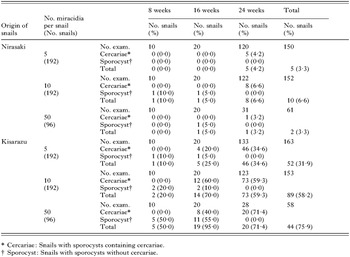
Table 3. Infection rates of two Japanese isolates of Oncomelania nosophora exposed to Schistosoma japonicum originating from Mindoro, the Philippines

Fig. 1. Histological observation of parasite larvae in the Nirasaki isolate of Oncomelania nosophora exposed to the Philippine Schistosoma japonicum. The snails were prepared for microscopy at 3 h (A) or 1 day (B, C) after exposure. Arrows in panels indicate the S. japonicum larvae in the snails.
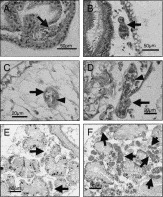
Fig. 2. Histological observation of parasite larvae in 2 isolates of Oncomelania nosophora exposed to the Philippine Schistosoma japonicum. (A, C, E) Sporocysts in O. nosophora from Nirasaki, Yamanashi Prefecture. (B, D, F) Sporocysts in O. nosophora from Kisarazu, Chiba Prefecture. The snails were prepared for microscopy at 1 day (A, B), 3 days (C, D), or 15 days (E, F) after exposure. Arrows in panels indicate the sporocysts in the snails. Arrowheads in C and D indicate typical germ cells in primary sporocysts.
 You have
Access
- 9
- Cited by