Introduction
Epidemiologic data and animal studies support the hypothesis that conditions in the womb during fetal development may program adult metabolic processes and contribute to adult disease. Long latency periods between environmental triggers and the onset of subsequent disease are well documented in the pathogenesis of certain cancers.Reference Wynn, Scumpia and Winfield1,Reference Santos, Camargo and Constantino2 Furthermore, substantial evidence indicates that early life events play a critical role in influencing susceptibility to chronic diseases later in life.Reference Wynn, Scumpia and Winfield1,Reference Hsu and Tain3,Reference Miller, Dodson and Mankouski4
Animal models have confirmed the association of fetal growth restriction with the development of adult metabolic syndromeReference Vickers, Breier, Cutfield, Hofman and Gluckman5 and have begun to elucidate the underlying mechanisms of fetal programming. In humans, low birth weight is associated with increased rates of coronary heart disease, stroke, type 2 diabetes mellitus, obesity, and osteoporosis in adulthood.Reference Barker and Osmond6–Reference Osmond, Barker, Winter, Fall and Simmonds8 Recent studies have also highlighted the role of maternal undernutrition in altering immune-related gene expression in the placenta and fetal liver, as well as in key fetal immune organs such as the thymus, further supporting the concept of fetal programming.Reference Muroya, Otomaru and Oshima9
Full-term neonates, and even more so premature neonates, exhibit various immaturities in their humoral and cellular immunologic mechanisms, particularly during the first days of life.Reference Tsafaras, Ntontsi and Xanthou10 Fetal growth restriction exacerbates these deficiencies in neonatal host defense mechanisms by decreasing thymic hormone activity and reducing the numbers and activity of T- and B-lymphocytes in the peripheral blood.Reference Xanthou11 In a rat model of maternal protein restriction (8% protein instead of 20% in the control diet) throughout pregnancy and lactation, the expression of a large number of genes associated with the immune system and inflammation was significantly reduced.Reference Guan, Arany and van Beek12 We previously demonstrated that maternal undernutrition can induce long-term attenuation of immune responses in female offspring during adulthood.Reference Desai, Gayle, Casillas, Boles, Ross and Ross13 Recent evidence suggests that maternal undernutrition during pregnancy and lactation can also impair offspring immune function via epigenetic modifications, further emphasizing the importance of early-life nutrition.Reference Wu, Cheng, Bai and Ma14
In this study, we aimed to determine the effects of maternal undernutrition during pregnancy and/or lactation on various components of the male neonatal innate immune system, both under basal conditions and in response to inflammatory stimuli.
Methods
Maternal diet
The rat model utilized for maternal food restriction during pregnancy and lactation has been previously described.Reference Desai, Gayle, Casillas, Boles, Ross and Ross13 Briefly, first-time-pregnant Sprague Dawley rats (Charles River Laboratories, Hollister, CA) were housed in a facility with constant temperature and humidity and a controlled 12:12-h light/dark cycle. Pregnancy was confirmed by the presence of vaginal plugs, which were checked daily following overnight mating. The day of vaginal plug detection was designated as gestational day 0.
Maternal AdLib food intake was measured and found to be 20 ± 3 g/day. The FR dams were provided exactly one-half of this amount (50% food-restricted diet) each day, ensuring precise and consistent dietary restriction
At 10 d of gestation, rats were provided either an ad libitum (AdLib; n = 12) diet of standard laboratory chow (Lab Diet 5,001, Brentwood, MO) or a 50% food-restricted (FR; n = 12) diet determined by quantification of normal intake in ad libitum-fed rats. Gestational day 10 was chosen because implantation is complete and this stage represents the beginning of organogenesis, a critical period for fetal immune system development. These diets were provided from day 10 of pregnancy to term and throughout the 21-d lactation period.
Offspring
Pups born to FR dams were allocated to one of two groups of dams during lactation. Half of the pups born to FR dams (n = 6) were nursed after cross-fostering to ad libitum–fed dams (n = 6; FR/AdLib), whereas the other half were nursed by their own FR dams (n = 6; FR/FR). The pups born to ad libitum–fed dams continued to be nursed by these dams (n = 6; AdLib/AdLib). Thus, three groups were studied: (1) Control (AdLib/AdLib), (2) food restriction during pregnancy alone (FR/AdLib), and (3) food restriction during both pregnancy and lactation (FR/FR). At 3 weeks of age, 4 male offspring from each litter per group were studied.
Offspring studies
At day 1 (P1) of life, litters were limited to 8 pups (4 males and 4 females) to normalize rearing. Among the excess pups (5–7 excess pups per litter), one half of the males from AdLib (n = 6) and FR (n = 6) dams were decapitated, (6–12 -pups from each group), blood collected for determination of serum TNF-α levels, and body, spleen, and thymus weights obtained. The blood was pooled from pups from the same litter. The other half of excess day 1 pups (6–12 from each group) were injected with intraperitoneal LPS or saline, decapitated, and blood collected after 3 h for determination of serum TNF-α levels (as previously demonstrated in rat models showing peak cytokine responses following systemic LPS exposure 3–4 hours following LPS in rat neonates)Reference Gayle, Beloosesky, Desai, Amidi, Nuñez and Ross15.
At 3 weeks (P21) of age, one male from each of the six litters per group (total 6 from each group) was sacrificed. Body, spleen, and thymus weights for each offspring were obtained before pups were decapitated, and blood was collected via cardiac puncture for determination of white blood count and TNF-α levels. The remaining males of the litters were injected with either intraperitoneal LPS or intraperitoneal normal saline (control), decapitated, and blood collected after 3 h for determination of white blood count and serum TNF-α levels.
The group size of six litters per group was selected based on our prior studies using this model, which demonstrated that this number is sufficient to detect statistically significant differences in TNF-α and related immune parameters in neonatal offspring.Reference Desai, Gayle, Casillas, Boles, Ross and Ross13
LPS (E. coli., serotype 0111:B5, Calbiochem) was reconstituted in physiological saline and intraperitoneally administered at 100 μg/kg body weight at P1 and P21.
ELISA determinations
A commercial enzyme-linked immunosorbent assay (ELISA, R&D Systems, Minneapolis, MN) kit was used to determine neonatal plasma protein levels of TNF-α as previously described.Reference Gayle, Beloosesky, Desai, Amidi, Nuñez and Ross15 The minimum detectable concentration was <10 pg/ml, with intra-assay and inter-assay variations <10%.
Analysis of whole blood samples for WBC counts
Whole blood samples were collected in EDTA-coated tubes (Medical Supply 123, Cat. No. 07–6,013) and kept on ice for a minimum of 30 min but no more than 4 h to be analyzed on the HEMAvet machine (Drew Scientific; Model No. HV950) according to the protocol. The HEMAvet machine analyzes the counts of circulating WBCs, including lymphocytes, neutrophils, monocytes, eosinophils, and basophils. Prior to running samples, a species-specific, synthetic control blood (Drew Scientific; Cat. No. MULTI-1835–01) was run to ensure proper calibration of the machine.
Data analyses
Blood levels of TNF and morphometric parameters of newborns from FR and AdLib groups were compared using an unpaired t-test. Differences between the three groups (AdLib/AdLib, FR/AdLib, and FR/FR) of offspring on day 21 were compared using analysis of variance (ANOVA) with Dunnett post hoc tests, with FR/FR designated as the reference group for all statistical comparisons. Within-group comparisons of basal vs. post-LPS levels for lymphocytes, neutrophils, and monocytes were performed using the Wilcoxon signed-rank test. Values are expressed as means ± SE. Differences were considered significant at P < 0.05 compared with FR/FR.
Results
Newborn pups at day 1
On day 1 of life, male pups of FR (food restriction) dams were significantly smaller than AdLib pups (6.5 ± 0.76 vs. 7.4 ± 0.75 g). There were no differences in thymus and spleen weights between FR and AdLib pups (Thymus: 0.0023 + 0.001 vs 0.0021 + 0.001 g, Spleen: 0.0027 ± 0.0004 vs 0.0027 ± 0.0004 g respectively). Similarly, when adjusted for body weight, no significant differences were observed between the groups.
TNF Response to LPS: Basal TNF levels were undetectable in both groups. Three hours after LPS injection, pups of both FR and AdLib dams had similar increases in TNF-α levels (760.4 ± 28.1 vs. 761.5 ± 33.9 pg/ml). Saline injection had no effect on TNF levels in pups in the different groups.
Offspring at 3 weeks
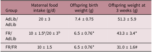
At three weeks of age, offspring exposed to maternal food restriction during pregnancy and/or lactation (FR/AdLib and FR/FR groups) exhibited significantly reduced body weights compared to offspring from normally nourished dams (AdLib/AdLib group), with the most pronounced growth restriction observed in the FR/FR group (FR/FR: 31.0 ± 1.6 g vs. FR/AdLib: 43.3 ± 3.4 g vs. AdLib/AdLib: 51.3 ± 5.9 g, p < 0.05). The thymus weight was significantly reduced in both FR/FR and FR/AdLib groups, with FR/FR offspring demonstrating the lowest weight compared to the AdLib/AdLib group (0.13 ± 0.02, 0.18 ± 0.02, and 0.22 ± 0.01 g, respectively, p < 0.05). However, differences were not demonstrated after adjustment to body weight. Splenic weight was also significantly decreased in FR/AdLib and FR/FR compared to AdLib/AdLib (FR/FR: 0.09 ± 0.01 g vs. FR/AdLib: 0.16 ± 0.02 g vs. AdLib/AdLib: 0.20 ± 0.05 g, p < 0.05). After adjustment to body weight, splenic weight for AdLib/AdLib and FR/AdLib were similar (0.0037 and 0.0036, respectively), but splenic weight in the FR/FR group was significantly reduced (0.0028 ± 0.0001; p < 0.05).
To facilitate group comparisons, maternal food intake and offspring biometric parameters at birth and at three weeks of age are summarized in Table 1.
Maternal food intake and offspring biometric data

Values are expressed as mean ± SE.
AdLib, ad libitum feeding; FR, 50% food restriction.
a During pregnancy (gestational days 10–21).
b During lactation (postnatal days 1–21).
* P < 0.05 compared with AdLib/AdLib.
# P < 0.05 compared with FR/AdLib.
Inflammatory response to LPS
Basal TNF levels were undetectable in all groups. At three hours after LPS challenge, AdLib/AdLib and FR/AdLib offspring had similar increases in pro-inflammatory TNF-α levels (484 ± 59 pg/ml and 511 ± 91 pg/ml, respectively), while the FR/FR offspring had a markedly reduced inflammatory response (206 ± 28 pg/ml) (Fig. 1). Saline injection had no effect on TNF levels in either group data not shown.
TNF-a level following IP LPS at 3 weeks. Suppressed TNF-a levels in FR/FR vs. FR/AdLib and AdLib/AdLib males. Values are mean ± SE; *P < 0.05 compared FR/FR.

WBC indices
There were no differences in baseline white blood count levels among the three groups: 1.38 ± 0.08 × 10³/μl, 1.45 ± 0.18 × 10³/μl, and 1.56 ± 0.16 × 10³/μl for AdLib/AdLib, FR/AdLib, and FR/FR groups, respectively. Monocyte levels were significantly lower in the FR/FR offspring group compared to the AdLib/AdLib group, though similar to the FR/AdLib group (1.6% ± 0.54, 3.09% ± 0.39, 2.4% ± 0.41, respectively). Neutrophil percentage was similar in the three groups (17.8% ± 1.2, 14.8% ± 1.5, and 18.1% ± 1.8), as was lymphocyte percentage (78% ± 1.4, 82% ± 1.9, and 79.9% ± 1.9 for AdLib/AdLib, FR/AdLib, and FR/FR groups, respectively) (Figs. 2–4).
Baseline and LPS-induced monocytes at 3 weeks. (a). Monocyte percentages were measured under basal conditions and 3 h after intraperitoneal LPS injection. FR/FR offspring exhibited a significantly higher percentage of circulating monocytes compared with FR/AdLib and AdLib/AdLib males. Data are presented as mean ± SE; p < 0.05 compared with FR/FR.

Baseline and LPS-induced Neutrophils at 3 weeks. Neutrophil percentages were determined under basal conditions and 3 h after LPS challenge. FR/FR offspring displayed a significant increase in neutrophil percentage following LPS compared with FR/AdLib and AdLib/AdLib groups. Data are presented as mean ± SE; p < 0.05 compared with FR/FR.

Baseline and LPS-induced lymphocytes at 3 weeks. Lymphocyte percentages were measured under basal conditions and 3 h after LPS injection. FR/FR offspring had a significantly lower lymphocyte percentage compared with FR/AdLib and AdLib/AdLib groups. Data are presented as mean ± SE; p < 0.05 compared with FR/FR.

In response to LPS injection, total WBC count at three hours was reduced in all groups compared to their basal levels, with no significant differences among the three groups (1.00 ± 0.25 × 10³/μl, 0.67 ± 0.06 × 10³/μl, and 0.92 ± 0.11 × 10³/μl for AdLib/AdLib, FR/AdLib, and FR/FR groups, respectively). There was a trend towards a higher percentage of monocytes in the FR/FR offspring compared to AdLib/AdLib and FR/AdLib (5.4 ± 1.1 vs. 2.0 ± 0.70, and 2.0 ± 0.57, respectively, p = 0.067). Monocytes in the FR/FR group significantly increased following LPS compared to the basal state (1.6 ± 0.54 vs. 5.4 ± 1.1%), while it decreased in the other two groups (3.1 ± 0.4 vs. 2 ± 0.7%, 2.4 ± 0.41 vs. 2 ± 0.57% for AdLib/AdLib, FR/AdLib, respectively). Neutrophil percentage was significantly increased following LPS injection in the FR/FR group compared to the AdLib/AdLib and FR/AdLib groups (50.6 ± 5.5, 20.0 ± 4.2, and 17.2 ± 2.4, respectively). However, lymphocyte percentage was significantly decreased in the FR/FR group compared to the AdLib/AdLib and FR/AdLib groups (43.2 ± 5.9, 77.2 ± 4.4, and 80.0 ± 2.4, respectively) (Figs. 2–4).
LPS-induced changes
To better illustrate the effect of LPS, we added an integrated (Fig. 5) presenting basal and post-LPS values for monocytes, neutrophils, and lymphocytes. This combined panel shows that marked basal-to-post-LPS changes occurred exclusively in FR/FR offspring, including large increases in neutrophils and monocytes and a substantial drop in lymphocytes. In contrast, AdLib/AdLib and FR/AdLib offspring exhibited minimal or no basal-to-post-LPS changes. These findings reinforce that the impaired inflammatory response becomes evident only after immune stimulation.
Basal and post-LPS levels of immune cell populations in male offspring at 3 weeks. White blood cell differential counts (monocytes, Panel a; neutrophils, Panel b; lymphocytes, Panel c) were measured under basal conditions (dark blue bars) and 3 h after intraperitoneal LPS injection (orange bars; 100 μg/kg). FR/FR offspring exhibited significantly altered immune cell populations following LPS challenge, with increased monocyte and neutrophil percentages and decreased lymphocyte percentage compared with FR/AdLib and AdLib/AdLib groups. Data are presented as mean ± SE; **p < 0.05 compared with FR/FR.

Discussion
In this study, we demonstrated that maternal undernutrition during pregnancy and lactation is associated with significant alterations in offspring development. Specifically, FR/FR offspring were markedly smaller at 3 weeks of age compared to AdLib/AdLib and FR/AdLib offspring, consistent with previous reports of growth restriction due to maternal undernutrition.Reference Desai, Gayle, Casillas, Boles, Ross and Ross13 Consistent with growth restriction, FR/FR offspring demonstrated reduced thymus and spleen weights. The thymus is a critical organ for T-cell development, and its reduced size in FR/FR offspring may reflect impaired thymopoiesis, a process essential for the establishment of a functional adaptive immune system. This is consistent with human studies linking prenatal undernutrition to reduced thymic function and increased susceptibility to infections.Reference McDade, Beck, Kuzawa and Adair16 Similarly, the smaller spleen size in FR/FR offspring may indicate impaired development of secondary lymphoid organs, further compromising immune competence. These findings highlight the importance of maternal nutrition in shaping not only the innate but also the adaptive immune system. These findings align with a growing body of evidence suggesting that early-life nutritional deficits can have profound and lasting effects on offspring health, including immune function.
Maternal food restriction significantly compromises the maternal immune system during gestation and lactation, characterized by reduced cytokine production, decreased immunoglobulin synthesis, and impaired antioxidant capacity.Reference Marques, O’Connor, Roth, Susser and Bjørke-Monsen17 This maternal immune dysfunction directly impacts offspring through reduced passive antibody transfer, altered maternal-fetal cytokine signaling, and epigenetic programming of immune-related genes. The compromised maternal immune environment during critical developmental windows likely contributes to the immune deficits we observed in FR/FR offspring, including reduced TNF-α responses and abnormal immune cell populations, highlighting the maternal immune system as a key mediator linking nutritional restriction to offspring immune programming.
One of the most striking findings of our study was the 50% reduction in serum TNF-α response to LPS in 3 week old FR/FR offspring compared to AdLib/AdLib and FR/AdLib neonates. This attenuated inflammatory response occurred despite higher monocyte counts in FR/FR offspring following LPS stimulation, suggesting that the defect lies not in the availability of immune cells but in their functional capacity. This is consistent with studies showing that maternal undernutrition can impair immune cell signaling pathways, such as the LPS-TLR4-NF-κB cascade, which is critical for mounting an effective innate immune response.Reference Levy18 For example, maternal protein restriction has been shown to alter the expression of genes involved in inflammation and immune regulation, potentially explaining the blunted TNF-α response observed in our study. These findings indicate that while monocyte numbers increase in FR/FR offspring following LPS stimulation, their ability to mount an effective inflammatory response is compromised. This reflects a state in which immune cells are numerically adequate but functionally deficient, a phenomenon linked to early-life nutritional restriction in prior studies.Reference Wu, Cheng, Bai and Ma14,Reference Levy18
Another line of evidence suggests that oxidative stress and inflammation act as critical mediators linking maternal undernutrition to adverse outcomes in offspring, further underscoring the need for targeted interventionsReference Fioretto, Colombelli and Da Silva19
Maternal food restriction is also known to activate the maternal–fetal stress axis, with increased glucocorticoid exposure to the fetus (partly via reduced placental 11β-HSD2), programming hypothalamic–pituitary–adrenal (HPA) reactivity in offspring. This glucocorticoid milieu can impair thymic growth and T-cell output and skew innate/adaptive responses, culminating in blunted cytokine production and altered leukocyte distributions. These HPA-mediated effects provide a biologically plausible link between maternal undernutrition and our findings of reduced LPS-stimulated TNF-α and perturbed monocyte/neutrophil–lymphocyte profiles in FR/FR pups, consistent with prior evidence that early-life undernutrition programs long-term immune dysfunction via HPA activationReference Lesage, Blondeau, Grino, Bréant and Dupouy20 and with human data relating prenatal undernutrition to reduced thymic functionReference McDade, Beck, Kuzawa and Adair16 and the known immaturity of neonatal immune responses.Reference Tsafaras, Ntontsi and Xanthou10In addition to the reduced TNF-α response, 3 week old FR/FR offspring exhibited abnormal lymphocyte and neutrophil counts following LPS stimulation. Specifically, we observed a shift from lymphocyte to neutrophil dominance in FR/FR offspring, a pattern not seen in the other groups. These differences were only apparent after LPS stimulation and not at baseline, indicating that the defect lies in the dynamic immune response rather than in steady-state immune cell populations. The exaggerated neutrophil increase combined with the lymphocyte decrease suggests a maladaptive inflammatory activation, potentially driven by altered cytokine production and stress-related hormonal pathways. The integrated visualization in Fig. 5 further highlights that only FR/FR offspring display substantial basal-to-post-LPS shifts across leukocyte subsets, reinforcing that the programmed immune deficit becomes evident only under inflammatory challenge. The minimal or absent changes in AdLib/AdLib and FR/AdLib offspring emphasize that the impaired response is specific to those exposed to nutritional restriction both prenatally and postnatally.
This suggests that maternal undernutrition may disrupt the balance between different immune cell populations, potentially compromising the offspring’s ability to respond appropriately to infections or other inflammatory challenges. These findings are supported by work showing that maternal undernutrition can induce long-term immune dysfunction through mechanisms involving oxidative stress and inflammation.Reference Fioretto, Colombelli and Da Silva19
These findings of abnormal immune response in offspring following maternal undernutrition confirm prior reports. Gotardo et al., focusing on the humoral immune response rather than the innate immune response in Wistar rats, demonstrated that food restriction between days 6–17 of gestation resulted in alterations in delayed-type hypersensitivity response and humoral immune response against sheep red blood cells in 21-day-old offspring.Reference Gotardo, Dipe, Hueza and Górniak21 Interestingly, in their study, the adjusted weight of the thymus was increased in FR as compared to AdLib pups, while in our study, thymus-adjusted weights were similar between groups. This discrepancy may reflect differences in the timing and duration of maternal undernutrition, as well as species-specific responses to nutritional stress. Badr et al. demonstrated that early undernutrition, particularly during the prenatal and lactation periods, attenuated T cell survival and the immune response in 8-week adult male rats.Reference Badr and Mohany22 Their findings align with our observation of reduced thymus weight in FR/FR offspring, suggesting that maternal undernutrition may impair thymic function and T-cell development. This is further supported by studies showing that maternal undernutrition can induce epigenetic modificationsReference Zelko, Zhu and Roman23 that alter immune-related gene expression, potentially explaining the long-term immune deficits observed in offspring.
The immune deficits observed in FR/FR offspring were not seen in FR/AdLib offspring, which exhibited responses similar to AdLib/AdLib neonates. This contrasts with our previous findings in female offspring at 9 months of age, where both FR/FR and FR/AdLib groups showed attenuated immune responses.Reference Desai, Gayle, Casillas, Boles, Ross and Ross13 This discrepancy may reflect differences in the timing of immune assessment (neonatal period vs. adulthood) rather than dose-dependent effects. Neonatal immune responses are inherently immature and differ significantly from those of adults, which may explain the observed differences.Reference Tsafaras, Ntontsi and Xanthou10 Additionally, sex-specific effects cannot be ruled out, as male and female offspring may respond differently to maternal undernutrition.
The immaturity of the neonatal immune system further complicates the interpretation of these findings. Neonates rely heavily on innate immunity during the first weeks of life, as their adaptive immune system is not fully developed.Reference Tsafaras, Ntontsi and Xanthou10 This immaturity makes them particularly vulnerable to the effects of maternal undernutrition, which may disrupt the delicate balance of immune cell populations and signaling pathways required for effective host defense. For example, maternal undernutrition has been shown to alter the gut microbiota, which plays a critical role in shaping neonatal immune development.Reference Jin, Zhang and Jia24 These early-life disruptions may have long-term consequences, increasing the risk of infections and inflammatory diseases later in life.Reference Lesage, Blondeau, Grino, Bréant and Dupouy20 Moreover, maternal low-protein diets can induce sex-specific developmental programming in offspring, highlighting the importance of evaluating both male and female progeny in future investigations.Reference Cavariani, de Mello Santos and Chuffa25
While some of our findings confirm previous reports of altered immune function following maternal undernutrition,Reference Desai, Gayle, Casillas, Boles, Ross and Ross13,Reference Badr and Mohany22 this study provides new insights into the early neonatal period and the role of postnatal nutrition. By assessing immune parameters at both day 1 and day 21 and using a cross-fostering design, we distinguished the effects of gestational versus combined gestational and lactational undernutrition. This novel approach revealed that prolonged maternal undernutrition throughout pregnancy and lactation leads to persistent defects in TNF-α production and immune cell distributions, changes not seen with gestational restriction alone. These findings expand our understanding of how the timing and duration of maternal undernutrition shape neonatal immune development
Limitations
While our study provides important insights into the effects of maternal undernutrition on offspring immune function, several limitations should be noted. First, our findings are specific to male offspring, and sex-specific effects warrant further investigation. Second, the mechanisms underlying the observed immune dysregulation, such as epigenetic changes or oxidative stress, require deeper exploration. Third, we focused on TNF-α as a sensitive marker of innate immune activation; measuring additional cytokines such as IL-1β and IL-6 would provide a broader view of the inflammatory response and will be included in future studies. Fourth, we did not assess body composition or metabolic parameters such as triglycerides, free fatty acids, or glucose. Future studies will incorporate these measurements to more fully characterize the relationship between growth restriction, metabolic status, and immune function. Finally, our study focused on the neonatal period, and longer-term follow-up is needed to determine whether these immune deficits persist into adulthood.
Conclusion
In conclusion, our study demonstrates that prolonged maternal undernutrition during pregnancy and lactation is associated with significant changes in the male offspring innate immune system, including reduced TNF-α responses, abnormal immune cell counts, and reduced absolute thymus and spleen weights, consistent with overall growth restriction. These findings highlight the critical role of maternal nutrition in shaping neonatal immune development and underscore the importance of early-life interventions to mitigate the long-term health consequences of maternal undernutrition. Future studies should explore the mechanisms underlying these effects and investigate whether similar changes occur in female offspring.
Acknowledgments
The authors acknowledge the use of AI language model for assistance with manuscript formatting and language editing. The authors remain fully responsible for all scientific content, data interpretation, and conclusions presented in this manuscript.
Funding statement
This work was supported by the National Institutes of Health Grants R01HD054751 and R01DK081756.
Competing interests
The author(s) declare(s) that there is no conflict of interest.
Ethical standards
The authors assert that all procedures contributing to this work comply with the ethical standards of the American Association for Accreditation of Laboratory Animal Care (AAALAC) and National Institutes of Health (NIH) guidelines and that the study has been approved by the Animal Research Committee of the Lundquist Institute at Harbor-UCLA.