INTRODUCTION
The dinoflagellate Kryptoperidinium foliaceum (Stein) Lindemann occurs in coastal waters, and is a common inhabitant of shallow water lagoons, estuaries and tidal creeks, where the species can tolerate a great range of salinities from fresh and brackish waters up to true oceanic salinity (e.g. Prager, Reference Prager1963; Johnston & Gilliland, Reference Johnston and Gilliland2000; Kempton et al., Reference Kempton, Wolny, Tengs, Rizzo, Morris, Tunnell, Scott, Steidinger, Hymel and Lewitus2002; López-Flores, Reference López-Flores2005; Figueroa et al., Reference Figueroa, Bravo, Fraga, Garces and Llaveria2009; Hallegraeff et al., Reference Hallegraeff, Bolch, Huisman, de Salas, Hallegraeff, Bolch, Hill, Jameson, LeRoi, McMinn, Murray, de Salas and Saunders2010; Konovalova & Selina, Reference Konovalova, Selina and Adrianov2010; Domingues et al., Reference Domingues, Anselmo, Barbosa, Sommer and Galvão2011). Although the diversity of plankton and benthic dinoflagellates of Kuwait's marine environment has been recently well documented (Al-Yamani et al., Reference Al-Yamani, Bishop, Ramadhan, Al-Husaini and Al-Ghadban2004; Al-Kandari et al., Reference Al-Kandari, Al-Yamani and Al-Rifaie2009; Saburova et al., Reference Saburova, Al-Yamani, Polikarpov, Krupp, Musselman, Kotb and Weidig2009; Al-Yamani & Saburova, Reference Al-Yamani and Saburova2010), K. foliaceum has never been reported previously from Kuwait's waters or intertidal sediments. The latest detailed survey of Kuwait's shore at a small hypersaline tidal creek in the north-western area has revealed a dense population of K. foliaceum.
Hypersaline environments are typical extreme habitats, in which the high salt concentration is the main environmental factor that may limit their biodiversity. The organisms that inhabit hypersaline environments are predominantly prokaryotes (cyanobacteria and a heterogeneous group of archaea and bacteria), whereas eukaryotic diversity is much lower and largely comprises photosynthetic flagellates belonging to the Dunaliella Teodoresco and Asteromonas Artari genera, some diatoms, not numerous Protozoa (e.g. ciliate Fabrea salina Henneguy) and fungi (e.g. Ben-Amotz et al., Reference Ben-Amotz, Katz and Avron1982; Rodriguez-Valera et al., Reference Rodriguez-Valera, Ventosa, Juez and Imhoff1985; Ventosa, Reference Ventosa, Logan, Lappin-Scott and Oyston2006). Dinoflagellates cannot be related to common inhabitants of extreme environment such as hypersaline water bodies. Records of dinoflagellates from hypersaline habitats are rare and poorly documented. Prorocentrum micans Ehrenberg was reported from salt lagoons in the Caribbean Islands at salinities above 90 psu (Steidinger & Tangen, Reference Steidinger, Tangen and Tomas1997); dinoflagellates Gymnodinium sp., Protoperidinium sp., Gonyaulax verior Sournia and Oxyrrhis marina Dujardin were abundant at salinity of 78.7 ± 8.8 psu in a costal solar saltern in Tunisia (Abid et al., Reference Abid, Sellami-Kammoun, Ayadi, Drira, Bouain and Aleya2008); survival of O. marina in cultures was reported at salinities ranged from 4 to 64 psu, however maximum growth rate was recorded at salinity of 16 psu (Droop, Reference Droop1959); an unidentified thecate dinoflagellate was found in low abundance at salinity of 52 ± 18 psu from a hypersaline pond in the British Virgin Islands (Jarecki et al., Reference Jarecki, Burton-MacLeod and Garbary2006).
Here we present the first report of Kryptoperidinium foliaceum from a hypersaline habitat within Kuwait's shore. The morphology of this dinoflagellate, its abundance and distribution along a tidal creek are examined.
MATERIALS AND METHODS
Sampling site
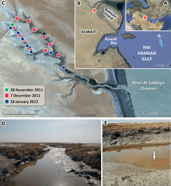
Kuwait's coastline stretches about 180 km in a north-north-westerly direction, occupying the north-west corner of the Arabian Gulf (Figure 1A, B) and providing a wide range of near-shore habitats for marine biota (e.g. Jones, Reference Jones1986; Al-Yamani et al., Reference Al-Yamani, Bishop, Ramadhan, Al-Husaini and Al-Ghadban2004). The examined small tidal creek lies throughout the broad low-gradient salt flat, which is partially inundated by spring tides and flows into the Khor Al-Sabbiya Channel (Figure 1C, D). The creek has an approximate length of 1.8 km and stretches from 29°54′32″N 48°00′18″E to 29°53′50″N 48°00′55″E in a south-easterly direction. The upper stream zone of the creek consists of two branches, which merge as they flow downstream into the sea (Figure 1C).
Sampling site: (A, B) satellite images of Arabian (Persian) Gulf and Kuwait shore (Google Earth) where the sampling area was located; (C) sampling points overlaid on satellite image of the tidal creek; (D) upper stream zone of the tidal creek during ebb tide, creek banks are colonized by glassworts; (E) middle stream zone of the tidal creek where bloom of Kryptoperidinium foliaceum associated with water discoloration (arrow) was recorded.

The creek experiences mixed semidiurnal tidal influence from the Khor Al-Sabbiya Channel with tidal range of over 1 m and very low to negligible freshwater input. The bottom of the creek is composed of interlaced sandy and muddy sediments. During ebb tide the creek becomes shallower and changes to a chain of small pools surrounded by uncovered bottom sediments. The surface of the muddy sediments is occupied by extensive diatom-dominated algal mats. Clay creek banks are colonized by glassworts (Figure 1D) and covered with numerous burrows of crabs and mudskippers. During the study period, water temperature ranged from 14.3 to 18.1°C.
Sampling
Water and sediment samples were collected monthly during November 2011–January 2012 (Figure 1C) at different tidal stages. Surface water was sampled by 0.5 l plastic bottles; surface layer (0–2 cm) of the bottom sediments was collected using a hand-made plastic tube. Several physical, chemical and biological variables were measured, including water temperature and salinity, concentration of dissolved inorganic macronutrients (ammonia, nitrites, nitrates, phosphates and silicates), and microplankton/microbenthic composition and abundance.
Sample processing
Water salinities were measured in situ by a hand held refractometer (Atago, Japan) as well as by a salinity probe (Orion model 140) in the laboratory in 100 ml aliquots of the 500 ml samples. All nutrient analyses were performed on GF/F pre-filtered water samples. Nutrients were determined according to the spectrophotometric methods (Grasshoff et al., Reference Grasshoff, Ehrhardt and Kreming1983). Nitrite, nitrate, phosphate and silicate were measured with a San 2 ++ segmented flow autoanalyser (Skalar BV, Netherlands), while ammonia was analysed using the phenol-hypochlorite method with optical density measured by Beckman DU-640 spectrophotometer (Beckman Coulter Inc., USA).
Unpreserved water samples were screened using an inverted microscope at 40–200× magnification (Leica DMIL, Wetzlar, Germany) to estimate the microplankton composition. Aliquots of samples were preserved with 4% Lugol's iodine solution for future evaluation. Subsamples (25 ml) were settled in Utermöhl chambers and cells were enumerated with a Leica DMIL inverted microscope. The appropriate area of the chamber was scanned, depending on the abundance of each species. Microbenthos was separated from the sediment samples by extraction using the frozen seawater method (Uhlig, Reference Uhlig1964) with a 110-µm mesh size for qualitative examination.
The morphology of the cells was examined by light and epifluorescence microscopy. For detailed observation, single cells were isolated by micropipetting in preparation for high-magnification photomicroscopy. Cells were examined using a Leica DMLM microscope (Leica, Wetzlar, Germany) at 630–1000× magnification. Thecal plates were observed after staining with Calcofluor White (Sigma Chemical Co, USA) in accordance with the method of Fritz & Triemer (Reference Fritz and Triemer1985). To visualize nuclei, a fluorescent stain, 4′, 6-diamidino-2-phenylindole (DAPI, Sigma) was applied to cells fixed in 2.5% glutaraldehyde solution. Light micrographs were made using the Leica DMLM microscope connected to a Leica DFC 320 colour digital camera (Leica, Wetzlar, Germany). Cell dimensions were measured from images using Leica Application Suite v. 3.7 (Leica Microsystems (Switzerland) Ltd) software.
RESULTS
SYSTEMATICS
Class DINOPHYCEAE Fritsch in West & Fritsch, 1927
Order PERIDINIALES Haeckel, 1894
Family PERIDINIACEAE Ehrenberg, 1831
Genus Kryptoperidinium Lindemann, Reference Lindemann1924
Kryptoperidinium foliaceum (Stein) Lindemann Reference Lindemann1924
(Figure 2)
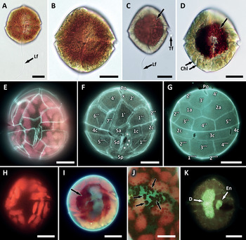
Light micrographs of Kryptoperidinium foliaceum: (A, B) cells from freshly collected sample in ventral view showing general appearance, cingulum and sulcus paths, longitudinal flagellum (Lf) and evenly distributed reddish-brown globules; (C, D) cells after exposure in laboratory in ventral view showing general appearance, transverse (Tf) and longitudinal (Lf) flagella, peripherally arranged chloroplasts and central spot of concentrated reddish-brown globules (arrows); (E) epifluorescent light microscopy (LM) image showing delicate theca with distinct sutures, ventral view; (F, G) epifluorescent LM images of the thecal plate arrangement in ventral (F) and dorsal (G) views, small sulcal plates are marked with asterisks; (H) epifluorescent LM image showing the chloroplast arrangement in ventral view; (I) epifluorescent LM image showing layer of reddish-brown globules concentrated around nucleus; (J) reddish-brown globules (arrows) visible against the blue background of nucleus, epifluorescence; (K) 4′-6-diamidino-2-phenylindole (DAPI)-stained cell in dorsal view showing dinokaryotic (D) and endosymbiont (En) nuclei. Scale bars: A–I and K, 10 µm; J, 5 µm.

DESCRIPTION
Vegetative motile cells of Kryptoperidinium foliaceum were examined using light and epifluorescence microscopy. Cells were approximately oval in ventral view (Figure 2A–D), strongly dorso-ventrally flattened, with concave ventral and convex dorsal sides; 27–40 µm long (33.9 ± 3.3 µm, N = 21), and 25–34 µm wide (30.3 ± 3.2 µm, N = 21). The cingulum was median and slightly displaced (Figure 2E, F). The sulcus slightly widened towards the posterior and reached the antapex (Figure 2A–C, E, F). The chloroplast were numerous, elongate, light golden-brown to greenish-brown, and arranged peripherally (Figure 2D, E, H). Nuclear staining by DAPI revealed a large, ellipsoidal to bean-shaped dinokaryon with distinct condensed chromosomes together with smaller rounded to irregularly shaped endosymbiotic nucleus, in which the chromosomes were not distinguishable (Figure 2K). All cells examined were binucleate. The cytoplasm contained numerous reddish-brown globules, which determined intensive cell coloration (Figure 2A–D, I, J). These globules were evenly distributed throughout the cytoplasm in freshly collected cells (Figure 2A, B), whereas they were concentrated in the central cell area surrounding the nucleus and forming a clearly visible large reddish-brown spot (Figure 2C, D, I) during further exposure of samples in the laboratory. The eyespot, which is often made reference to in K. foliaceum literature, was not observed for the Kuwaiti strain. Theca was delicate, with distinct sutures (Figure 2E). We determined the following thecal plate pattern for Kryptoperidinium foliaceum isolated from Kuwait's tidal creek: Po, X, 4′, 2a, 7″, 4c, >5s, 5″′, 2″″ (Figure 2F, G).
Non-motile resting stages were observed in the natural population of K. foliaceum from the surface layer of the bottom sediments and during short term (2–5 days) exposure of the water samples in the laboratory. These cells were round to ovoid, slightly flattened, variable in size, 23–31 µm long, and 23–34 µm wide. Rounded cytoplasmic part was covered by hyaline wall, which was thicker than the theca in motile cells, smooth and not divided into plates. Non-motile cells were filled with numerous small reddish-brown globules that masked other cytoplasm content, resulting in the intense dark reddish-brown cytoplasm.
Hypersaline tidal creek environment
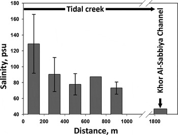
Average tidal creek salinity was 105.5 ± 36.7 psu and ranged from 63.7 up to 178.5 psu during the study period depending on the tidal cycle. Highest salinities coincided with the upper stream zone at ebb tide, and concentrations were gradually depleted in lower creek stations down to 47 psu in Khor Al-Sabbiya Channel (Figure 3).
The average (±standard deviation (SD)) salinity distribution during different tidal stages along the tidal creek. The distance axis runs from 0 m at the upper stream zone of the tidal creek to 1800 m at its confluence with the Khor Al-Sabbiya Channel. The average and SD were calculated from the values for each 200 m interval (n0–200 = 11, n200–400 = 4, n400–600 = 3, n600–800 = 1, n800–1000 = 3).

The nutrient concentrations showed a wide fluctuation along the tidal creek and were generally at low levels except for silicates. The lowest concentration was recorded for nitrates that varied between undetectable values within the creek upper stream zone and 0.58 µM−1 in the lower creek zone with an average value of 0.1 ± 0.21 µM−1. A little higher concentration was detected for nitrites ranging between 0.06 and 0.61 µM l−1 with an average value of 0.15 ± 0.20 µM l−1. Higher values were observed for phosphates ranging from 0.17 to 0.47 (average value of 0.29 ± 0.12 µM l−1) and for ammonia (from 0.53 to 6.34; average value of 1.65 ± 2.10 µM l−1). Highest concentrations were recorded for silicates varying between 13.37 and 22.71 µM l−1 with an average value of 17.24 ± 3.40 µM l−1.
Kryptoperidinium foliaceum abundance and distribution
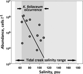
Kryptoperidinium foliaceum was first detected on 28 November 2011 in the upper stream part of the right branch of the tidal creek at salinity of 95 psu and in high abundance (7.3 × 10³ cells l−1). The second survey of both creek branches from upper to middle stream zone was conducted on 7 December 2011 during ebb tide, which revealed the presence of K. foliaceum within the right branch. High concentrations (5.4–6.2 × 10⁴ cells l−1) of K. foliaceum were observed in the middle stream zone of the creek at salinities of 80–96 psu. Detailed re-examination of the creek system during rising tide (18 January 2012) indicated the presence of an association of K. foliaceum with the right creek branch where a bloom was detected. Kryptoperidinium foliaceum was present in high concentrations with an average value of 3.9 ± 3.8 × 10⁵ cells l−1, coinciding mainly with the right branch at salinities ranging from 63.7 to 109 psu. Lower concentration of K. foliaceum cells (2.0 × 10⁴ cells l−1) was observed in a sample collected from the middle stream zone just after the confluence of the creek branches. Moreover, bloom of this species (1.1 × 10⁶ cells l−1) which contributed to water discoloration (Figure 1E) was recorded in the middle stream zone within the right creek branch.
The density of the Kryptoperidinium foliaceum population and its distribution were strongly correlated with a salinity pattern (Figure 4), which was determined by the tidal cycle. The development of this species was limited within salinities ranging from 63.7 to 109 psu and restricted to the right tidal branch. The major part of the K. foliaceum population was represented by vegetative motile cells and was associated with the water column. However, swimming cells and non-motile resting stages of this species were observed from the bottom sandy sediments as well but in much lower density.
The log-transformed (base 10) abundance of Kryptoperidinium foliaceum along the salinity gradient in the tidal creek. Solid line indicates linear regression of the data (coefficient of determination R2 is 0.78).

Among the rather dense population of Kryptoperidinium foliaceum, cells of the dinoflagellates Durinskia baltica (Levander) Carty & Cox and Oxyrrhis marina were observed in water samples at lower concentrations (1.3 × 10³–5.5 × 10⁴ and 2.5 × 10³–3.8 × 10⁴ cells l−1, correspondingly). Amphidinium carterae Hulburt sporadically occurred in diatom-dominated algal mats at the upper stream creek zone at salinities of 90 ± 22 psu but in low abundance as well as Amphidinium scissum Koifoid & Swezy, which was occasionally recorded in the bottom sandy sediments in the middle stream zone at salinities ranged from 47 to 73 psu. Additionally, well-developed diatom-dominated algal mats covering the surface of the bottom mud were observed throughout the tidal creek and were mainly composed of very dense population of the benthic diatom Nitzschia longissima (Brébisson) Ralfs. Among the algal mat, cells of the benthic diatoms Nitzschia reversa Smith, Petrodictyon gemma (Ehrenberg) Mann, Entomoneis sp., Amphora cf. lineolata Ehrenberg and Navicula sp. were found in lower densities. Rather high concentrations of these benthic diatoms within the shallow water column were observed as well due to the mixing effect of strong tidal currents.
DISCUSSION
The small dinoflagellate Kryptoperidinium foliaceum has attracted heightened scientists' attention and has been extensively studied due to its ecological plasticity, rare genome type and high variability among strains from different localities (e.g. Dodge & Crawford, Reference Dodge and Crawford1969; Withers & Haxo, Reference Withers and Haxo1978; Kempton et al., Reference Kempton, Wolny, Tengs, Rizzo, Morris, Tunnell, Scott, Steidinger, Hymel and Lewitus2002; Horiguchi, Reference Horiguchi, Mawatari and Okada2004; Wolny et al., Reference Wolny, Kempton, Lewitus, Steidinger, Landsberg, Tomas and Vargo2004; Figueroa et al., Reference Figueroa, Bravo, Fraga, Garces and Llaveria2009). Kryptoperidinium foliaceum is a taxon with knotty taxonomic history. This species was originally described by Stein in 1883, and it was assigned to the genus Glenodinium Ehrenberg as G. foliaceum Stein. After the initial record of Stein, this species has been repeatedly revised and reclassified (e.g. Paulsen, Reference Paulsen, Brandt and Apstein1908; Lindemann, Reference Lindemann1924; Lebour, Reference Lebour1925; Biecheler, Reference Biecheler1952; Prager, Reference Prager1963). Lindemann (Reference Lindemann1924) revised this species and reclassified it under the new genus Kryptoperidinium Lindemann, whereas it was reassigned to the genus Peridinium Ehrenberg by Biecheler (Reference Biecheler1952), while other taxonomists continued to assign this species to the genus Glenodinium (e.g. Schiller, Reference Schiller1937; Prager, Reference Prager1963; Dodge & Crawford, Reference Dodge and Crawford1969), which caused taxonomic and nomenclatural confusion. Moreover, according to Sournia (Reference Sournia1986), the generic name Phyllodinium Conrad may be considered as a questionable taxonomic junior synonym of Kryptoperidinium. Following Sournia (Reference Sournia1986), we consider Kryptoperidinium to be the taxonomic senior synonym of Glenodinium and Peridinium.
The morphology of the dinoflagellate isolated from a hypersaline tidal creek in Kuwait conforms closely with those previously reported for Kryptoperidinium foliaceum (= Glenodinium foliaceum Stein, Peridinium foliaceum (Stein) Biecheler) by Biecheler (Reference Biecheler1952), Dodge (Reference Dodge1982), Sournia (Reference Sournia1986), Kempton et al. (Reference Kempton, Wolny, Tengs, Rizzo, Morris, Tunnell, Scott, Steidinger, Hymel and Lewitus2002) and Wolny et al. (Reference Wolny, Kempton, Lewitus, Steidinger, Landsberg, Tomas and Vargo2004), and hence, Kuwait's strain can be attributed to K. foliaceum. Variability in number and shape of epithecal and cingular plates has been described for this species, and the integrated plate formula can be determined as Po, X, 4(3)′, 2a, 7(6)″, 4(5)c, 4–5s, 5″′, 2″″ (Biecheler, Reference Biecheler1952; Dodge, Reference Dodge1982; Sournia, Reference Sournia1986; Kempton et al., Reference Kempton, Wolny, Tengs, Rizzo, Morris, Tunnell, Scott, Steidinger, Hymel and Lewitus2002; Horiguchi, Reference Horiguchi, Mawatari and Okada2004; Wolny et al., Reference Wolny, Kempton, Lewitus, Steidinger, Landsberg, Tomas and Vargo2004; Figueroa et al., Reference Figueroa, Bravo, Fraga, Garces and Llaveria2009). Kuwait's strain generally possesses the typical plate arrangement for K. foliaceum, although the peculiar arrangement of sulcal plates (Figure 2F) was not noticed in previous descriptions of the species and needs to be described thoroughly.
Kuwait's strain of Kryptoperidinium foliaceum differs from the majority of the previously described strains of this species in the absence of an eyespot and in possessing small reddish-brown globules in the cytoplasm in great number. Conspicuous red eyespot was described as the characteristic feature of K. foliaceum (Biecheler, Reference Biecheler1952; Dodge, Reference Dodge1982; Sournia, Reference Sournia1986; Kempton et al., Reference Kempton, Wolny, Tengs, Rizzo, Morris, Tunnell, Scott, Steidinger, Hymel and Lewitus2002; Horiguchi, Reference Horiguchi, Mawatari and Okada2004; Wolny et al., Reference Wolny, Kempton, Lewitus, Steidinger, Landsberg, Tomas and Vargo2004). However, Prager (Reference Prager1963) recorded the variability in shape, size and localization of the eyespot depending on the stage of life cycle in this species, and several strains without the eyespot have been detected recently (Kempton et al., Reference Kempton, Wolny, Tengs, Rizzo, Morris, Tunnell, Scott, Steidinger, Hymel and Lewitus2002). Moreover, one of these strains initially possessed the eyespot that was lost later in a long-term culture (Dodge & Crawford, Reference Dodge and Crawford1969; Kempton et al., Reference Kempton, Wolny, Tengs, Rizzo, Morris, Tunnell, Scott, Steidinger, Hymel and Lewitus2002; Figueroa et al., Reference Figueroa, Bravo, Fraga, Garces and Llaveria2009). As described in the present study Kuwait's strain is one more example of K. foliaceum without the eyespot. Our results support the conclusion of Figueroa et al. (Reference Figueroa, Bravo, Fraga, Garces and Llaveria2009) that the presence of an eyespot could be considered as a variable feature of Kryptoperidinium. Although the composition of small reddish-brown globules in the cytoplasm of Kuwait's strain was not determined in this study, these cytoplasm inclusions are similar to those described for K. foliaceum previously as extraplastidic carotenoid-rich lipid globules (Withers & Haxo, Reference Withers and Haxo1978, as Peridinium foliaceum) and resemble large reddish spots in the non-motile stage of K. foliaceum which was described and illustrated by Figueroa et al. (Reference Figueroa, Bravo, Fraga, Garces and Llaveria2009). Withers & Haxo (Reference Withers and Haxo1978) have examined the composition of extraplastidic lipid globules and found that higher content of these carotenoid-rich lipid globules can be associated with nutrient depletion. Similar phenomena of carotenoid accumulation have been observed in other microalgae, such as halotolerant species of Dunaliella (Chlorophyceae), which thrive in hypersaline environment and can produce great quantities of β-carotene in lipid globules within chloroplasts (e.g. Ben-Amotz et al., Reference Ben-Amotz, Katz and Avron1982). High content of reddish-brown globules within the cytoplasm in Kuwait's strain of K. foliaceum can be considered as adaptation to a combination of extremely high salinity with low nutrient status within its environment in Kuwait.
Kryptoperidinium foliaceum is known as a widely distributed species with a great tolerance range to salinity. It has been reported from almost fresh waters with salinity <0.5 psu (Domingues et al., Reference Domingues, Anselmo, Barbosa, Sommer and Galvão2011), and from brackish waters of coastal lagoons, marshes and estuaries (Johnston & Gilliland, Reference Johnston and Gilliland2000; López-Flores, Reference López-Flores2005; Figueroa et al., Reference Figueroa, Bravo, Fraga, Garces and Llaveria2009), where the species bloomed regularly. It has been recorded in Tunisian coastal waters with salinities ranging from 38.9 up to 44 psu (Turki et al., Reference Turki, Balti and Ben Salah2007). Cultures of K. foliaceum were maintained at salinities of 24, 31 and 36 psu (Kempton et al., Reference Kempton, Wolny, Tengs, Rizzo, Morris, Tunnell, Scott, Steidinger, Hymel and Lewitus2002) and 6–56 psu (Prager, Reference Prager1963).
Our recent observations of Kryptoperidinium foliaceum in hypersaline conditions in appreciable concentrations extend the known range of salinity tolerance for this species up to over 100 psu. Occupying the small hypersaline tidal creek of Kuwait's shore, this dinoflagellate plays an important role in this small unique system. Whereas the bottom sediments of the creek are occupied by diatom-dominated algal mats, high productivity of the water column is contributed mainly by this dinoflagellate. Together with diatoms, K. foliaceum is a key food source for the diverse and dense protozoan community of the examined tidal creek. The observations of K. foliaceum from this study can be considered as a new record for Kuwait's flora.
Kryptoperidinium foliaceum is not a toxic species but can be categorized as harmful for the marine environment. Bloom-associated fish mortality events have been reported for this species from South Carolina estuaries (Lewitus et al., Reference Lewitus, Schmidt, Mason, Kempton, Wilde, Wolny, Williams, Hayes, Hymel, Keppler and Ringwood2003; Wolny et al., 2004) and from Tunisian coastal waters (Turki et al., Reference Turki, Balti and Ben Salah2007). As reported here the occurrence of K. foliaceum in Kuwait's coastal area, its capacity to form bloom in hypersaline conditions and bloom-associated fish kills documented for this species elsewhere can be assessed as a weighty reason to include this dinoflagellate into the list of potentially harmful microalgae for Kuwait's marine environment. Further coastal surveys are needed to detect the distribution range for K. foliaceum on Kuwait's shoreline and its seasonal dynamics.
ACKNOWLEDGEMENTS
We acknowledge the help of Mr Alan Lennox, Dr Valery Skryabin and Mr Shajim Sainulabdeen (Kuwait Institute for Scientific Research, Kuwait) in field sampling. The authors thank Mrs Maryam Al-Enezi, Hadeel Al-Mansouri and Mr Hani BenZedan (Kuwait Institute for Scientific Research, Kuwait) for their contribution to the hydrochemical analysis. We also acknowledge the help of Mr Alok Nigam (Behbehani United General Trading Co, Microscopy Products, Kuwait) in providing the light microscopy service. The authors are grateful to the MBDR editorial team, especially Dr Cynthia Trowbridge, for their kind help and an anonymous referee for constructive comments. This study was supported by the Kuwait Institute for Scientific Research.