Introduction
Radiation therapy (RT) is one of the most common and effective tools in cancer therapy. Current RT practice relies entirely on computers, starting from simulation (e.g., computed tomography (CT) based simulation) to delivery of treatment. After simulation on a CT scanner, acquired CT images are exported to a treatment planning system (TPS) where radiation oncologist delineates tumour and nearby critical organs-at-risk (OARs). Available commercial TPSs employ different methods to calculate volume of contoured RT structure such as grid sampling or random sampling method with calculation of volume up to the last contoured slice or up to mid of last contoured and successive slice.1 Further these contoured images are used to generate treatment plans for a given patient. In this era where we are aiming at precise RT treatment delivery with sub-millimetre accuracy, we must aim to eliminate systematic errors and minimise random errors to the maximum possible extent. More often, these errors are taken care of by adding a greater margin to the clinical target volume (CTV) which results in a bigger planning target volume (PTV) and hence the irradiated volume of the patient is enlarged.Reference Herk2
Moreover, while evaluating an RT plan for its clinical use, we look for adequate coverage (at least 95% volume) of PTV by prescribed dose. Similarly, for OARs, dose constraints depend on volume of OARs not only in the case of parallel organs but also in the case of serial organs. If the dose exceeds the maximum tolerable dose value of an organ (e.g., 54 Gy for brainstem), one looks for the actual volume which receives this dose.Reference Marks, Yorke and Jackson3 Dose–volume histogram (DVH) supports such crucial evaluations with an ease and is an age-old RT plan evaluation tool. DVH represents frequency of radiation dose distribution within a volume (structure) of interest. Even though DVH lacks in providing spatial information about the dose distribution, it is still widely used and relied upon for clinical plan evaluation.
Accuracy of contoured structure volume is of high importance since it has a direct impact on the accuracy of the DVH and hence on plan evaluation. Studies have shown that volume of a given contoured structure may differ among different TPS, if it is drawn on one TPS and is exported to other.Reference Sharma, Passi, Devi and Sood4,Reference Prabhakar, Rath and Haresh5
Many studies have been done on the Monaco TPS evaluating different features like system tools, optimisation process, various planning techniques and dose calculation accuracy but verification of the accuracy of structure volume has never been reported earlier.Reference Clements, Schupp, Tattersall, Brown and Larson6–Reference Mohandass, Khanna, Manigandan, Bhalla and Puri9
We initiated a systematic study to assess the accuracy of RT structure volume provided by Monaco TPS (version 5.00.00) where objects of known dimensions were scanned and contoured on this TPS. The study was conducted using three different thicknesses of CT slice. In this study, we have also tried to verify which of the two volume is more accurate, those shown under structures table or under DVH statistics (total volume) table (i.e., DVH table) as provided by Monaco TPS. In addition, practical influence of this difference in structure volume had been studied for brain or head and neck (H&N) patients.
Materials and Methods
Monaco TPS
Monaco is a versatile commercial TPS (Elekta Medical Systems, Stockholm, Sweden). It facilitates RT structure contouring, image fusion, plan generation and review. When an RT structure is contoured on Monaco TPS, it calculates the volume of structure by using the grid sampling method. Volume of an RT structure is given by the number of points that exist inside the RT structure times the cube of the sampling resolution (i.e., volume per point times number of points). For intensity modulated RT (IMRT) or volumetric modulated arc therapy planning, a two-stage optimisation process is followed. The first stage generates ideal fluence via finite size pencil beam (FSPB) algorithm since it is quite fast and accurate algorithm for IMRT optimisation. Monte Carlo (MC) dose calculation algorithm is used in the second stage where ideal fluence is converted to deliverable radiation beams, since it models multi leaf collimator details with better accuracy than FSPB. Hence, MC calculations are generally more accurate. Also, Monaco TPS provides the freedom of using both biological and physical dose constraints in a single plan. Due to statistical noise in MC calculated dose, accuracy of prescribed point dose cannot be assured. Hence, collapsed cone dose calculation algorithm is used for 3D conformal RT planning because the dose is prescribed at a point in this treatment technique.
The cumulative DVH is generated by summation of the number of dose points from high to low dose. The dose is computed as per user-defined calculation grid spacing within system-defined calculation volume. If an RT structure is not fully contained within calculation volume or study set (SS), it will be misrepresented in the DVH. Usually, the sampling resolution is different than the dose calculation resolution, or the sampling grid is offset from the calculation grid. In such a case, the dose at sampled points is computed by trilinear interpolation from the calculated dose matrix. These sample points are further grouped into dose bins and are used in generation of cumulative DVH.
Accuracy of volume calculation by Monaco TPS
Since PTV and OARs have a variety of shapes, hence six objects of different shapes (including rectangular, spherical, cylindrical and hexagonal) of known volumes were used in this study. Images of these objects were acquired using the CT scan machine (Somatom Definition AS + 128 slice, Siemens Healthineers, USA) with slice thickness of one mm, three mm and five mm. The digital imaging and communication in medicine (DICOM) files of these images were exported to Monaco TPS where contouring was done by a single user to eliminate the possibility of inter-observer variation.Reference Vinod, Jameson, Min and Holloway10 The TPS calculated volumes of these objects were recorded, both from structures table (V s ) and DVH table (V d ) and compared with their real volumes (V).
Correlation between volumes shown under different tables in Monaco TPS
In a clinical situation, it is difficult to compare the RT structure volumes given by the TPS with the volumes of the organs/tissues contoured because the volumes of the real organs/tissues are not known. Also, for a clinical site like brain or H&N which include small volume structures such as eye lens and optic nerve, if we compare volumes shown at different places in Monaco TPS either by taking percentage or absolute difference, it will be a futile exercise, since it is obvious to get small absolute difference with large percentage difference for small volume structures. Hence, this study tries to find correlation between volumes that are shown at different places in Monaco TPS (i.e., V s and V d ).
For this part of the study, we have taken a random sample of 40 patients (including brain or H&N cases) and the method of a previous study was followed to find out the correlation between these volumes.Reference Sharma, Passi, Devi and Sood4 Accordingly, we plotted scatter diagrams for all the target structures and OARs, using volumes recorded from structures table (V s ) and the volumes recorded from DVH table (V d ). Using the method of least square, approximation curves of V d on V s were generated using Microsoft Excel software and equation estimating the value of V d (i.e., V de ) from V s was formulated using the same software for each type of structure (e.g., PTV, brainstem, optic nerve). Finally, correlation coefficient ‘r’ was calculated for each type of structure using the following equation:Reference Spiegel and Stephens11
 $$r = \;\sqrt {{{\sum {{\left( {{V_{de}} - \,{{\mathop V\limits^ - }_d}} \right)}^2}} \over {\sum {{\left( {{V_d} - \,\mathop {{V_d}}\limits^ - } \right)}^2}}}} $$
where, V
d
is volume of structure recorded from DVH table, V
de
is estimated value of V
d
calculated from approximation curve using equation of estimation, and
 $\mathop V\limits^ - $
d
is the mean value of V
d
.
Correlation between difference in volumes of OARs that exist in pair
On a finer observation of the collected data, it had been noticed that within a single patient, for organs of comparable volume, value of V d is sometimes higher and sometimes lower than V s . This behaviour of data motivated us to further investigate correlation between difference in volume of OARs that exists in pair, since their volumes are symmetric (comparable). The meaning of difference here is percentage difference between V d and V s , although it is not going to make any difference even if we use absolute difference since we are exploring correlation only by using this difference. As discussed earlier, we plotted approximation curves using the magnitude of percentage difference of left-side organ (d L ) and magnitude of percentage difference of right-side organ (d R ). ‘r’ was calculated for each organ that exists in pair.
These results are dependent on data sample taken for analysis and directed us to find out the correlation for a larger sample size. Hence, ‘sampling theory of correlation’ was applied to estimate the values of ‘theoretical population correlation coefficient’ (ρ) for each category of OAR that exists in pair. To find out the value of ρ for a particular category, we made the assumptions, H0: ρ = 0, and H1: ρ ≠ 0. According to ‘sampling theory of correlation’,Reference Spiegel and Stephens11 hypothesis H0 can be rejected at 99% confidence limit (CL) if the value of statistic t (Equation 2) < t 99 for ν = (N – 2) degrees of freedom:
 $$t= r\surd \left( {N-{\rm{ }}2} \right)/\surd \left( {1{\rm{ }}-{r^2}} \right)$$
where, t is the statistic that follows student’s t distribution, r is the correlation coefficient of sample and N is number of data points (i.e., paired values) that are used to find ‘r’.
If H0 is rejected, we can conclude that ρ has non-zero value (ρ 0 ) for those categories. For all such cases, Fisher’s Z transformation equation is used,
 $$Z=0.5{\rm ln}\left( {\left( {1+ r} \right)/\left( {1{\rm{ }}-r} \right)} \right)$$
where, r is sample correlation coefficient.
with mean µ z and standard deviation σ z :
 $${\mu _z} = 0.5{\rm ln}((1+ {\rho _0})/(1{\rm{ }}-{\rho _0}))$$
 $${\sigma _z} =1/\surd \left( {N-{\rm{ }}3} \right)$$
where, ρ 0 is theoretical population correlation coefficient and N is number of data points (i.e., paired values) that are used to find ‘r’ respectively. Equations (3)–(5) were used together in Microsoft Excel to obtain the value of ρ both at 95 and 99% CL.
Results
Accuracy of TPS calculated volumes of known volume structures
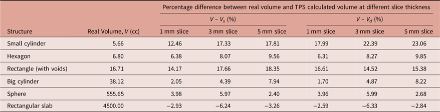
A total of six objects were scanned, contoured and named as per their geometrical shapes, namely small cylinder, hexagon, rectangle (with voids), big cylinder, sphere and rectangular slab. Real volume (V) of these objects are given in Table 1. Table 1 also contains the difference of real volume and volume recorded from structure table of TPS (V − V s ) as well as difference of real volume and volume recorded from DVH table (V − V d ) of TPS. It can be seen from Table 1 that volumes provided by Monaco TPS are overestimated contour volumes except for rectangular slab, where TPS is underestimating the volume. Also, most of the time V s is more accurate than V d . Moreover, volumes of contoured objects using CT of 1 mm slice thickness are more accurate than those contoured using CT of 3 and 5 mm slice thicknesses except for sphere, where most accurate volume corresponds to CT with 5 mm slice thickness.
Percentage difference between real volume (V) of structures used in this study and their volumes recorded from structure table (V s) and DVH statistics table (V d) of Monaco TPS for different computed tomography slice thicknesses

Correlation between volumes shown under different tables in Monaco TPS
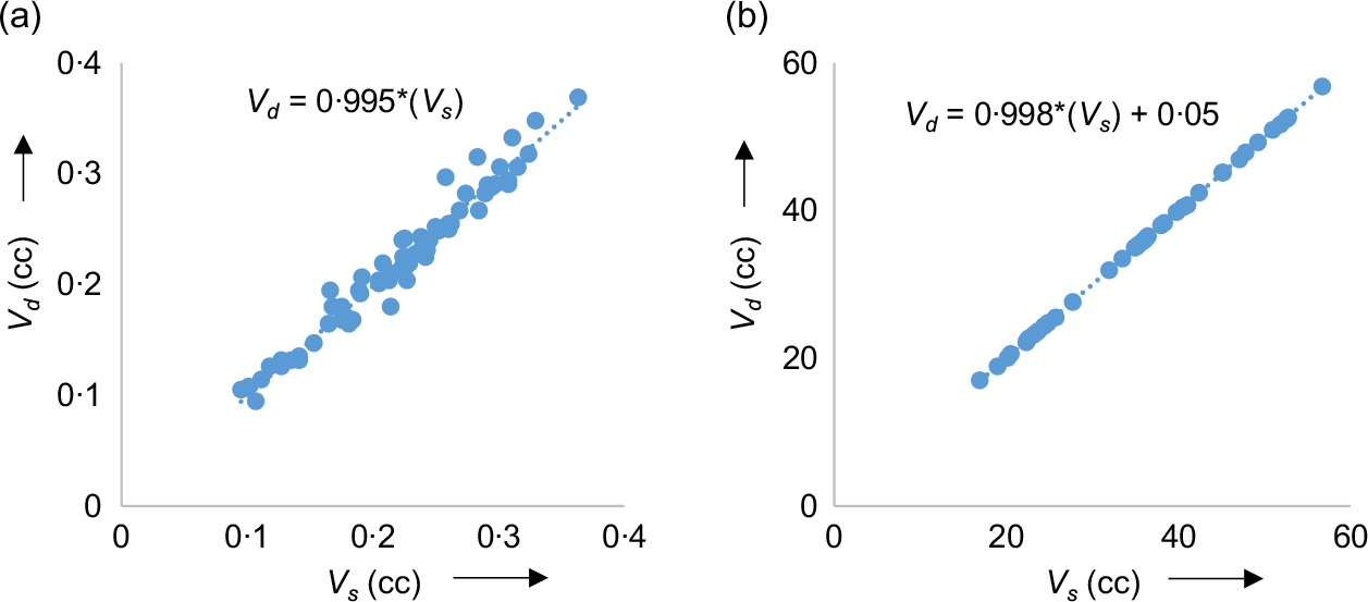
Figure 1 shows approximation curves for eye lens and brainstem along with equations of estimation after least square fitting (LSF) of data. ‘r’ was found to be ≈1 for all the categories of structure.
Approximation curves for (a) eye lens and (b) brainstem with respective equation of estimation.

Correlation between difference in volumes of OARs that exist in pair
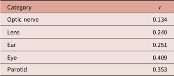
Values of ‘r’ for paired OARs are shown in Table 2. It is observed from Table 2 that the value of ‘r’ is far different from unity indicating poor correlation between differences in volumes of OARs.
Correlation coefficient ‘r’ for paired organs-at-risk

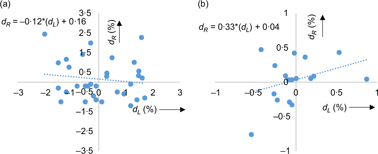
Approximation curves along with equation of estimation from LSF of the data for optic nerve and parotid are shown in Figure 2 for illustration purpose. After viewing the results of selected sample (of paired OARs), study was extended to theoretical population, which revealed that there does not exist any linear correlation between d L and d R as recommended by ρ = 0 at 99% CL from Student’s t test for all the paired structures. However, at 95% CL, the hypothesis that ρ = 0 was rejected for two organs (i.e., optic nerve and eye). Hence, for these organs, values of ρ at 95 and 99% CL was further calculated using Fisher’s Z transformation equation and are shown in Table 3.
Approximation curves for (a) optic nerve and (b) parotid with respective equation of estimation.

Theoretical population correlation coefficient ‘ρ’ for organs in pair

Discussion
The user guide of the Monaco TPS states that the volume of a structure depends on sampling resolution hence sampling resolution for volume calculation was not modified throughout the study. As discussed earlier, if an RT structure is not fully contained in structure set (SS), it will be misrepresented in DVH. This can be confirmed from ‘Is in SS’ column of DVH table. This includes the situation when a contour is drawn on the extreme end images (i.e., first or the last CT slice). This disclaimer by the vendor indicates that there may be different volumes of structure body shown under different tables and hence volume V d of RT structure body may be incorrect. Since external contour may be drawn from the first CT slice to the last CT slice and further might be much larger than the area of interest (calculation volume) in some cases. But in the clinical situation, we generally do not look DVH for body contour since it merely indicates existence of hot-spot (if any) without its spatial location. But in spite of being fully contained inside SS volume, V d of other contours were also found to be different than V s .
The Monaco TPS was found to overestimate structure volumes except for the structure with sharp corners. Although, it has very little or no impact in clinical situation since there does not exist sharp corner organs in the human body, also gross tumour has an irregular type of shape most of the time. Further, planning target volumes being an expansion of CTV cannot have sharp corners since expansion of a sharp corner object results in slightly curved corner object, not only in Monaco TPS but also in other TPSs. Hence, overall result suggests an overestimation of structure volume in clinical scenario with V s more accurate than V d . Although, it is V d which is used in DVH generation and hence used for final plan evaluation. Also, volumes of objects obtained using CT of 1 mm slice thickness were consistently found to be more accurate than those contoured using CT of 3 and 5 mm slice thickness. However, in the case of the sphere, there is an exception to this observation which may be due to its large volume.
Related to the study for obtaining the correlation between volumes of the same object (or organ) shown under different tables of Monaco TPS, the value of ‘r’ varies from −1 to +1 where negative and positive sign indicates inverse and direct correlation, respectively. Our results indicate perfect linear correlation between V s and V d (as expected) for all the structures. The same can be observed from Figure 1 and confirmed from equation of estimation (which is equation of a straight line) since nearly all the data points lie close to this straight line.
Although, r = −1 and r = +1 indicate perfect linear correlation, r = 0 indicates that there does not exist any linear correlation between two data sets and this is what we observed in case of the difference in volumes of OARs that exist in pair. Results of this study indicate that d L and d R are absolutely uncorrelated to each other for all the paired OARs. Even for parotid, we cannot conclude any linear correlation between d L and d R . This conclusion is further supported by approximation curve of parotid, where data points are not showing a linear pattern but rather scattered away from LSF equation of estimation in a random manner.
This study and its results are limited to a particular treatment planning system (i.e., Monaco TPS) only. As discussed earlier, prescribed point dose accuracy cannot be assured due to presence of statistical noise in MC calculated dose. Moreover, results have shown that volume of structures given under DVH table are less accurate than those shown under structure table. We suggest that while evaluating a clinical plan, particularly for serial organs with small volume, in addition to DVH evaluation, one must also review isodose distribution of tolerance dose near the OAR (e.g., 54 Gy isodose line near optic nerve) to make sure that the tolerance dose is not exceeded.
Conclusion
A systematic study was conducted to assess the accuracy of volumes provided by Monaco TPS in the structure table as well as in DVH table. The accuracy of DVH is of prime importance as it affects the decision process of selecting one plan over the other which has direct impact on the quality of patient treatment. Ideally, a single value of volume must be shown at all places in a TPS, but on the contrary, we came across ‘different values at different places’ situation in Monaco TPS. Even if, a TPS is doing such conversion, then it must utilise uniform method for all the structures, but it has been found that there does not even exist any linear correlation between the method(s), using which this TPS converts structure volume into DVH volume for a single organ that exists in pair in the same patient. Moreover, user guide of Monaco TPS does not clarify anything over this issue. Our study suggests that this issue must be reviewed and resolved by the supplier.
Acknowledgements
None.
Financial Support
This research received no specific grant from any funding agency, commercial or not-for-profit sectors.
Conflicts of interest
None.