Introduction
Despite advances in surgical techniques over recent decades, radiation technology and chemotherapy, the long-term survival of patients with advanced head and neck carcinoma has not improved significantly. There is a particular need to address lymph node metastasis, as this is the most important factor influencing the prognosis of head and neck cancer patients.
Host immune status also plays a significant role in the treatment and prognosis of patients with malignant tumours. Dendritic cells in the stroma of the cancer area and pericancer area are known to be the most important antigen-presenting cells, and their role has been studied in a variety of cancers. Failure of antitumour immunity may be due to a reduction in the function of such dendritic cells. Some dendritic cell phenotypic antigens have been reported to play an important role in the immune reaction process. However, no information has been previously been reported on dendritic cell phenotypic antigens in the cervical lymph nodes of head and neck cancer patients.
The S-100 protein is currently the most frequently used marker to detect dendritic cells, including immature dendritic cells (Langerhans' cells) and interdigitating reticulum cells.Reference Banchereau and Steinman1 Cluster of differentiation 1a glycoprotein is a marker of immature dendritic cells. Cells positive for this marker are mainly interdigitating reticulum cells, which are thought to have a greater engulfing activity, while also inducing B cell maturity. On the other hand, cluster of differentiation 83 glycoprotein is a phenotypic marker for mature dendritic cells, and cells positive for this marker generally exist in lymphatic organs. These two phenotypic antigens have different roles in stimulating helper T cells. Cluster of differentiation 1a glycoprotein positive dendritic cells assist the differentiation of helper T cells, while cluster of differentiation 83 glycoprotein positive dendritic cells are more likely to induce differentiation of Th0/Th2 cells.Reference Caux, Massacrier, Vanbervliet, Dubois, de Saint-Vis, ezutter-Dambuyant, Jacquet, Schmitt and Banchereau2
In the present study, we investigated dendritic cell positivity for S-100 protein and for cluster of differentiation 1a and 83 glycoproteins, in order to determine the status of dendritic cell phenotypic antigens in the cervical lymph nodes of patients with hypopharyngeal and laryngeal cancer. In addition, antibodies to CD45RO (a member of the CD45 family that includes CD45, CD45RA, and CD45RB, recognizes a 180-kilodalton (kd) isoform of the leucocyte common antigen (LCA)) were used to identify memory T cells in the lymph nodes, and the role of dendritic cells in tumour immunity was assessed.
Materials and methods
Approval for the study was obtained from the institutional review board of Kurume University School of Medicine (Kurume University, Kurume, Japan).
Patients and samples
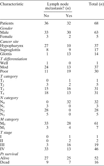
We studied 68 patients (37 with hypopharyngeal, 17 with supraglottic and 14 with glottic squamous cell carcinoma) who were operated upon at Kurume University Hospital between January 1998 and January 2003 (Table I). They comprised 63 men and five women, with ages ranging from 43 to 95 years (mean, 66 years). All patients underwent a total laryngectomy or pharyngectomy, either with or without a radical or modified radical neck dissection, according to their original diagnosis.
Patient characteristics, by lymph node metastasis

Data represent number of patients. Mod = moderately; T = tumour; N = node; M = metastasis; pt = patient
The patients' tumour–node–metastasis (TNM) stages were determined based on their clinical data and histopathological examination results, according to the 2002 TNM classification. None of the patients had previously received radiation therapy.
The resected cervical lymph nodes were separated from other surgically resected material as far as possible, and were fixed in 10 per cent formalin and processed for histopathological examination. From 36 patients diagnosed as positive for cervical lymph node metastasis, 178 nodes were obtained (58 with cancer cells and 120 without). From 32 patients diagnosed as node-negative but undergoing an elective neck dissection, 96 lymph nodes were obtained.
Immunohistochemical staining
Four-micrometer thick sections of lymph node were placed on glass microscope slides, deparaffinated and dehydrated in xylol with graded alcohol. After rinsing with phosphate-buffered saline, the slides were placed in hot 10 mmol/l citrate buffer with a pH of 6.0 for antigen retrieval and were then (except for the slides stained for S-100 antibody) heated in a microwave oven for 5 minutes. Using a Dako TechMate TM Horizon automated immunostainer (Dako, Glostrup, Denmark), the slides were incubated with primary antibodies for 60 minutes. Dendritic cell phenotypic antigens were detected using the following antibodies: anti-S-100 (1:800; Dako); anti cluster of differentiation 1a glycoprotein (1:25; Dako); and anti cluster of differentiation 83 glycoprotein (1:25; Novo, Castra Newcastle, UK). Anti CD45RO (1:50; Dako) was employed to detect memory T cells. The Dako EnVisionTM System-Peroxidase was used during immunohistochemical staining. Consecutive sections were also stained with haematoxylin and eosin for routine histopathological examination.
The extent of immunostaining was determined, using a light microscope (Nikon, Tokyo, Japan), by an investigator blinded to the patient's history. Dendritic cell numbers were counted in areas of greatest staining intensity. Counts were obtained in three high-power fields (original magnification ×400).
Statistical analysis
Study data are presented as mean ± standard deviation. Any correlations between immunohistochemical scores and patients' various clinicopathological features were assessed using Student's t-test and the chi-square test. Any correlations among the clinical parameters were evaluated by linear regression analysis and the Spearman test. A p value of less than 0.05 (two-sided) was considered to indicate statistical significance. Actuarial survival curves were generated using the Kaplan–Meier method. Patients were classified into different groups according to the median number of dendritic cells, dichotomised to above and below the median. The outcomes in different groups were compared using the log-rank chi-square test.
Results
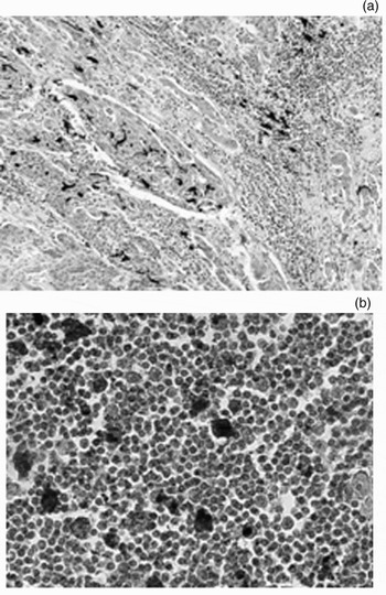
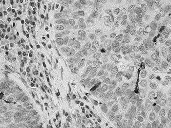
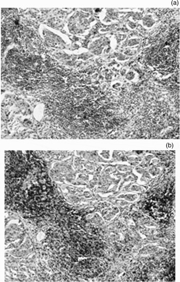
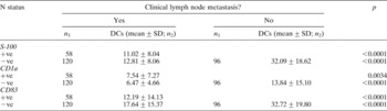
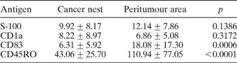
Dendritic cells positive for S-100 stained brown and had long ‘feet’ (Figure 1). They were detected in all lymph nodes and were mainly distributed around the lymphoid follicle, in both non-metastatic and metastatic cervical lymph nodes. A similar morphology was seen for cluster of differentiation 1a glycoprotein positive dendritic cells (Figure 2). However, less of these cells were present in peritumour and normal lymph nodes, compared with S-100-positive dendritic cells. Cluster of differentiation 83 glycoprotein positive dendritic cells were distributed diffusely in the lymphoid follicles of both metastatic and non-metastatic nodes (Figure 3). In order to assess the role of cluster of differentiation 83 glycoprotein positive dendritic cells, a consecutive section was stained with CD45RO antibody. CD45RO positive memory T cells were observed to be distributed around dendritic cells expressing cluster of differentiation 83 glycoprotein (Figure 3b). In general, the number of dendritic cells expressing each phenotypic antigen was significantly larger in non-metastatic lymph nodes compared with metastatic nodes (Table II). The observed number of cluster of differentiation 1a glycoprotein positive dendritic cells was generally less than that of dendritic cells positive for S-100 or cluster of differentiation 83 glycoprotein. In lymph nodes containing metastasis, cluster of differentiation 1a glycoprotein positive dendritic cells were predominantly detected within the cancer ‘nests’ (Table III). Mature dendrites stained by cluster of differentiation 83 glycoprotein antibody were prominent in non-metastatic lymph nodes. In metastatic lymph nodes, the degree of infiltration of cluster of differentiation 83 glycoprotein positive dendritic cells was significantly greater in peritumour areas than in cancer nests (in contrast to the distribution pattern of cluster of differentiation 1a glycoprotein positive dendritic cells).
Photomicrographs showing immunohistochemical distribution of S-100-positive dendritic cells in the cervical lymph node. (a) S100-positive dendritic cells visible in both the cancer ‘nest’ and peritumour areas, staining dark with long ‘feet’ (original magnification ×10). (b) S100-positive dendritic cells in normal cervical lymph node (original magnification ×40).

Photomicrograph showing immunohistochemical distribution of cluster of differentiation 1a glycoprotein positive dendritic cells within a cancer ‘nest’ in a cervical lymph node. Their morphology was similar to that of S-100-positive dendritic cells. (Original magnification ×100)

(a) Photomicrograph showing immunohistochemical distribution of cluster of differentiation 83 glycoprotein (CD83) positive dendritic cells; these were scarce in cancer ‘nests’ within the cervical lymph node but numerous in peritumour areas (original magnification ×10). (b) Photomicrograph showing immunohistochemical distribution of CD45RO positive T cells in a consecutive section to that shown in part (a); these cells were found to be distributed around dendritic cells expressing CD83 (original magnification ×10).

Expression of various dendritic cell phenotypic antigens in lymph nodes, by clinical lymph node metastasis

N status = histopathological node status of tumour; n 1 = number of lymph nodes examined; n 2 = number of DCs; DCs = dendritic cells expressing antigen; SD = standard deviation; CD1a = cluster of differentiation 1a glycoprotein; CD83 = cluster of differentiation 83 glycoprotein; +ve = cancer cell positive nodes (have cancer cell in lymph nodes); −ve = cancer cell negative nodes (no cancer cell in lymph nodes, but from metastasis patients)
Expression of dendritic cell phenotypic antigens and T Cell CD45RO in cancer ‘nest’ vs peritumour area, for metastatic lymph nodesFootnote *

Data represent numbers of positive cells, expressed as mean ± standard deviation, unless otherwise specified.
* n = 58. CD1a = cluster of differentiation 1a glycoprotein; CD83 = cluster of differentiation 83 glycoprotein; CD45RO = a member of the CD45 family that includes CD45, CD45RA, and CD45RB, recognizes a 180-kilodalton (kd) isoform of the leucocyte common antigen (LCA). The CD45 antigen is a protein tyrosine phosphatase
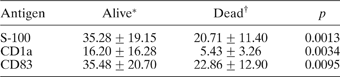
We also examined the distribution of dendritic cell phenotypic antigens according to patient survival status, with or without lymph node metastasis. In patients without lymph node metastasis, we observed a significantly greater number of dendritic cells positive for S-100 and for cluster of differentiation 1a and 83 glycoproteins, comparing surviving patients with deceased patients (Table IV). Likewise, in patients with lymph node metastasis, we also observed a significantly greater number of dendritic cells positive for S-100 and for cluster of differentiation 1a and 83 glycoproteins in cancer nests, comparing surviving patients with deceased patients. Even in the peritumour area of nodes from patients with lymph node metastasis, the numbers of dendritic cells positive for S-100 and cluster of differentiation 83 glycoprotein were greater, comparing surviving with deceased patients (Table V).
Expression of dendritic cell phenotypic antigens by survival status, in patients without lymph node metastasis
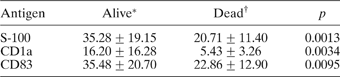
Data represent patient numbers, expressed as mean ± standard deviation, unless otherwise specified. *n=75; †n = 21. CD1a = cluster of differentiation 1a glycoprotein; CD83 = cluster of differentiation 83 glycoprotein
Expression of dendritic cell phenotypic antigens in cancer ‘nests’ vs peritumour area, by survival status, in patients with lymph node metastasis

Data represent numbers of positive cells, expressed as mean±standard deviation, unless otherwise specified. *n = 42; †n = 16
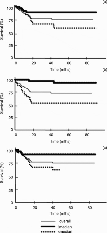
When patients were classified according to the median number of dendritic cells expressing the different types of phenotypic antigens (as having either the same or more than the median number (i.e. the majority group) or less than the median number (i.e. the minority group)), a significant difference in survival status was observed for each antigen: for S-100, χ2 = 6.46 and p < 0.05; for cluster of differentiation 1a glycoprotein, χ2 = 16.41 and p < 0.05; and for cluster of differentiation 83 glycoprotein, χ2 = 6.89 and p < 0.05 (Figure 4).
Analysis of patient survival according to median number of dendritic cells expressing various phenotypic antigens (overall three-year survival = 77.94 per cent, overall five-year survival = 75.71 per cent). (a) Survival curves according to the number of S-100-positive dendritic cells (median = 23; S100-positive dendritic cells ≥ 23/high power field (HP), n = 36; S100-positive dendritic cells < 23/HP, n = 32). (b). Survival curves according to the number of cluster of differentiation 1a glycoprotein positive (CD1a +) dendritic cells (median = 9; CD1a+ dendritic cells ≥ 9/HP, n = 37; CD1a+ dendritic cells < 9/HP, n = 31). (c) Survival curves according to the number of CD83+ dendritic cells (median = 19; CD83+ dendritic cells ≥ 19/HP, n = 33; CD83+ dendritic cells < 19/HP, n = 35). Mths = months

Discussion
Development of immunity against cancer cell infiltration involves three steps: (1) presentation of tumour-related antigen; (2) activation of specific T cells in the peritumour area; and (3) migration of the activated T cells to draining lymph nodes, where they are believed to activate the antitumour immune response.Reference Chang, Wright and Punnonen3–Reference Sogn5 Dendritic cells are considered to be the most potent antigen-presenting cells. In peripheral tissue, immature dendritic cells pick up and transport antigens to draining lymphatic tissue and then differentiate into mature dendritic cells. As a result, the antigen is efficiently presented, innate T cells are activated and these activated T cells play an important role in humoral immunity.Reference Schuler and Steinman6–Reference Steinman8 It has also been found that the grade of the dendritic cells infiltrating solid tumour is inversely correlated with histological differentiation, metastasis and prognosis.
The reason why tumour cells are not effectively identified and killed by T cells is that the soluble component released by tumour cells may act as a marker on dendritic cells, changing their molecular configuration and disabling their function of capturing and presenting tumour cells and activating T cells, and thus causing immune evasion.Reference Menetrier-Caux, Montmain and Dieu9, Reference Pawelec, Zeuthen and Kiessling10 Lymphatic tissue is generally thought to be the place where mature dendritic cells activate T cells. However, thus far there have been few reports on the delineated expression of dendritic cell phenotypic antigens within host lymph nodes. The infiltration of dendritic cells in head and neck tumours has also not been fully investigated.
The results of the present study showed that S-100-positive and cluster of differentiation 1a and 83 glycoprotein positive dendritic cells were present in every lymph node, although the site of infiltration varied. Specifically, S-100-positive and cluster of differentiation 83 glycoprotein positive dendritic cells tended to be scattered diffusely within non-metastatic lymph nodes and to be present in the peritumour area in metastatic lymph nodes, whereas cluster of differentiation 1a glycoprotein positive dendritic cells were predominantly distributed within the cancer nests of metastatic lymph nodes. These differences in distribution may be due to the differing functions of each phenotypic antigen. Cluster of differentiation 1a glycoprotein positive dendritic cells appeared to be more closely related to the engulfment process. Cluster of differentiation 83 glycoprotein positive dendritic cells, found outside the tumour nest, seemed to directly correlate with the dendritic cells' antigen-presenting capability, leading to T cell activitation.
In the lymph nodes obtained from laryngeal or hypopharyngeal cancer patients, whether metastatic or not, the level of cluster of differentiation 83 glycoprotein positive dendritic cells was found to be much higher than that of cluster of differentiation 1a glycoprotein positive dendritic cells. These results indicate that the dendritic cells existing in lymph nodes become mature and play an important role in the establishment of immune defence against tumours. Indeed, the detected CD45RO T cells appeared in clusters around cluster of differentiation 83 glycoprotein positive dendritic cells. This finding supports the idea that the lymph node is a very important site of tumour-specific immunity and that it is closely related to dendritic cells, with immature and mature dendritic cells playing different roles in different areas. Mature dendritic cells may release a variety of chemotaxins to intensify the stimulation of T cells by enhancing their aggregation; in this way, cluster of differentiation 83 glycoprotein positive dendritic cells may reflect the state of the general immune response, especially as regards antitumour immunity.Reference Wolenski, Cramer, Ehrlich, Steeg, Grossschupff, Tenner-Racz, Racz, Fleischer and von Bonin11, Reference Xia and Kao12
The grade of dendritic cells involved in lymph node infiltration has been proposed to correlate with the prognosis of patients with hepatic and gastrointestinal tumours.Reference Peh, Shek, Ng, Lo, Fan and Ngan13, Reference Tsujitani, Kakeji and Watanabe14 In head and neck squamous cell carcinoma, the distribution of dendritic cells has mainly been investigated in the mucosal tissue of laryngeal tumours.Reference Maluccio, Rao, Sharma, Lagman and Suthanthiran15–Reference Dong, Li, Zhu, Yu, Lu, Sun and Wang17 A recent report by Yilmaz et al. indicated that a higher degree of Langerhans cell infiltration was significantly associated with a lower rate of cervical lymph node metastasis, longer disease-free survival, less loco-regional recurrence and less clinical node-positivity.Reference Karakök, Bayazit, Ucak, Ozer, Kanlikama, Mumbuc and Sari18, Reference Yilmaz, Gedikoglu, Celik, Onerci and Turan19 A predominantly dendritic cell infiltration, with prognostic significance, was also identified in tumour-containing sentinel lymph nodes in cases of breast cancer.Reference Nancy, Aysegul, Kelly and Elizabeth20 However, there have been few reports regarding the effect of such changes on the prognosis of patients with head and neck tumours. The current study supports such an effect; a greater degree of infiltration of S-100-positive and cluster of differentiation 1a and 83 glycoprotein positive dendritic cells was found in the lymph nodes of patients without metastases and in patients with better survival. Further studies are now underway regarding the role of dendritic cells, not only within lymph nodes but also at the primary tumour site.
Acknowledgements
This work was supported in part by a Grant-in-Aid from the Ministry of Education, Science and Culture, Japan, and the Society for the Promotion of International Oto-Rhino- Laryngology. And also be supported in part by Shanghai Pujiang fellowship foundation (07pj14064).