Introduction
Advances in neonatal intensive care have brought about dramatic improvements in the prognosis of preterm infants.Reference Itabashi, Horiuchi and Kusuda 1 Preterm birth occurs at a time of rapid fetal growth and nutrient accumulation, and establishing postnatal nutrition in preterm infants is therefore essential. The time of introduction and the rate of advancement of enteral milk feeds for preterm infants can influence prognosis by, for example, reducing the risk of necrotizing enterocolitis as well as improving subsequent neurodevelopmental and growth outcomes. Early nutritional status is an important determinant of the risk of developing metabolic syndrome later in life.Reference Singhal, Farooqi and O’Rahilly 2 – Reference Ten and Maclaren 4
The two known incretins, glucagon-like peptide-1 (GLP-1) and glucose-dependent insulinotropic polypeptide (GIP), are produced in duodenal K cells and intestinal L cells, respectively;Reference Baggio and Drucker 5 they are released in response to enteral intake.Reference Ambati, Duan and Hartzell 6 Although there is some information on incretin levels during the neonatal period in preterm infants,Reference Kawamata, Suzuki and Yada 7 , Reference Kawamata, Suzuki and Yada 8 these studies did not assess insulin secretion. The present study, therefore, assessed serum GIP and GLP-1 levels in the early postnatal period in order to assess the influence of prematurity and insulin on the relationships of these incretins with insulin secretion.
Methods
Subjects
A pilot study was conducted in the neonatal intensive care unit of Juntendo University Hospital, Tokyo, Japan. A total of 30 (15 female and 15 male) singleton preterm infants who were born at the hospital between November 2011 and November 2014 were included in the study. Infants with major congenital abnormalities or metabolic disorders and those born to women with diabetes requiring insulin and/or dipeptidyl peptidase IV (DPP-IV) inhibitors, chronic hypertension or intrauterine infections were excluded to minimize the influence of maternal glucose–insulin metabolism. Gestational age was estimated from the mother’s last menstrual period, and confirmed through fetal ultrasound measurements. Sex- and gestational age-independent s.d. scores for birth weight were calculated by comparing with the Japanese standard birth weight curve.Reference Itabashi, Miura, Uehara and Nakamura 9
According to our nutritional protocol, feeding was typically initiated within the first 8 h after birth (20 ml/kg divided over eight feedings per day), and breast milk was the preferred type of feeding. However, when breast milk was unavailable due to mother’s unwillingness or inability to provide breast milk, the infants received a cow’s milk-based premature formula (GP-P®; Morinaga Milk Industry Co., Ltd, Tokyo, Japan). Milk intake was increased by ~20 ml/kg daily to 120 ml/kg, at which time the human milk fortifier (HMS-1, nutrient composition/1 g: calories 3.37 kcal, protein 0.26 g; Morinaga Milk Industry) was added to human milk. The manufacturer recommends that 30 ml of human milk be mixed with 0.8 g of HMS-1 before enteral feeding.
The infants were divided retrospectively into two groups according to the gestational age: the early group (12 infants; six boys and six girls) included infants with a gestational age of <30 weeks, and the late group (18 infants; nine boys and nine girls) included infants with a gestational age of ε 30 weeks.
This study was approved by the Institutional Review Board of Juntendo University Hospital (14-164). Written informed consent was obtained from parents of infants before study participation.
Biochemical measurements
Heel lancing was used to draw blood in the infants at birth (before the first glucose infusion or first feeding) and at 2 and 4 weeks of age (before feeding, following at least 2 h of fasting). Blood glucose concentrations were measured immediately using OneTouch Ultra Glucometer (Johnson & Johnson, New Brunswick, NJ, USA). Blood samples (~600 μl) were collected in BD Microtainer brand tubes with clot activator (Ref No. 365967), but not with protease inhibitors. Samples were centrifuged at 6000 g for 90 s. Serum was immediately separated by centrifugation at 4°C and stored at 80°C until further use.
We measured serum GIP [total; form (1–42) and form (3–42)] and GLP-1 [active form (7–36) amide; major form in circulation] levels using the suspension beads array system, Bio-Plex 100 system with Bio-Plex Manager 6.1 (Bio-Rad Laboratories, Hercules, CA, USA). Bio-Plex filter plates were pre-wetted with buffer before the addition of samples. Antibody-coupled beads for GIP and GLP-1 were premixed before the performance of assays according to the manufacturer’s protocol. After loading of the bead solutions and samples, plates were incubated for 30 min at room temperature with agitation. After washing three times, plates were incubated with detection antibodies for 30 min, followed by streptavidin-PE for 10 min. Beads were resuspended by agitation. GIP and GLP-1 were measured using the Bio-Plex 100 system.
Serum insulin levels were measured with electrochemiluminescence immunoassays using a Modular Analytics E170 Analyzer (Roche Diagnostics K.K., Tokyo, Japan). The quantitative insulin sensitivity check index (QUICKI) was used to estimate insulin sensitivity and calculated according to the following formula: QUICKI=1/[log plasma insulin (μU/ml)+log plasma glucose (mg/dl)].
Statistical analyses
Results are presented as mean±s.d.. Differences between the groups were analyzed using the Mann–Whitney’s U-test. Difference from baseline was tested by two-way repeated measures ANOVA and the post-hoc Games–Howell test. Correlations among variables were evaluated using Spearman’s rank correlation coefficient analysis. Values of P<0.05 were considered to be statistically significant. All statistical analyses were performed using StatView 5.0 software (Abacus Concepts, Berkeley, CA, USA).
Results
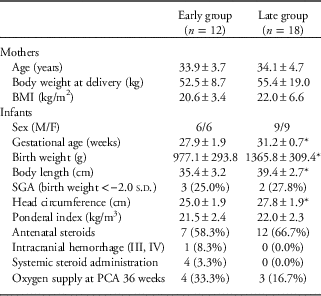
Clinical characteristics and anthropometric indices of the mothers and infants included in this study are summarized in Table 1. Significant differences were observed in anthropometric indices at birth between the early and the late groups. The mothers were 25–42 years of age, and none were vegetarian.
General characteristics of mothers and preterm infants at birth

BMI, body mass index, M/F, male/female; SGA, small for gestational age, PCA, post-conceptional age.
Data are presented as the mean±s.d. or the number (percentage) of subjects.
*P<0.01 compared with the early group.
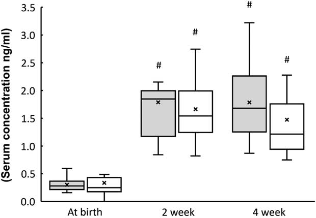
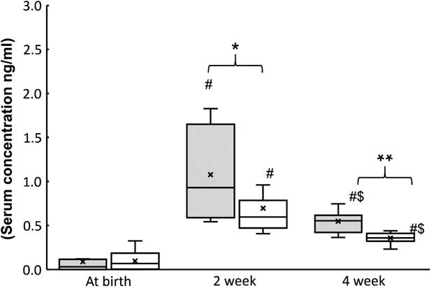
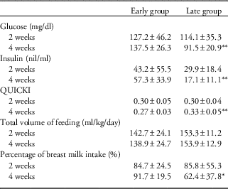
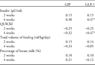
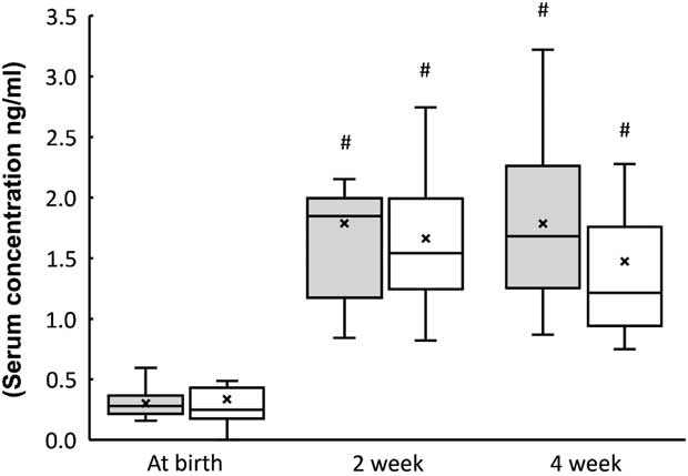
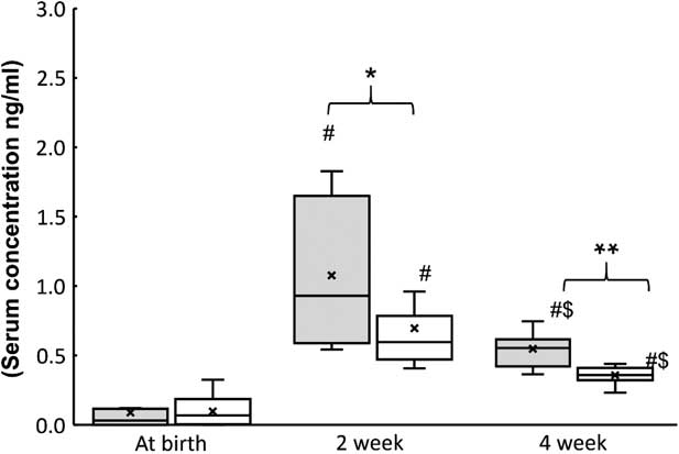
The levels of GIP at 2 and 4 weeks in both groups were significantly higher than those at birth, but intergroup differences were not significant (Fig. 1). The levels of GLP-1 levels at 2 weeks of age were significantly higher than those at birth and 4 weeks of age. The levels of GLP-1 at 2 and 4 weeks of age were significantly higher in the early group (1.08 and 0.55 ng/ml) than those in the late group (0.70 and 0.36 ng/ml) (Fig. 2). Serum insulin level was significantly higher and QUICKI was lower in the early group at 4 weeks of age than those in the late group (Table 2). At 4 weeks of age, GLP-1 levels correlated with QUICKI (P<0.01) and the serum insulin level (P<0.01) in all 30 infants (Table 3).
Postnatal change in glucose-dependent insulinotropic polypeptide levels in preterm infants (mean±s.d.). #P<0.01 compared to at birth.
Postnatal change in glucagon-like peptide-1 levels in preterm infants (mean±s.d.). *P<0.05, **P<0.01, #P<0.01 compared to at birth; $P<0.01 compared to 2 weeks.
Glucose, insulin, quantitative insulin sensitivity check index (QUICKI) and feeding data at 2 and 4 weeks of age

Data are presented as the mean±s.d.
*P<0.05, **P<0.01 compared with the early group.
Correlations between glucose, quantitative insulin sensitivity check index (QUICKI), feeding data and glucose-dependent insulinotropic polypeptide (GIP), glucagon-like peptide-1 (GLP-1) at 2 and 4 weeks of age in preterm infants

*P<0.05.
Discussion
To the best of our knowledge, this is the first study to assess the relationship of incretin levels and insulin secretion in preterm infants in the early postnatal period. The results indicate that serum GLP-1 levels are associated with insulin secretion and are higher in more premature babies, that is, born before 30 weeks.
In this study, serum GLP-1 levels peaked at 2 weeks of age and serum GIP levels significantly increased from 2 weeks of age and were consistently high until 4 weeks of age. Previously, Kawamata et al.Reference Kawamata, Suzuki and Yada 8 reported that fasting serum GIP and GLP-1 levels in preterm infants peaked at 3–4 weeks after birth, and the peak levels were much higher than the normal range in adults. Other studies also reported similar findings.Reference Padidela, Patterson, Sharief, Ghatei and Hussain 10 , Reference Berseth, Nordyke, Valdes, Furlow and Go 11 Kawamata et al.Reference Kawamata, Suzuki and Yada 8 speculated that these differences between infants and adults in GIP or GLP-1 levels result from a difference in the amount of enteral feeding. In preterm infants, enteral feeding is usually every 3 h, which may stimulate GIP and GLP-1 secretion to a greater level in neonates than in older children and adults. Padidela et al.Reference Padidela, Patterson, Sharief, Ghatei and Hussain 10 also reported that the high plasma GLP-1 levels in newborn infants might reflect immaturity of the DPP-IV enzyme system or reduced clearance by the kidneys. A previous murine study reported very low levels of intestinal DPP-IV enzyme activity at 2 weeks of age, before significantly increasing until 2 to 3 months of age.Reference Detel, Baticic and Varljen 12
The primary actions of GLP-1 on glucose homeostasis involve enhancement of glucose-dependent insulin secretion and subsequent reduction of hepatic glucose production.Reference Hvidberg, Nielsen, Hilsted, Orskov and Holst 13 Other studies also provided evidence of a beneficial effect on ®-cell proliferationReference Buteau, Foisy and Rhodes 14 , Reference Buteau, Foisy, Joly and Prentki 15 and reduced ®-cell apoptosis.Reference Farilla, Hui and Bertolotto 16 GIP also has an insulinotropic effect on the ®-cell in healthy adult subjects.Reference Flint, Raben, Astrup and Holst 17 GIP and GLP-1 also play important roles in regulating lipid metabolism and storage partly through their effects on insulin secretion.Reference Ambati, Duan and Hartzell 6 , Reference Small and Bloom 18 Elevated insulin levels in infants stimulate glycogen storage in the liver, and increase lipid synthesis and fat accumulation. In this study, GLP-1 secretions may have been higher from intestinal cells in the more premature infants. GLP-1 stimulates insulin secretion from ®-cells and may affect fat accumulation (catch-up fat) in the early postnatal period. In fact, mean body weight and ponderal index at discharge (mean 39.4 weeks of corrected age) in the early group was significantly (P<0.01) higher than that in the late group (30.8 v. 26.8). High GLP-1 as well as high insulin and low QUICKI levels in the early postnatal period in very preterm infants may contribute to obesity in the future. We assessed the influence of not only feeding volume, but also feeding types. Lucas et al.Reference Lucas, Boyes, Bloom and Aynsley-Green 19 showed that in 6-day-old term infants, formula induced greater insulin and GIP responses than breast milk, but similar postprandial blood glucose. In this study, the levels of GIP and GLP-1 did not correlate with the percentage of breast milk intake at 2 and 4 weeks of age. Serum levels of glucose and insulin were significantly higher, and the QUICKI index was significantly lower, in the early group at 4 weeks of age. These findings may be attributable to the greater insulin resistance of earlier preterm infants, as previously described by Wang et al.Reference Wang, Divall and Radovick 20
The present study had some limitations. The sample size was relatively small. The influence of maternal glucose and insulin metabolism could not be eliminated. We were unable to measure incretin levels in term infants. Further studies with more subjects are required in the future in order to confirm the present results.
In conclusion, the results of the present study suggest that enteral feeding to premature intestine may be associated with GLP-1 secretion. GLP-1 production in preterm infants is related to the stimulation of insulin secretion in early postnatal period.
Acknowledgments
The authors are indebted to Mutsuko Hara, Juntendo Atopy Research Center, for her technical support.
Financial Support
This work was supported in part by JSPS KAKENHI Grant Number (22791039).
Conflicts of Interest
None.
Ethical Standards
This study was approved by the Institutional Review Board of Juntendo University Hospital (14-164).