Implications
Hair fibres are products with value depending on quality and quantity produced. We require information to optimise breeding and husbandry of fibre-producing animals. This includes better understanding of the hair follicles in skin, which produce fibre. This report reviews results from in vitro studies on follicles removed from skin of sheep and goats. It describes how: (i) variation in fibre growth by different animal types relates to cells and protein production, (ii) important regulating chemicals affect follicle behaviour and (iii) certain nutrients affect fibre growth and viability of follicles. Such results advise on selection and nutrition to optimise fibre production.
Introduction
Despite the commercial importance of animal fibres to farmer-producers, processors and end-users, important gaps remain in knowledge of underlying biology of production by hair follicles. These follicles are located in skin and have properties in common with other components of the mammalian integument (outermost layers of tissue of the body). The current background to research on hair follicles in commercial animal species indicates limited funding and associated activity in Europe and elsewhere including Australia (Rogers, Reference Rogers2006). This is despite evidence of some diversification of study on the science of integumental tissues of farm animals into areas such as foot lameness biology in cattle (Galbraith and Scaife, Reference Galbraith and Scaife2008). The production of hair by follicles, which have epidermal and dermal constituents similar to other tissues of the integument, is a complex process. This has also been the subject of review in this symposium (Galbraith, Reference Galbraith2010). It has parallels in fundamental regulatory mechanisms involving stem cells, and programmed cellular proliferation and differentiation, which occurs in typical development from conception to maturity of multicellular animals. The production of a commercial fibre product requires the anatomical construction of the follicle pre- and post-natally and the initiation and maintenance of active growth of the hair shaft and supporting structures. These processes, and those including subsequent follicle cycling, are regulated by a range of signalling systems derived from external environment interacting with those of systemic and locally determined origins. Examples of signalling molecules included in this report are the hormones melatonin and prolactin (Santiago-Moreno, Reference Santiago-Moreno2004) and growth factors such as epidermal growth factor (EGF) (Panaretto et al., Reference Panaretto, Leish, Moore and Robertson1984). The nutritional status of the animal is also important (i) indirectly, in affecting systemic and locally produced hormonal and other signalling systems which act on the hair follicle and (ii) directly in providing chemical components required for effective function of enzyme systems and for synthesis of follicular structures (Galbraith, Reference Galbraith2000; Hynd, Reference Hynd2000). Nutritional influences include minerals and vitamins, which contribute to catalytic function and amino acids necessary for synthesis of the range of proteins expressed in the follicle. These include enzymes and proteins with roles in the structure of cell membranes, and cystoskeleton particularly in epidermal keratinocytes of hair shaft and fibroblasts and extracellular matrix in the dermis. The essential water soluble vitamin biotin is important because of its general role in providing a prosthetic group in biochemical activities such as carboxylation in carbohydrate metabolism and synthesis of long chain fatty acids (Wolf and Raetz, Reference Wolf and Raetz1983). Interest in the sulphur-containing amino acid methionine arises from knowledge of its roles in function in mammalian integument. These include polyamine synthesis, protein synthesis and the provision of cysteine, which contributes essentially, via disulphide bonding, to structural integrity of cytoskeleton in keratinocytes of tissues such as hair and hoof horn (Marshall and Gillespie, Reference Marshall and Gillespie1989; Galbraith and Scaife, Reference Galbraith and Scaife2008).
In terms of available in vitro methodology to investigate hair follicle biology, the approach developed by Philpott et al. (Philpott et al., Reference Philpott, Green and Kealey1990; Kealey et al., Reference Kealey, Philpott and Guy1997) for human and rodent skin is of particular interest since it permits direct and dynamic investigation of behaviour of isolated intact follicles. Similar methodology has been successfully applied to investigations of sheep wool follicles (Ansari-Renani and Hynd, Reference Ansari-Renani and Hynd2004; Bond et al., Reference Bond, Wynn and Moore1996). This current study reviews selected results from the application of this in vitro technique in our research programmes that studied the chemical regulation and nutrition of pelage secondary hair follicles from major fibre-bearing ruminant animal species. The studies compared growth and protein synthesis in follicles of different genetic origins and the responses to signalling molecules, melatonin, prolactin and EGF and nutrients, biotin and sulphur amino acids in isolated follicles of sheep and Angora and cashmere-bearing goats.
Collection and preparation of hair follicles
Systems for collection and incubation of follicles are based on the techniques described by Ibraheem et al., Reference Ibraheem, Galbraith, Scaife and Ewen1994 and Tahmasbi et al., Reference Tahmasbi, Galbraith and Scaife2007a. Briefly, small samples of skin (ca. 1.0 cm2) are collected post-mortem, or under local anaesthetic, by ethically approved and licensed technique. Individual, or groups of, anagen secondary follicles are separated at the level of the sebaceous gland by micro-dissection and allocated to multi-well plates containing 500 μl Williams’ E medium supplemented typically with 1.0 mM l-glutamine, insulin (10 mg/l), hydrocortisone (10 μg/l), penicillin (64 mg/l) and streptomycin (100 mg/l). To these are added chemical regulators and test nutrients and the follicles are maintained, free-floating, at 37°C in an unhumidified atmosphere of 5% CO2/95% air. Fresh media are provided at 24 h intervals and measurements made on follicles removed from media, washed carefully and subjected to the required analytical procedures. Additional details are provided as footnotes to figures and tables. A typical pattern of elongation for an anagen sheep wool follicle during a 96 h time period is shown in Figure 1.
Image of an isolated mohair follicle incubated in Williams E medium with changes in length (elongation), shown from 0 h and at 24 h and 96 h (modified from Tahmasbi et al., Reference Tahmasbi, Galbraith and Scaife2007a). Magnification × 25.

Comparison of growth and protein synthetic activity in cashmere and mohair follicles
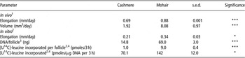
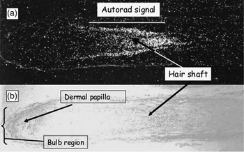
A useful prerequisite to investigating mechanisms responsible for differences in fibre production between, or within genotypes, is to define physical properties and synthetic capacities of active hair follicles. Data provided in Table 1 show differences in hair shaft elongation and volume of clean fibre produced in vivo by representative cashmere and mohair follicles with average diameters of 18.4 and 33.7 μm (s.e.d. = 3.0) respectively. Both rates of elongation (+0.28 fold) and volume (+4.2 fold) were greater for mohair. These effects were associated with a greater cellularity as shown by greater DNA concentration per follicle (+4.7 fold) and greater deposition of [U-14C]-leucine-labelled perchloric acid precipitable protein per follicle (+9.0 fold) and per ug of DNA (+2.0 fold). The results suggest that the greater production of mohair compared with cashmere may be attributed (i) to a greater number of follicular cells and (ii) to apparently greater protein synthetic activity per cell. More precise description of sites of synthesis and migration of labelled amino acids in deposited proteins in follicle cells may be obtained by use of autoradiography such as shown in Figure 2 for mohair. This image shows the location of [3H]-leucine after 72 h incubation following a pulse of exposure at time 0 for 1 h. The signal is concentrated distal to the bulb region predominantly in hair shaft and inner root sheath. It is associated with movement of cells from the basal layer of the matrix, which exhibits strong signalling after 1 h (not shown). Similarly, sites and degree of cellular proliferation as evidenced by synthesis of DNA may be further investigated using isotopically labelled methyl thymidine or BrdU (Figures 3 and 5) or other nuclear staining systems. Quantification of number and size of proliferative cells in addition to identification of positioning of these cells has potential to aid identification and phenotypic properties of follicles of different origins. Such an approach was recently used to define cellularity of basal keratinocytes by Galbraith et al. (Reference Galbraith, Rae, Omand, Hendry, Knight and Wilde2006) for epidermis of the bovine claw.
Comparison of growth and protein synthetic activity (mean values with pooled s.e.d.) in vitro of secondary follicles of Angora and cashmere-bearing genotypes

1Physical measurements are made on 30 random fibres per animal from mid-rib patch sample in time period from ca. 2 months preceding slaughter from June to July at 57 degrees North.
2Mid-rib pelage intact follicles collected at slaughter with numbers per treatment for cashmere = 24 (4 animals × 6: 15 follicles per replicate) and for Angora goats = 18 (3 animals × 6: 10 follicles per replicate). Elongation measurements of viable follicles for 120 h were made for cashmere = 21 (3 animals × 7) and for Angora = 30 (2 animals × 15).
3DNA was measured by the method of Fiszer-Szafarz et al. (Reference Fiszer-Szafarz, Szafarz and De Murillo1981).
4Protein deposition was measured in freshly isolated follicles after incubation and homogenisation by the incorporation of 18.5 MBq[U-14C]-leucine/500 μl into the perchloric acid-precipitable protein containing fraction after 3 h incubation. Horizontal comparisons were made by Student’s t-test (Lee and Galbraith, unpublished).
(a) Autoradiographic image of a mohair follicle pulsed with [H3]-leucine (370 MBq/0.5 ml) at time 0 h for 1.0 h, rinsed three times in media containing 10 mM leucine and incubated in an isotope-free medium for a further 71 h; dark field illumination: (b) light illumination, haematoxylin. wax-embedded sections at 5 μm thickness. Autoradiographs prepared in Ilford K5 dipping emulsion with development after 2 weeks. Magnification × 100 (Lee and Galbraith, unpublished).

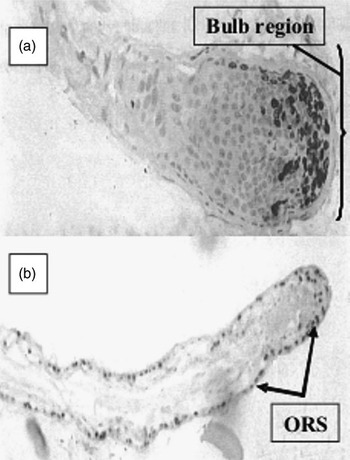
Effect of epidermal growth factor (0 (a) or 10 μg/l (b)) for 0 to 72 h on incorporation of BrdU incubated for hours 72 to 78, by isolated mohair follicles. Wax-embedded sections at 5 μm thickness. BrdU detected by imunohistochemistry (Mouse anti-BrdU monoclonal antibody: rabbit anti-mouse labelled streptavidin-biotin-horseradish peroxidase complex (Dako) with diaminobenzidine (Sigma) visualisation of signal. Magnification × 200 (a) and × 100 (b). ORS = outer root sheath (Souri and Galbraith, unpublished).

Molecular mechanisms responsible for the observed differences in phenotype and synthetic capacity of hair follicles and production of differences between the cashmere-bearing and Angora genotypes remain poorly understood.
Response of isolated mohair and cashmere follicles to melatonin and prolactin treatment
Mechanisms which signal changes in photoperiod to physiological processes in receptive animals are known to include melatonin and prolactin. Such processes include the growth of hair. While there is good evidence to conclude a role for prolactin via fibroblasts in the dermal papillae of, for example, the wool follicle in sheep (Nixon et al., Reference Nixon, Ford, Wildermoth, Craven, Ashby and Pearson2002), direct effects of melatonin, as opposed to modulations of other hormonal systems, are now becoming better understood (Fischer et al., Reference Fischer, Slominski, Tobin and Paus2008).
Again, studies on isolated hair follicles have provided a model to test for such direct effects (Table 2). Results obtained from such an approach showed significant increases in both hair shaft elongation and apparent protein synthesis compared with unsupplemented control media for cashmere follicles for prolactin (200 μg/l) and melatonin (150 ng/l). Positive responses for mohair were also obtained for prolactin and for melatonin, although at a concentration of 50 ng/ml, (not shown). These results, at physiological concentrations, suggest that both of these hormones may contribute directly in both genotypes for the regulation of growth and protein synthetic activity in the follicles studied.
Effect of prolactin and melatonin on mean values (with pooled s.e.d. or ±s.e.m.) for elongation (mm) and protein synthetic activity in mohair and cashmere secondary hair follicles in vitro

Follicles (prepared and treated essentially as described for Table 1) were incubated for 48 h in culture containing prolactin (ovine: NIDDK-oPrl-19: NIH, Baltimore, USA) and melatonin (Sigma) and measurements of elongation made. Follicles were then placed in fresh media containing [U-14C]-leucine for a further 3 h followed by washing, homogenisation and measurement of incorporation of radiolabel in the perchloric acid-precipitable protein fraction.
1[U-14C]-leucine incorporation (pmoles/μg DNA per 3 h). Horizontal comparisons were made by Student’s t-test against the 48 h control.
*Significance at P < 0.05 (Lee, Ibraheem and Galbraith, unpublished; Lee, Galbraith and Scaife, 1994).
Response of isolated mohair follicles to EGF
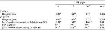
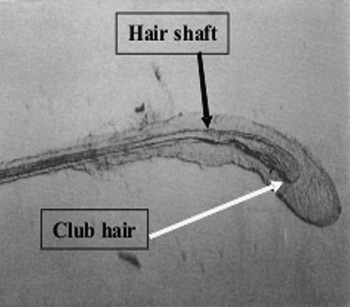
EGF is one of a number of signalling molecules which are known to affect growth of fibre in sheep and has been used as a systemic treatment to assist in harvesting of wool (Rogers, Reference Rogers2006). Such a treatment using mouse-derived EGF was tested at two concentrations in mohair follicles (Table 3). Results obtained showed significant positive responses after 48 h incubation in hair shaft elongation and incorporation of [U-14C]-leucine per follicle at 1.0 μg/l, but not 10.0 μg/l concentration. There was evidence of a greater [U-14C]-leucine incorporation at the 10.0 μg/l concentration when measured per μg follicular DNA and in the context of an apparently reduced concentration of DNA compared with that recorded for 1.0 μg/l EGF treatment. These responses were associated (i) with a greater apparent cell proliferation in the outer root sheath with reductions in matrix when measured by BrdU incorporation (Figure 3a and b) and (ii) increased incidence of club hair formation (Figure 4). These results are consistent with those described by Philpott and Kealey (Reference Philpott and Kealey1994) in human hair follicles with the response resembling the anagen-catagen transition described in sheep follicles (Panaretto et al., Reference Panaretto, Leish, Moore and Robertson1984). Responses to EGF in sheep wool follicles have been reported by Bond et al. (Reference Bond, Wynn and Moore1996) and Ansari-Renani and Hynd (Reference Ansari-Renani and Hynd2004). The data presented thus indicate concentration-dependent effects on mohair follicles and confirm, and taking into account the activity of mouse-derived growth factor, an apparent conservation of biological activity of EGF across species.
Effect of EGF on mean values (with pooled s.e.m.) for growth and protein synthetic activity of isolated mohair secondary follicles in vitro

EGF = epidermal growth factor.
Follicles were isolated and prepared as described as for Table 1. Approximately 70 anagen follicles per treatment were incubated for 48 h in the presence of mouse EGF (Sigma) at final concentrations of 0, 1.0 and 10.0 μg/ml. Measurements for DNA and protein deposition were determined as for Table 1. Values in the same row with dissimilar subscripts are significantly different (P < 0.05) (Adapted from Souri et al., Reference Souri, Galbraith and Scaife1997).
Isolated mohair follicle following treatment with 10.0 μg/l epidermal growth factor for 72 h. Magnification × 40 (Souri and Galbraith, unpublished).

Response of isolated sheep hair secondary follicles to supplementation with biotin
Biotin deficiency in farm animals is characterised by a range of lesions in skin and integumental products such as hair and hoof horn. The vitamin is water soluble and may be provided by microorganisms in the digestive tract such as occurs in ruminants and which complicates the determination of relationships between supply in the diet and requirement of individual tissues. While data are available to describe its role in carboxylation and other metabolic processes, information is limited which relates quantitative biotin supply and function in hair follicles of fibre-producing species (Tahmasbi et al., Reference Tahmasbi, Galbraith and Scaife2007a and Reference Tahmasbi, Galbraith and Scaife2007b). The study reported here again utilises isolated secondary follicles as a means of studying targeted responses to biotin and focuses on important properties of maintenance of viability, elongation, apparent protein synthesis and ATP production. Follicles were obtained by approved technique from skin of male castrate Suffolk sheep aged approximately 1 year, and prepared, and incubated in the presence of varying concentrations of biotin. The results in Table 4 show essentially progressive reductions in viability in all treatments with increasing time of incubation. However, significant effects of both 0.25 and 0.50 mg/l were recorded in maintaining viability and in stimulating production of ATP (Table 5), with the higher concentration being most effective. Rates of elongation of viable follicles were not significantly affected by biotin supplementation (Table 5). Comparisons of treatments indicated trends towards increased DNA concentration per follicle. There was also evidence of increases in apparent protein synthesis per follicle although not as a function of DNA concentration (Table 6). These results clearly indicate an important role for biotin in the maintenance of viability and potentially in synthesis of proteins in the follicles studied. Other study (not shown) (Tahmasbi et al., Reference Tahmasbi, Galbraith, Scaife and Finn1999) has shown reductions in apoptosis and increases in the apoptosis inhibitor Bcl-2 in mohair follicles in biotin-deficient media when supplemented with biotin. These results may also be considered in the context of responses to biotin supplementation in vivo. For example, combed hair loss, indicative of loss of viability of follicles, was increased in Angora and cashmere-bearing goats when provided with liquid diets deficient in biotin supply and when compared with biotin-supplemented controls (Tahmasbi et al., Reference Tahmasbi, Galbraith and Scaife2007b).
Effect of biotin concentration in incubation media on mean values for proportion of isolated sheep follicles remaining viable for up to 120 h in vitro

Follicles were collected as for experiment 1 in the time period from September to February. Follicles were incubated in the absence or presence of biotin for up to 120 h. Results are expressed as proportion of viable follicles (showing elongation in previous 24 h); n = 3 sheep: 30 to 40 follicles/treatment per sheep.
Comparison of means made by χ 2 test with χ 2 value in brackets (d.f. = 1); *=P < 0.05, **=P < 0.01 ***=P < 0.001 (Adapted from Tahmasbi et al., Reference Tahmasbi, Galbraith and Scaife2007a).
Mean values (±s.e.m.) for elongation of, and ATP concentration in, sheep wool secondary follicles incubated in culture medium containing biotin at concentrations of 0, 0.25, 0.5 or 1.0 mg/l

Follicles were prepared as from Table 4; n = 3 sheep and 4 to 6 replicates of each containing 4 follicles. ATP was analysed by the method of Spielmann et al. (Reference Spielmann, Jacob-Muller and Schulz1981). Data analysed by one-way ANOVA values in the same row with dissimilar subscripts are different (P < 0.01) (Adapted from Tahmasbi et al., Reference Tahmasbi, Galbraith and Scaife2007a).
Effect of biotin concentration (mean ± s.e.m.) on DNA concentration and apparent protein deposition in isolated sheep secondary follicles

Follicles (n = 4 sheep; 7 assays/treatment; 10 follicles/assay) were pre-incubated for 48 h in medium containing biotin and incubated for a further 4 h with 18.5 MBq/ml [U-14C]-leucine with incorporation and DNA concentration measured as for Table 1.
Statistical analysis by one-way ANOVA. Values in the same row with different superscripts are significantly different (P < 0.05) (Adapted from Tahmasbi et al., Reference Tahmasbi, Galbraith and Scaife2007a).
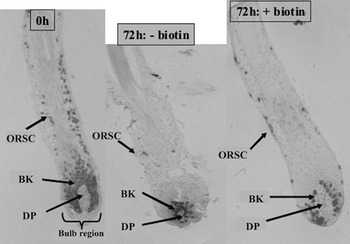
Figure 5 shows the uptake of BrdU into basal keratinocytes and cells of surrounding root sheaths in representative biotin-supplemented sheep follicles at 0 h and 72 h. Biotin deficiency, at 72 h, reduced uptake in matrix and inner root sheath cells and produced apparent disaggregation of the dermal papilla. The results confirm the essential nature of biotin in maintaining viability of sheep hair follicles in vitro and suggest target concentrations of 0.5 to 1.0 mg/l may usefully be aimed at, in the local circulatory environments of follicles.
Longitudinal sections of sheep secondary wool follicles at time 0 and 72 h following incubation without (−) biotin or with (+) 0.5 mg/l biotin. Synthesis of DNA and proliferation of cells determined by uptake of 0.05 M BrdU during a further 6 h incubation followed by conventional immunohistochemical detection (see Figure 3). ORSC = outer root sheath cells; BK = basal keratinocytes; DP = dermal papilla. Magnification × 200 (modified from Tahmasbi et al., Reference Tahmasbi, Galbraith and Scaife2007a).

Effect of l-methionine and l-cysteine on growth and viability of isolated mohair anagen follicles
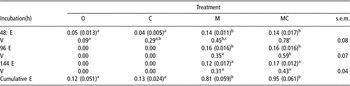
Hair fibres contain protein at concentrations typically in excess of 900 g/kg dry matter (e.g. Galbraith, Reference Galbraith2000). They also contain certain protein/peptide fractions with disproportionately higher concentrations of certain amino acids than are typically supplied in metabolisable protein arising from the diet or from digestible rumen microbial protein. There is particular interest in the contribution of sulphur-containing amino acids l-cysteine and l-methionine, which are present in whole fibre at concentrations of, for example, 90 to 120 and 4.0 to 6.0 g/kg, respectively. Cysteine has a particular presence at higher concentrations in the non-filamentous keratin-associated proteins which combine with the filamentous keratins to form the cytoskeleton in suprabasal differentiating keratinocytes. The inter- and intra-molecular disulphide bonding provided by cysteine has a major influence in determining the physical properties of the fibre product. Since the exposed sulphydril group is readily oxidisable, most in vivo studies investigating l-cysteine have utilised supplements of protein or rumen-protected l-methionine which may contribute to post-absorptive cysteine synthesis via the transulphuration pathway (Radcliffe and Egan, Reference Radcliffe and Egan1974). However, responses to l-cysteine may be investigated directly in vitro. Results from such a study which determined the relative importance of supply of l-cysteine and its dimer l-cystine (cyst(e)ine) and l-methionine at the level of individual secondary follicles of the Angora goat, are given in Table 7. These results show that l-methionine and l-cyst(e)ine-depleted culture media significantly reduced both viability and growth of remaining viable follicles (results shown for 48 to 144 h of incubation). These effects were not reversed by supplementation with l-cyst(e)ine, but by l-methionione, which indicates the nutritional essentiality of l-methionine. There was, however, some evidence of greater responses for example in viability at 144 h, when l-methionine and l-cyst(e)ine were combined compared with l-methionine given alone. These results are consistent with other results (Souri et al., Reference Souri, Galbraith and Scaife1998) which presented evidence of transfer of the sulphur moiety from l-methionine to l-cysteine typical of the transulphuration process in mammalian tissues. These data thus indicate that l-methionine is an essential nutrient and that requirements may be met by its inclusion at approximately 100 μM concentration in culture media although lower concentrations in excess of 10 μM appear not to have been tested. Comparisons may be made with the study of Hepburn et al. (Reference Hepburn, Knight, Wilde, Hendry and Galbraith2008) in which l-methionine uptake was described in dermal-epidermal containing explants of bovine foot integument. Results from this study indicated optimal concentrations of 50 μM to support cell proliferation and protein synthesis. The localisation of 35S-label from 35S-methionine was principally in cornifying epidermis which is similar to that observed in cornifying hair shaft (Souri and Galbraith, unpublished not shown) which may suggest some commonality in uptake characteristics among keratinising integumental tissues. Interestingly, Thomas et al. (Reference Thomas, Tivey, Penno, Nattrass and Hynd2007) have recently described the kinetics of uptake of cysteine and other amino acids (although not methionine) into Corriedale sheep hair follicles in strips of skin. The results obtained suggested a high capacity, low-affinity uptake system for cysteine with a K M value of 1.01 mmol/l. This system appeared to have similar characteristics to that described for methionine (K M of 3.61 mmol/l for l-methionine) in the bovine foot integumenatal tissue system of Hepburn et al. (Reference Hepburn, Knight, Wilde, Hendry and Galbraith2008). These authors suggested that rates of protein synthesis were not limited by uptake capacity of the epidermal-dermal unit. This observation appears yet to be confirmed for the ovine or caprine hair follicle. Results from such studies on individual or groups of follicles provide information on the importance of supply of nutrients in the immediate environment of the follicle.
Effect of inclusion of l-methionine and/or l-cysteine and l-cystine in otherwise deficient culture medium on hair shaft elongation (E: mm, mean ± s.e.m.) and viability (proportion V: means with pooled s.e.m.) of isolated mohair follicles

Follicles from Angora goats (n = 3): approximately 20 follicles/goat per treatment were incubated in medium without l-methionione, l-cysteine and l-cystine and unsupplemented (0) or supplemented with 330 μM l-cysteine + 83 μM l-cystine (C); 100 μM l-methionine alone (M) or 100 μM l-methionine + 330 μM l-cysteine + 83 μM l-cystine (MC); Elongation measured over previous 24 h or cumulatively. Viability measured by presence of elongation in previous 24 h. Comparisons by one-way ANOVA. Values with dissimilar subscripts in rows are significantly different (P < 0.05) (Souri and Galbraith, unpublished).
Conclusion
The study reviewed here provides a number of examples in which the biology of isolated pelage anagen hair follicles of caprine and ovine origin were studied in an in vitro research programme. Major focus was on intrinsic differences between hair follicles derived from different genotypes and responses to certain growth factors/hormones (prolactin, melatonin and EGF) and nutrients (l-methionine, l-cysteine and biotin). The approach utilised simple elongation measurement, histology and analysis of follicle extracts to provide information on relative responses in hair shaft growth rate, keratinocyte proliferation, and apparent DNA and protein synthesis. The data obtained describe responses to the signalling molecules and nutrients tested and provide information on the effects of different concentrations, which were present in the local environment of the follicles. For individual nutrients this latter information may have importance in defining optimal concentrations at the level of follicular tissue. The challenge of applied nutrition in vivo would appear to be to create conditions in the digestive tract and post-absorptively to deliver such concentrations to the cells and extracellular components of the individual follicle.
In terms of relevance to other studies in the symposium such in vitro methodology may have potential to advance knowledge of hair follicle biology of South American camelids and to explain the basis for differences across genotypes which include T’amphullis and Q’aras (llama) and Suri and Huacaya (alpaca) as described by Antonini(Reference Antonini2010). For alpaca, there is particular interest in the crimp-type (Huacaya) and longer ‘cork screw’ curl-type (Suri) fleece patterns which suggest differences in the degree of bilateral asymmetry of hair shaft cortex. The in vitro approach coupled with histological, immunohistochemical and in situ hybridisation methodology may be applied to facilitate investigation of intrinsic morphology and hair shaft elongation in such follicles. Of particular interest will be the patterns of keratinocyte proliferation and differentiation and role of candidate regulatory molecules (Galbraith, Reference Galbraith2010) in producing the recognised hair growth phenotypes.
Acknowledgement
This study is based on an invited presentation at the 59th Annual Meeting of the European Association for Animal Production held in Vilnius, Lithuania during August 2008.