Introduction
Deoxyribonucleic acid (DNA) is considered to be the primary target of radiation. The helices of the DNA strands are wound around the histone protein, known as H.2AX. In the event of DNA damage, structural changes in these proteins occur.Reference Kuo and Yang 1 These proteins are phosphorylated on residue serine 139 as a response to DNA double-strand breaks (DSBs).Reference Rogakuo, Pilch, Orr, Ivanova and Boriner 2 The formation of the phosphorylated H.2AX (γ-H.2AX) is very rapid and is a known cellular response to DNA DSB from ionising radiation. The histone H.2AX not only becomes phosphorylated but also believed to recruit DNA repair factors to sites of DNA DSBs.Reference Paull, Rogakou, Yamazaki, Kirchgessner, Gellert and Bonner 3 The normal repair time of the DSBs is typically within 2–6 hours following radiation, but in some experiments with several cultured cell lines, the maximum expression of the phosphorylated protein occurred within 1 hour after irradiation.Reference Fuhrman, Kilgore and LaCoursiere 4 , Reference MacPhail, Banáth, Yu, Chu, Lambur and Olive 5 Once the damage is repaired γ-H.2AX is dephosphorylated and the cells join back for progression in the cell cycle.
A direct quantification by H.2AX assay helps in assessing the severity of reactions induced by radiation and also monitor the efficiency of treatment by looking at its role in DNA damage.Reference Goutham, Mumbrekar and Vadhiraja 6 HeLa cell line that harbours the oncogene human papilloma virus-18 was used to study the phosphorylation of the core protein, H.2AX, as a response to DNA DSB incited by radiation, chemotherapeutic agent and a combination of both chemotherapeutic agent and radiation. The treatment of cervical cancer follows a more ‘one-size-fits-all’ rather than a specific one. The presentation of a complex individual heterogeneity of the body in terms of composition and function poses a hindrance to accurately and effectively deliver the desired optimal treatment. Since DNA damage affects the progression of the normal cell cycle, damage response and activation of repair proteins may vary from individual to individual. Identifying the response of this protein as a marker of DNA DSB can help in understanding intrinsic radiosensitivity and individualise the treatment module by earlier assessment and evaluation.
Materials and Methods
Cell cultures and preparation
The commercially available HeLa cells initially stored in liquid nitrogen were routinely thawed and cultured. Cells were grown in Dulbecco’s modified Eagle’s medium (DMEM) supplemented with 10% fetal bovine serum in addition to 1% penicillin/streptomycin. The control set were also cultured and kept in CO2 incubator.
Assessment of clonogenic ability
Assessment of the clonogenic potential of the cell line was done by seeding the cells in 60 mm dishes (100 cells/dish) and allowed to adhere at 37°C (with 5% CO2) for 24 hours. The cells were then exposed to 0·2, 0·5, 0·8, 1, 2, 4, 6, and 8 Gy γ-radiation at a dose rate of 400 cGy/minute and re-incubated at 37°C. After allowing for 1 week of repair, colonies were washed with phosphate buffered saline 1×(pH 1·4), fixed in 0·25% glutaraldehyde and stained with 0·5% crystal violet. The plates were dried at room temperature, and colonies were counted manually under the stereomicroscope microscope (OLYMPUS CKX41). This assay is also done for cells treated with different doses of radiation, chemotherapeutic drug (paclitaxel) and combination of both paclitaxel and radiotherapy. The concentration of paclitaxel considered was 1 nM. A group of cells above 50 is considered as colony. This is known as the hay flick’s limit. Cells less than 50 were not taken into consideration.
Protein estimation by Lowry’s method
The concentration of proteins in each plate irradiated with different doses of radiation is determined by Lowry’s method.Reference Lowry, Rosebrough, Farr and Randall 7 This method is linear over the range of 1–100 µg protein. A standard best-fit plot was obtained by using a standard housekeeping protein bovine serum albumin. The absorbance value obtained from the spectrophotometer for each sample irradiated at different doses of radiation was used to find the protein concentration in each sample for all experiments.
Western blotting
Western blotting is a multi-step technique used to detect proteins. Initially, cell lysates were prepared so that the cell membranes are broken up and cellular contents are released. The proteins are then solubilised with the help of sodium dodecyl sulphate and separated based on their size by the action of an electric field. On applying voltage, the protein migrates towards the bottom of the glass slab and bands are formed in parallel with varying intensity. The gels from the slabs are blotted carefully on polyvinyl dine fluoride membrane and the pattern of the protein in the gel were transferred to the membrane. Blocking reagents were used to reduce antibodies non-specific binding on the membrane. Samples were incubated in blocking buffer solution (5% bovine serum albumin) for 1 hour and primary antibody was raised against the γ-H.2AX protein followed by secondary antibody dilution with IgG HRP conjugate to allow visualisation of γ-H.2AX.
γ-H.2AX assay
HeLa cells were suspended in DMEM medium before irradiation. Post irradiation, the cells were incubated at 5% CO2, 37°C. Since the H.2AX phosphorylation declines with time, the DNA damage was quantified at different time points: 1 hour, 3 hours after irradiation. The cells were irradiated with 50, 80 and 200 cGy. In another experiment, 1 nM paclitaxel was added 1 hour before radiation to the cells and incubated for 3 hours after irradiation. The later experiment was done to compare the intensity for radiation alone and intensity after the addition of paclitaxel. The DNA DSBs quantification was carried out by H.2AX assay by Western-blot technique. Protein estimation after radiation for each plate was carried out by Lowry’s method. The membrane was exposed to light in the gel documentation system. Analysis of the protein bands obtained from Western blotting was done with the help of the imageJ software developed by the National Institutes of Health. 8
Results
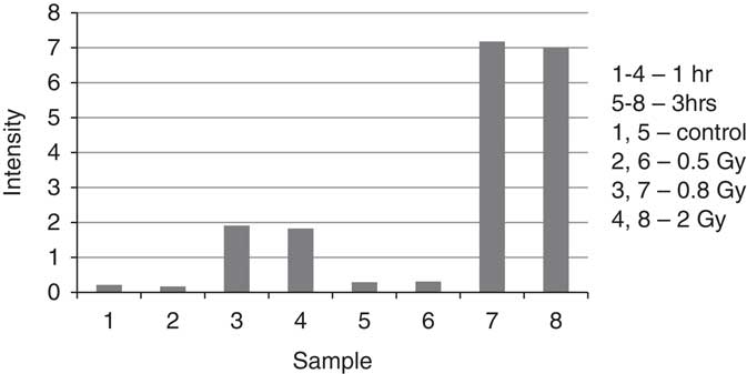
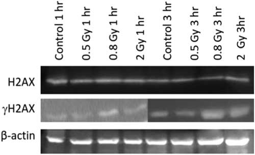
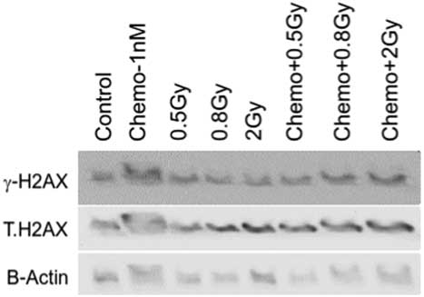
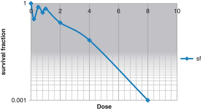
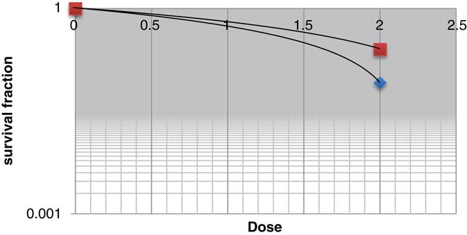
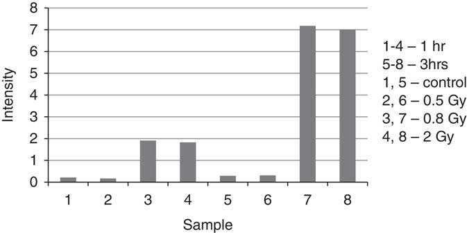
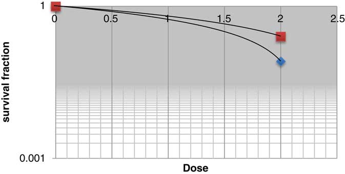
Residual γ-H.2AX is used to identify radiosensitivity of cells and also the efficiency of the cells to repair DSBs. The intensity of γ-H2AX increases as the dose increases. There is more elevation of the γ-H.2AX expression at 3 hours as compared with 1 hour (see Figure 1). The intensity of γ-H.2AX increased linearly with dose. Higher expression of these proteins indicates that the DSBs are still left unrepaired. The protein expressions are blotted on the membrane at different doses of radiation and analysis at different time points. Protein expression as observed in the membrane after 10 minutes exposure to light is shown in Figure 2. Protein expression after 3 hours of radiation and combined treated cells is shown in Figure 3. We observed that the combined treated cells at 0·5 Gy gives the highest expression. Previous studies have shown that HeLa cells are known to exhibit hyper-radiosensitivity at radiation dose <1 Gy.Reference Das, Singh, George, Vijaykumar and John 9 This correlates with our study in Figure 4. In addition, the cell survival curve for cells irradiated with radiation in conjunction with chemotherapeutic drug is steeper than the curve for cells with radiation alone. Survival curve of HeLa cells with combination of chemo-radiation and radiation alone is shown in Figure 5.
The intensity of γ-H.2AX increased linearly with dose.
Protein expression as observed in the membrane after 10 minutes exposure to light.

Protein expression after 3 hours of radiation and combined treated cells.

Cell survival curve of HeLa cells with radiation.

Survival curve of HeLa cells with combination of chemo-radiation and radiation alone.
Conclusion
The infrared treated cells showed that γ-H.2AX significantly increased at 90 minutes and subsequently declined with time.Reference Fuhrman, Kilgore and LaCoursiere 4 However, in our study, we observed that the expression of γ-H.2AX gradually increased with a peak at 3 hours. In γ-H.2AX foci analysis done by Goutham et al,Reference Goutham, Mumbrekar and Vadhiraja 6 the residual γ-H.2AX increased with the severity of reactions. The expression of γ-H.2AX may vary depending on the radioresistance of the cells. Understanding the expression of these proteins is of fundamental importance as it gives us a clue of the repair mechanism and treatment efficacy. It has potential clinical application in monitoring the effects induced by radiation therapy and assess the individual response to the treatment delivered.
Acknowledgements
We would like to thank Department of Neurochemistry for opening their lab for research and the Medical Physics team for their valuable inputs.
Financial support
The Institutional Review Board partially funded this project. while individual contributions from co-authors were our main source of support.
Conflict of interest
None.