Introduction
The genus Deletrocephalus was erected by Diesing in 1851 for a single species, D. dimidiatus Diesing, Reference Diesing1851. In 1861, based on the morphology of nematodes, Molin identified two varieties of the genus Deletrocephalus: D. dimidiatus var. minor and D. dimidiatus var. major. This classification scheme considers the major variety to be the species described by Diesing (Freitas & Lent, Reference Freitas and Lent1947). Years later, in 1936, Vaz described the third species of the genus, D. cesarpintoi Vaz, Reference Vaz1936. Freitas & Lent (Reference Freitas and Lent1947) later analysed the study and drawings of D. dimidiatus var. minor in Chitwood & Chitwood (Reference Chitwood and Chitwood1937) and created a new genus for this species, Paradeletrocephalus Freitas & Lent, Reference Freitas and Lent1947, with P. minor as the unique species in this genus (Freitas & Lent, Reference Freitas and Lent1947). Thus, the family Deletrocephalidae consists of three species from two genera: D. dimidiatus, D. cesarpintoi and P. minor, which differ from one another in terms of the buccal capsule structure, the dorsal ray from the male copulatory bursa and the length of the vagina in the female (Freitas & Lent, Reference Freitas and Lent1947). All of the species in this family are parasites of the gastrointestinal tract of the rhea, Rhea americana Linnaeus, 1758. Of these, only D. dimidiatus was reported to be a parasite of the other rhea species, Darwin's rhea, R. pennata (D'Orbigny, 1834) (Ewing et al., Reference Ewing, Yonzon, Page, Brown and Davidson1995). Currently, there are no studies on the ultrastructure and biology of these parasites or their pathogenicity in these birds. Thus, the present study aimed to examine the ultrastructure of these worms in greater detail.
Materials and methods
Collection and examination of nematode samples
Four adult rheas (two males and two females) from a conservation breeding facility located in the city of Cachoeiro de Itapemirim in the state of Espírito Santo, Brazil, were necropsied, and the gastrointestinal tract was collected and examined for the presence of parasites. Intestinal contents were passed through a sieve with 75-μm mesh, and the mucosa was observed under a stereomicroscope. Helminths that were found were washed in saline solution (0.09% NaCl). A number of the specimens were processed for light microscopy, and the remaining specimens were prepared for scanning electron microscopy.
Nematodes were fixed in hot AFA (70°GL ethanol, 93%; formaldehyde, 5%; glacial acetic acid, 2%) (70°C) overnight, transferred to a solution containing 70% ethanol and 5% glycerin, cleared with lactophenol (one part distilled water, two parts glycerin, one part lactic acid and one part phenic acid) and mounted on semi-permanent slides. Nematodes were then observed under a light microscope.
Measurements were performed to the nearest micron (mean ± SD (range)) and were conducted on mature adult specimens (20 males and 20 females), 200 eggs in the morula stage and 22 larvated eggs in utero from multiple birds. Measurements were conducted with an Axioplan Zeiss light microscope (Carl Zeiss, Jena, Germany) equipped with a Canon Power-Shot A640 digital camera (Canon, Beijing, China) and Zeiss AxionVision Sample Images Software (Carl Zeiss) for image analysis. Drawings were performed with the aid of an Axioplan Zeiss light microscope (Carl Zeiss) equipped with a camera lucida and were digitized using Adobe Photoshop Elements 8.0 software with the aid of an Intuos4 Wacon® pen tablet (Wacon Co. Ltd, Saitama, Japan). Transverse sections from the oesophageal, midbody and posterior regions of a male and a female were cut by hand with a curved bistoury blade and were prepared in glycerin jelly to allow the determination of the presence or absence of a synlophe. The description of the copulatory bursa and the numbering of the bursal rays followed Chabaud et al. (Reference Chabaud, Puylaert, Bain, Petter and Durette-Desset1970) and Durette-Desset (Reference Durette-Desset, Anderson and Chabaud1983). Representative specimens were deposited in the Harold W. Manter Parasite Collection at the University of Nebraska-Lincoln (UNL/USA); the specimens were given the registration number HWML 67093.
Scanning electron microscopy
Nematodes were fixed for 2 h in 2.5% glutaraldehyde, 4% freshly prepared paraformaldehyde, 5 mm calcium chloride in 0.1 m cacodylate buffer, pH 7.2 and post-fixed in 2% osmium tetroxide in 0.1 m cacodylate buffer. The samples were dehydrated in an acetone series, critical-point dried with CO2, sputter-coated with gold and examined in a Zeiss 962 scanning electron microscope (SEM) operating at 15 kV.
Results
General morphology
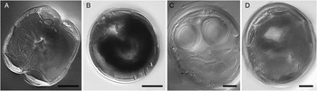
Filiform body, white or pale yellow in life. Females larger than males (ratio of 1.08:1). Cuticle with strong striations, synlophe absent (fig. 1), oesophagus claviform, reaching larger diameter at rear end (figs 2A and 3A).
Transverse sections of Deletrocephalus dimidiatus from Rhea americana, showing the absence of synlophe. Sections at the level of: (A) oesophagus; (B) midbody; (C) ovejector; and (D) spicules. Scale bars: 50 μm.

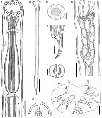
Deletrocephalus dimidiatus from Rhea americana. (A) Anterior region of a female, showing the structure of the oesophagus, the position of the nerve ring, excretory pore, buccal capsule with teeth and lancets, cervical papilla and oesophageal–intestinal junction, scale bar: 125 μm; (B) spicules, ventral view, scale bar: 150 μm; (C) apical view, drawing from SEM, showing the structure of the oral aperture, lips and distribution of cephalic papillae and amphids, scale bar: 25 μm; (D) ovejector in ventral view, scale bar: 150 μm; (E) posterior extremity of a female showing anus and vulva aperture, scale bar: 150 μm; (F) gubernaculum with accessory structure in ventral view; scale bar: 25 μm; (G) differential dorsal ray pattern, and the external dorsal ray from the copulatory bursa, ventral view, scale bar: 60 μm; (H) copulatory bursa showing the organization of bursal rays, scale bar: 120 μm.

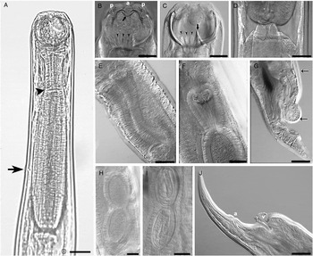
Females of Deletrocephalus dimidiatus from Rhea americana. (A) Anterior region showing the structure of the oesophagus, position of the nerve ring (arrow) and cervical papilla (arrowhead), scale bar: 100 μm; (B) buccal capsule, showing corona radiata (arrow), lancets (arrowheads), papillae (p) and amphidia (a), scale bar: 50 μm; (C) buccal capsule, showing tooth (arrow) and lancets (arrowheads), scale bar: 50 μm; (D) oesophagus–intestinal valve, scale bar: 50 μm; (E) ovejector, scale bar: 50 μm; (F) ovejector infundibulum, scale bar: 50 μm; (G) posterior extremity with cuticular inflation at the level of the vulva, showing the vagina (between arrows), lateral view, scale bar: 50 μm; (H) eggs in morula stage inside the ovejector, scale bar: 50 μm; (I) embryonated eggs inside the ovejector, scale bar: 50 μm; (J) posterior extremity showing vulva (v) and anus (a) aperture, scale bar: 100 μm.

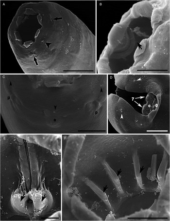
Oral aperture with a slight hexagonal shape, bounded by a corona radiata, six external leaves, radially striated (figs 2C and 4A). Four rounded cephalic papillae, a pair of amphids and three pairs of diminute papillae, being one papilla near each cephalic papilla and one near each amphid, seen only by scanning electron microscopy (figs 2C, 4A and C). Subglobular buccal capsule (figs 2A and 3A–C) with an inner undulated corona radiata, with six protrusions and six depressions with numerous chitinized denticuliform elements. Just below the undulated corona radiata, an undulated chitinous structure accompanies the corona radiata (figs 2A, 3B and 4B). Around the bottom of the buccal capsule, there are 11 lancets (figs 2A, 3B–C and 4F) and two teeth, larger in size, dorsally located (figs 2A, 3C and 4E). A discrete cuticular ring is present at the cephalic end (fig. 4A). Small and conical cervical papilla, difficult to visualize (figs 2A and 3A). Excretory pore near the nerve ring, also difficult to visualize, located in the narrow portion of the oesophagus (Fig. 2A). Oesophageal–intestinal valve well developed, formed by three units (figs 2A and 3D).
Scanning electron microscopy of Deletrocephalus dimidiatus from Rhea americana. (A) Oral aperture, cephalic papillae (arrow) and amphidia (arrowhead), scale bar: 25 μm; (B) undulated chitinous projections inside the buccal capsule (arrow), scale bar: 20 μm; (C) anterior extremity showing three diminutive papillae (arrowheads), cephalic papillae (p) and amphidia (a), scale bar: 50 μm; (D) copulatory bursa of a male of D. dimidiatus from R. americana, showing two papillae at the copulatory bursa membrane (arrowheads), structure of the genital cone with cloaca aperture (c), papilla seven (7) and papilla zero (0), scale bar: 50 μm; (E) pair of prominent teeth at the dorsal region of the buccal capsule (arrows), scale bar: 10 μm; (F) lancets around the base of the buccal capsule (arrows), scale bar: 20 μm.

Male
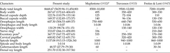
Males (table 1) smaller than females; total body length approximately 8668.67 ± 1749.92 (5639.94–11,454.80) with a greater width at the midline of the body, approximately 267.67 ± 78.58 (172.63–404.43). Body width at the anterior end 138.92 ± 10.38 (117.39–157.30). Buccal capsule approximately 122.76 ± 7.47 (110.79–133.14) deep and 149.57 ± 9.44 (132.85–173.57) wide. Oesophageal infundibulum measuring 57.98 ± 8.45 (43.59–73.11) long and 110.76 ± 8.77 (98.14–133.24) wide, stoma measuring 21.37 ± 3.32 (16.58–28.73) long by 31.71 ± 4.09 (26.71–43.32) wide. Oesophagus length approximately 607.30 ± 52.20 (504.53–689.57), with the largest diameter of the oesophagus at the level of the bulb, 130.29 ± 12.50 (98.74–151.15). Body width measured at base of oesophagus 211.84 ± 19.18 (184.05–245.22), ratio of oesophagus length to total body length 1:0.07. Oesophageal–intestinal valve 47.71 ± 11.27 (33.93–71.29) long by 130.29 ± 12.50 (98.74–151.15) wide. Distance from nerve ring to anterior end 333.07 ± 44.96 (266.11–458.89), body width at level of nerve ring 188.19 ± 20.61 (159.14–227.47). Distance from excretory pore to anterior end 367.77 ± 69.04 (167.72–437.63), body width measured at level of excretory pore 198.08 ± 61.62 (159.33–397.83). Distance from cervical papilla to anterior end 600.82 ± 42.20 (547.12–651.71).
Morphometrics (μm) of the male of Deletrocephalus dimidiatus Diesing, Reference Diesing1851, from rheas, Rhea americana Linnaeus, 1758 for comparison with previous descriptions; range in measurements given in parentheses.

a Description of Quasistrongylus rheae (synonym for D. dimidiatus).
b Measured from anterior end.
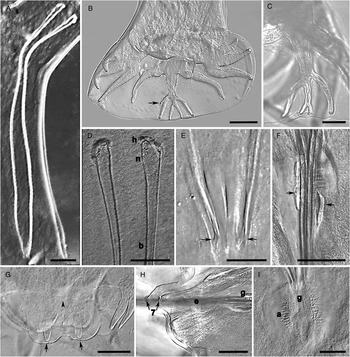
Posterior extremity ending in a well-developed type 2–3 campanuliform copulatory bursa, closed ventrally (figs 2H, 4D and 5B). Pre-bursal papillae absent. Rays two to six arising from a common trunk, separating later; rays two and three remaining in a common trunk; rays four to six in another common trunk. Rays two and three short, similar in length and oriented to anterior region, with ray three slightly shorter than ray two (figs 2H and 5B). Ray five larger than ray four. Rays four and five oriented to anterior region. Ray six departing backward, arising from the posterior half of rays four and five, similar in length to ray five, almost reaching the edge of the copulatory bursa. External-dorsal ray (ray eight) 51.35 ± 15.11 (27.98–83.42) in length, not touching the edge of the membrane of the copulatory bursa, arising within one-third of the dorsal ray length, with globular and irregular projections present or absent (figs 2H and 5B). Dorsal ray long, measuring 251.70 ± 49.39 (132.30–317.56) in length, with the first branch at 153.65 ± 34.02 (86.34–202.94) and the second branch at 207.51 ± 46.09 (120.36–243.76) from the base of the dorsal ray. Last branch measuring 35.34 ± 9.31 (19.11–42.67) in length. Several specimens showed a small lateral rudimentary branch on the first right branch. This rudimentary branch exhibited different patterns (figs 2H and 5B). Such branches were only present in two specimens (figs 2G and 5C).
Males of Deletrocephalus dimidiatus from Rhea americana. (A) Total view of spicules (dark-field contrast), scale bar: 100 μm; (B) copulatory bursa showing a lateral rudimentary ramification (arrow), scale bar: 100 μm; (C) dorsal ray without the lateral rudimentary ramification, scale bar: 50 μm; (D) anterior portion of spicule showing head (h), neck (n) and part of the body (b), scale bar: 25 μm; (E) spicules posterior extremity (arrows), scale bar: 25 μm; (F) gubernaculum (arrow), lateral view, scale bar: 50 μm; (G) genital cone, ventral view, showing double papilla seven (arrows) and simple papilla zero (arrowhead), scale bar: 50 μm; (H) genital cone, lateral view, showing papillae seven (arrows), spicule (e) and gubernaculum (g), scale bar: 50 μm; (I) gubernaculum (g) with accessory structure (a), ventral view, scale bar: 50 μm.

Spicules slender, well chitinized, similar in length, with proximal end larger and distal end slightly pointed (figs 2B, 5A, D, E). Left spicules: total length 1160.07 ± 171.59 (934.59–1526.34), maximum width 8.13 ± 1.12 (6.63–10.75), spicule neck width 16.14 ± 3.62 (11.53–27.72), spicule head 22.07 ± 4.84 (15.08–32.56) long by 27.44 ± 4.07 (20.53–37.49) wide, distal end 25.31 ± 3.93 (16.75–28.48) long by 5.07 ± 1.04 (4.19–7.44) wide. Right spicules: total length 1111.22 ± 147.29 (913.15–1395.83), maximum width 8.52 ± 1.23 (6.65–11.44), spicule neck width 17.03 ± 2.54 (11.45–21.54), spicule head 22.69 ± 5.16 (15.55–33.90) long by 27.77 ± 4.94 (18.79–39.82) wide, distal end 24.83 ± 3.79 (19.81–30.82) long by 5.03 ± 1.28 (3.68–7.53) wide.
Ratio of total body length to length of spicules approximately 1:0.14. Gubernaculum short, weakly chitinized (figs 2F, 5F, I), 48.57 ± 13.81 (27.79–79.30) long. Well-developed genital cone, 104.41 ± 20.33 (55.46–133.51) long by 134.96 ± 42.88 (64.66–205.58) wide (figs 5G, H). Genital cone papillae well developed, papilla zero simple, papilla seven double (figs 4D and 5G). Body width at cloaca level 226.61 ± 55.29 (126.72–310.11).
Female
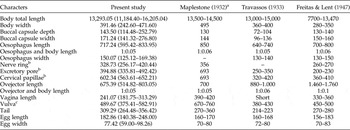
Females larger than males (table 2), total length approximately 13,293.05 ± 1515.48 (11,184.40–16,205.04), maximum body width at middle body region 391.46 ± 72.93 (242.60–471.60). Body width at anterior end 160.00 ± 15.15 (138.70–188.60). Buccal capsule 143.50 ± 32.83 (114.48–252.79) deep by 171.24 ± 34.03 (141.32–276.80) wide. Stoma 22.16 ± 3.60 (14.45–28.76) deep by 31.32 ± 6.37 (19.18–39.58) wide. Oesophageal infundibulum 55.13 ± 9.26 (40.67–68.68) long by 7.81 ± 116.34 (107.21–131.95) wide. Oesophagus length 717.24 ± 62.72 (595.42–833.95), maximum width 150.07 ± 12.49 (125.12–169.38). Body width at level of oesophagus base 235.16 ± 28.50 (196.78–289.87), ratio of total body length to oesophagus length 1:0.05. Oesophageal–intestinal valve 55.49 ± 16.39 (34.24–83.13) long by 106.79 ± 25.54 (84.54–164.82) wide. Distance from nerve ring to anterior end 328.73 ± 43.85 (256.17–420.44), body width at level of nerve ring 209.78 ± 21.67 (175.61–239.35). Distance from excretory pore to anterior end 394.88 ± 44.36 (335.81–492.42), body width at level of excretory pore 215.65 ± 26.02 (177.95–274.52). Distance from cervical papilla to anterior end 602.34 ± 28.62 (563.61–652.21).
Morphometrics (μm) of the female of Deletrocephalus dimidiatus Diesing, Reference Diesing1851 from rheas, Rhea americana Linnaeus, 1758 for comparison with previous descriptions; range in measurements given in parentheses.

a Description of Quasistrongylus rheae (synonym for D. dimidiatus).
b Measured from anterior end.
c Measured from posterior end.
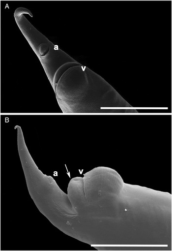
Didelphic ovaries and parallel prodelphic uterus (figs 2D and 3E). Ovejector total length approximately 675.39 ± 98.10 (514.82–803.05), with sphincter, vestibule and infundibulum measuring 130.86 ± 17.88 (106.33–179.23), 398.66 ± 96.30 (234.07–568.29) and 150.89 ± 35.06 (75.78–217.28), respectively. Ratio of total body length to total ovejector length 1:0.05. Vagina short (figs 2E and 3G), 241.07 ± 39.72 (181.75–313.29) long. Vagina width at median region approximately 68.82 ± 12.26 (51.80–98.04). Vulva with prominent lips, opening in a transverse slot near the anus, 489.67 ± 57.95 (375.41–582.91) from posterior end (figs 2E, 3G, J and 6), body width at level of vulva 229.74 ± 45.23 (131.65–290.88). Conical tail (fig. 6), approximately 309.29 ± 22.67 (264.48–356.42) long. Body width at level of anus 121.56 ± 8.40 (110.30–140.19). Morula stage eggs 182.86 ± 22.00 (140.38–248.00) by 77.24 ± 8.98 (59.00–98.26) (fig. 3H). Several female specimens contained embryonated eggs inside the ovejector and the uterus (fig. 3I). These eggs were 175.65 ± 11.91 (152.79–198.44) by 80.59 ± 8.67 (64.88–96.20).
Scanning electron microscopy of Deletrocephalus dimidiatus from Rhea americana. (A) Posterior end of a female with absence of cuticular inflation, showing anus (a) and vulva (v) apertures, scale bar: 200 μm; (B) posterior end of a female with a cuticular inflation at the level of the vulva (arrow), showing anus (a) and vulva (v) apertures, scale bar: 200 μm.

Discussion
Nematodes collected in the rhea intestine were identified as D. dimidiatus according to the morphology described by Maplestone (Reference Maplestone1932), Travassos (Reference Travassos1933) and Freitas & Lent (Reference Freitas and Lent1947).
According to Freitas & Lent (Reference Freitas and Lent1947), the subfamily Deletrocephalinae is distinguished from Strongylinae by an external corona radiata, which consists of few elements; the localization of the vulva in an extreme posterior position; the stretching of the sphincter; and by the divergence of the ventral rays from the copulatory bursa. In the present study, we used the term corona radiata, like Freitas & Lent (Reference Freitas and Lent1947), to designate the structure that Vaz (Reference Vaz1936) described as cuticular membranes which delimit the mouth. Travassos (Reference Travassos1933) used the term ‘leaf’ to designate this structure and compares it with those observed in the species of the genus Oesophagostomum. The subfamily Deletrocephalinae is closer to Oesophagostominae, but differs from this subfamily in the appearance of the buccal capsule, the absence of a ventral cephalic groove, the appearance of sphincters and the divergence of the medial-lateral and posterior-lateral rays (Freitas & Lent, Reference Freitas and Lent1947).
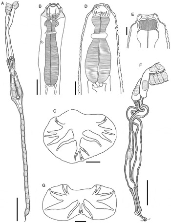
Freitas & Lent (Reference Freitas and Lent1947) described the buccal capsule of D. dimidiatus as a structure bearing 13 ridges, two larger and with a tooth-like shape and 11 smaller and laterally arranged. By observing the morphology of these structures using scanning electron microscopy, we verified that the highest ridges are morphologically characterized as dorsal teeth and that the 11 smaller ridges are similar to lancets, with a similar size and a symmetrical distribution in the base of the buccal capsule. Deletrocephalus dimidiatus differs from another species of the genus, D. cesarpintoi, in the number and shape of ridges present in the oral capsule. Deletrocephalus cesarpintoi has 23 thin ridges that are serrated on the inner edge and are of a variable length. One of these ridges is ventral, and the remaining 22 are lateral, with 11 ridges on each side (Vaz, Reference Vaz1936; Freitas & Lent, Reference Freitas and Lent1947) (fig. 7B).
Deletrocephalus cesarpintoi (A–C) and Paradeletrocephalus minor (D–G), parasites of Rhea americana. (A) Female reproductive system, note the long vagina, scale bar: 500 μm; (b) anterior region, note the buccal capsule ridges and the presence of a cap at the anterior region of the oesophagus, scale bar: 400 μm; (C) copulatory bursa, scale bar: 600 μm. (D) Anterior region, note its shorter length and buccal capsule ridges, scale bar: 100 μm; (E) buccal capsule, note the presence of longitudinal striations, scale bar: 50 μm; (F) female reproductive system, scale bar: 400 μm; (G) copulatory bursa, scale bar: 200 μm. All images were re-drawn from Freitas & Lent (Reference Freitas and Lent1947).

Maplestone (Reference Maplestone1932) described the buccal capsule of Quasistrongylus rheae (synonymous to D. dimidiatus) as containing a pair of dorsally long teeth, ending in a simple point, and three or four pairs of lateral and subventral teeth that are smaller in size, with a bifid tip, which was not observed in the ultrastructural images (SEM) and was not described by other authors (Travassos, Reference Travassos1933; Freitas & Lent, Reference Freitas and Lent1947). Moreover, D. cesarpintoi has the anterior portion of its oesophagus dilated and coated by a thick, chitinous lining that arises from the base of the buccal capsule and forms a cap on the anterior portion of the oesophagus with an irregular surface, small projections and depressions (Vaz, Reference Vaz1936; Freitas & Lent, Reference Freitas and Lent1947). This feature is not present in D. dimidiatus from the present study.
Females can be distinguished according to vagina length. The vagina is considerably larger and weakly muscular, 2.27–2.95 mm long, in D. cesarpintoi, and shorter (0.18–0.31 mm) in D. dimidiatus (Freitas & Lent, Reference Freitas and Lent1947). Males can be distinguished by the presence of striations in the copulatory bursa, which are present in D. dimidiatus and absent in D. cesarpintoi, and by the morphology of the dorsal ray of the copulatory bursa (Freitas & Lent, Reference Freitas and Lent1947).
Freitas & Lent (Reference Freitas and Lent1947) created the genus Paradeletrocephalus, which re-categorized the species described by Molin in 1861 as D. dimidiatus var. minor as the current P. minor (Molin, Reference Molin1861). This new genus differs from the genus Deletrocephalus by its outer corona radiata, which consists of non-striated elements, a non-sinuous inner corona and a distinctly lengthwise-striated buccal capsule (fig. 7D and E). The buccal capsule included ridges that were located in its background and that did not adhere to its wall. These structures formed three sets of ridges, totalling nine. These ridges are relatively large and exceed half the length of the buccal capsule. The oesophageal infundibulum is absent, the oesophagus is shorter and the lateral rays of the copulatory bursa are adjacent to the lateral median rays. Furthermore, on the dorsal trunk of the copulatory bursa, the external-dorsal ray extends beyond the level of the dorsal ray tip, presenting small globular projections with variable aspects on its basal portion.
The measurements obtained in our study are consistent with those reported by Maplestone (Reference Maplestone1932), Travassos (Reference Travassos1933) and Freitas & Lent (Reference Freitas and Lent1947), in that one of these authors described the species as Quasistrongylus rhea, now considered a synonym for D. dimidiatus. However, several characteristics described in the current study differ from those reported by these previous authors. In particular, the distance from the excretory pore to the anterior end was described differently in Freitas & Lent (Reference Freitas and Lent1947), who stated that the structure was anterior to the nerve ring. This placement was not observed in our study or by other authors (Maplestone, Reference Maplestone1932; Travassos, Reference Travassos1933). We observed the excretory pore posterior to the nerve ring.
Another difference was the distance from the cervical papillae to the anterior end. In our study, this distance was approximately double the distance reported by Travassos (Reference Travassos1933) and Freitas & Lent (Reference Freitas and Lent1947). The total length of the spicules also varied in comparison to that reported by previous authors; however, the ratio between the spicule length and the total length of the body did not vary substantially.
Moreover, the length of the spicules in D. cesarpintoi is greater than that in D. dimidiatus (approximately five times the size) (Vaz, Reference Vaz1936). For this reason, the ratio between the total length of the spicules and the total body length is a characteristic that helps to distinguish these species (Vaz, Reference Vaz1936; Freitas & Lent, Reference Freitas and Lent1947). The females described by Freitas & Lent (Reference Freitas and Lent1947) were smaller than those described by other authors, most likely due to the study of juvenile specimens or due to the fixative medium from the Oswaldo Cruz Institute Helminthological Collection (Rio de Janeiro, Brazil) used for the specimens in their study. Moreover, Freitas & Lent (Reference Freitas and Lent1947) reported that the ovejector was relatively large, almost twice the size found in our study and by Maplestone (Reference Maplestone1932) and Travassos (Reference Travassos1933).
Maplestone (Reference Maplestone1932) also described a slight difference in the distance from the vulva to the posterior end. Several females exhibited a swelling at the vulva level with no internal structure, similar to that described for Codiostomum struthionis, a nematode parasite of the caecum of ostriches, Struthio camelus (Ederli et al., Reference Ederli, Oliveira, Lopes and Rodrigues2008).
Freitas & Lent (Reference Freitas and Lent1947) re-described the three species of the subfamily Deletrocephalinae. According to their description, the females of D. dimidiatus showed a pair of small lateral papillae 0.10–0.13 mm from the posterior end. These papillae were not observed in the specimens described by optical and scanning electron microscopy in the present study. Furthermore, the description of the three species of the subfamily Deletrocephalinae by Freitas & Lent (Reference Freitas and Lent1947) states that these species have an opistodelphic-type female reproductive system (i.e. parallel uteri directed toward the posterior end). In our study, however, we verified that the female reproductive system is of the prodelphic type (i.e. parallel uteri that are directed toward the anterior region), in accordance with the description of D. cesarpintoi given by Vaz (Reference Vaz1936). Travassos (Reference Travassos1933) states that, although a prodelphic nematode was specified in the description of Molin (Reference Molin1861), he observed an evident opistodelphy, according to Freitas & Lent (Reference Freitas and Lent1947). However, Travassos (Reference Travassos1933) contradicts himself in his own work, describing nematodes as having parallel rectilinear uteri that are directed forward, a characteristic of prodelphy. Most likely, this description is a result of the nomenclature used by Travassos (Reference Travassos1933) and the descriptions of the specimens revised by Travassos (Reference Travassos1933) and Freitas & Lent (Reference Freitas and Lent1947), as their images of the female reproductive system show a prodelphic-type structure.
The occurrence of nematodes of the genus Deletrocephalus has not been reliably reported. Several papers cite the presence of these nematodes in birds (Ewing et al., Reference Ewing, Yonzon, Page, Brown and Davidson1995; Taylor et al., Reference Taylor, Hunt, Smith and Otter2000; Monteiro et al., Reference Monteiro, Flores, Segabinazi and Lagaggio2002); however, the characters were not clearly described, and clear descriptions of characters are essential for the identification of nematode species because they exhibit similar morphological characters and careful analysis is needed for specific diagnosis. The genus Paradeletrocephalus was reported without accompanying detailed descriptions of its characters. Moreover, the scanning electron microscopy images by Acomolli et al. (Reference Acomolli, Ocayo, Cruz, Milano and Roux2006) are of low definition and do not permit the observation of the absence of striations on the corona radiata. Furthermore, Acomolli et al. (Reference Acomolli, Ocayo, Cruz, Milano and Roux2006) illustrate scanning electron microscopy images of the copulatory bursa without adding consistent information to identify the parasite. That paper also includes optical microscopy images of females. However, the light microscopy image of the anterior end, although it lacked clear definition, showed features that suggest P. minor. These features included the buccal capsule shape and the structure of the oesophagus, which were not clearly described in the text.
The species D. cesarpintoi and P. minor were not found in the four rheas analysed in our study. However, Zettermann et al. (Reference Zettermann, Nascimento, Tebaldi and Szabó2005) compared helminth species found in R. americana reared in captivity in the city of Jaboticabal, São Paulo, Brazil with helminths found in free-living rheas of the same species from Pantanal, Mato Grosso do Sul, Brazil. They found nine species of helminths, seven of which were nematodes (D. dimidiatus, D. cesarpintoi, P. minor, Sicarius uncinipenis, Dicheilonema rheae, Torquatoides crotophaga and Capillaria venteli); the remaining two species were cestodes (Houttuynia struthionis and Chapmania tauricolis). According to these authors, T. crotophaga and C. venteli are known to parasitize the spoonbill (Ajaia ajaia) and Guira cuckoo (Guira guira). These observations of the presence of these parasites indicate that cross-infection occurs due to the occurrence of various birds in the same habitat.
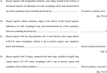
A key to the subfamily Deletrocephalinae is given in fig. 8.
Key to species of the subfamily Deletrocephalinae.

Financial support
This study was supported by Coordenação de Aperfeiçoamento de Pessoal de Nível Superior (CAPES), Conselho Nacional de Desenvolvimento Científico e Tecnológico (CNPq), and Fundação Carlos Chagas Filho do Rio de Janeiro (FAPERJ).
Conflict of interest
None.