Introduction
Chondrosarcoma of the larynx is an uncommon malignancy, representing 1 per cent of all laryngeal tumours, with the cricoid ring being the most common laryngeal site.Reference Ferlito, Nicolai, Montaguti, Cecchetto and Pennelli1 The treatment of choice is complete surgical excision, usually performed via an ‘open’ approach (e.g. laryngofissure) or by partial or total laryngectomy. The goal of this study was to evaluate the feasibility and short-term results of transoral, endoscopic resection of macroscopically localised, low grade, subglottic chondrosarcoma.
Methods
The institutional review board of the Stanford University Medical Center approved the study. We reviewed the charts of patients with laryngeal tumours diagnosed between 2002 and 2007, to locate patients diagnosed with low grade, subglottic laryngeal chondrosarcoma. These patients' post-operative course, vocal fold function, airway patency and oncological results were evaluated.
Surgical technique
The patient is placed supine on the operating table. General anesthesia is maintained by jet ventilation via the adult Dedo laryngoscope.
The laryngoscope is introduced to the level of the glottis and suspended from a Mayo stand. Zero° and 70° degree endoscopes are used to asses the full extent of the tumor. Under visualization with a microscope and using an endoscopic knife, the mucosa along the base of the mass is incised. The endoscopic knife is then passed between the wall of the cricoid ring and the mass itself. Using multiple, serial, axial passes with the knife, the tumor is dissected away from the cricoid ring. Further dissection is performed with micro-scissors, using the blades in an open position to skive along a plane between the mass and the cricoid ring. The tumor is grasped with forceps and the final cuts are made, delivering the tumor through the glottis. The patient is then intubated with a 5.0 or 5.5 microlaryngeal endotracheal tube. The glottis is lightly packed with cottoinoid pledgets soaked in 1% lidocaine and epinephrine 1:100,000 for several minutes, for hemostasis. The procedure is terminated.
The patient is awakened and extubated. The patient is observed for several hours to ensure adequate recovery from anesthesia and is then discharged home with prescriptions for pain medication, proton pump inhibitor and per-oral antibiotics.
Results
Two patients were diagnosed with low grade subglottic chondrosarcoma and underwent tumour resection in the manner described above.
Patient one
A 60-year-old man developed increasing hoarseness over an 18-month period.
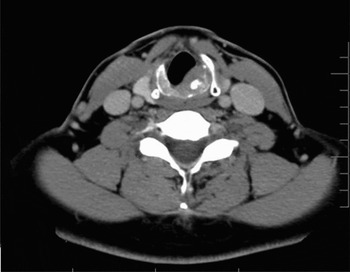
Fibre-optic laryngoscopy demonstrated bilateral true vocal fold bowing. Incidental notation was made of a well circumscribed, subglottic mass arising from the left posterolateral aspect of the cricoid ring, which was confirmed by computed tomography (Figure 1).
Pre-operative, axial computed tomography image of the subglottic larynx of patient one. A discreet mass is seen at the left side of the posterior table of the cricoid cartilage.

A direct laryngoscopy and biopsy was performed under general anaesthesia. Histopathological examination revealed a cartilaginous neoplasm composed of lobules of mildly hypercellular, well differentiated hyaline cartilage with focal areas of ossification and bone marrow formation. The chondrocytes showed minimal cytological atypia, with rare binucleated chondrocytes. The specimen was judged to be consistent with a low grade cartilaginous neoplasm, probably chondrosarcoma.
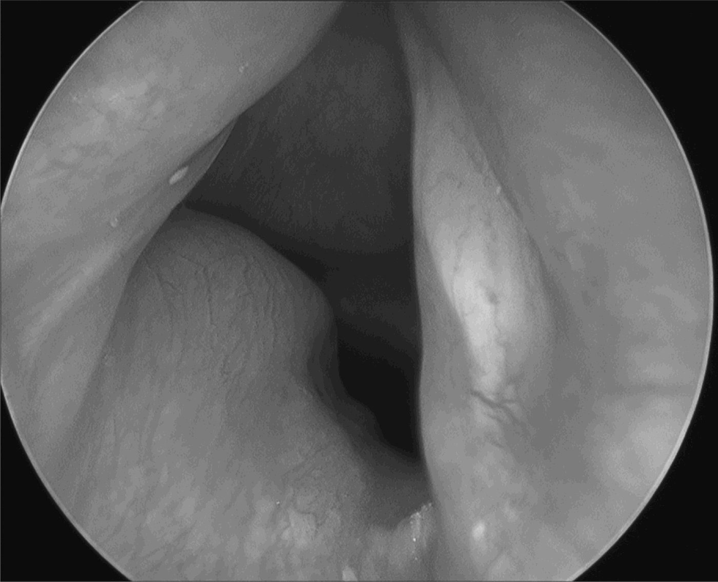
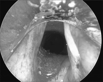
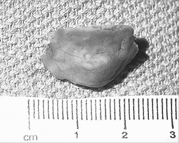
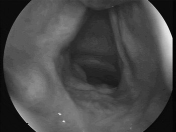
The patient subsequently underwent transoral, endoscopic excision of the lesion (Figures 2 to 4). The patient was followed up in the clinic by recurrent indirect fiber-optic rhinolaryngoscopies (Figure 5).
Intra-operative, rigid endoscopic image of the subglottis of patient one. A well circumscribed, submucosal, subglottic mass is seen arising from the left posterolateral aspect of the cricoid cartilage.
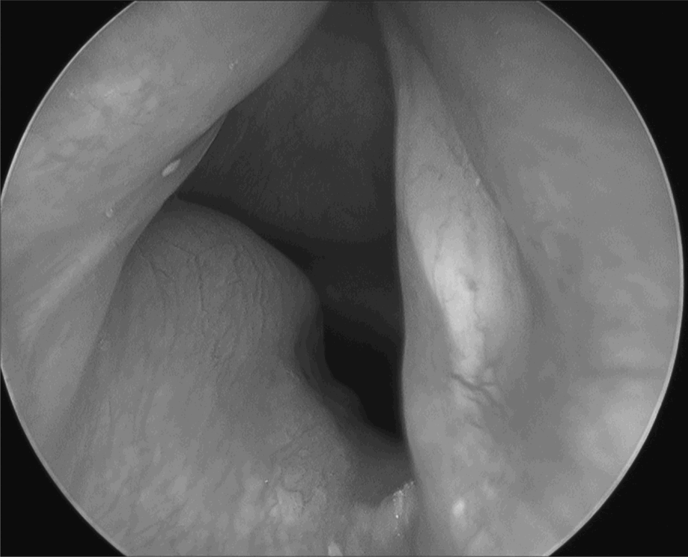
Intra-operative, rigid endoscopic image of the subglottis of patient one after excision of the subglottic mass.

Macroscopic view of the excised low grade chondrosarcoma of patient one.

Follow-up, indirect, fibre-optic rhinolaryngoscopic image of the subglottis of patient one, one month post tumor resection. The subglottis is widely patent and no tumour is seen. Some granulation tissue is seen in the left subglottis.

One year after endoscopic tumour resection, indirect laryngeal fibre-optic examination showed no evidence of recurrence.
Patient two
A 49-year-old man with a history of Charcot-Marie-Tooth syndrome presented with a two-year history of increasing hoarseness and dyspnoea.
Fibre-optic rhinolaryngoscopy demonstrated left vocal fold paralysis.
On direct laryngoscopy, the patient was found to have a subglottic mass projecting from the right posterolateral aspect of the cricoid, with approximately 50 per cent narrowing of the airway. He underwent debulking of the mass. Histopathological examination revealed low grade chondrosarcoma.
Computed tomography of the larynx revealed a residual, 3.2 × 2.6 × 3.5 cm mass arising from the cricoid cartilage. The patient subsequently underwent transoral, endoscopic resection.
Nine months after endoscopic tumour resection, direct microlaryngoscopic examination revealed no evidence of tumour recurrence.
Both procedures were completed on an out-patient basis, with an average operative time of 90 minutes. Histopathological examination demonstrated low grade chondrosarcoma in both cases. Neither patient experienced dyspnoea or airway compromise post-operatively. Vocal fold function was unchanged. Both patients subsequently underwent injection laryngoplasty for glottic insufficiency due either to vocal fold bowing (patient one) or vocal fold paralysis (patient two). Follow-up examination (by indirect, fibre-optic laryngoscopy at 12 months post-operatively for patient one and by direct laryngoscopy at nine months post-operatively for patient two) revealed a patent subglottis in both cases, with no evidence of tumour recurrence.
Discussion
Surgical excision is considered the treatment of choice for low grade laryngeal chondrosarcoma.Reference Ferlito, Nicolai, Montaguti, Cecchetto and Pennelli1 Traditional treatment options include local excision via laryngofissure, and partial or total laryngectomy. The choice of treatment depends on the tumour extent and the severity of airway obstruction.Reference Kozelsky, Bonner, Foote, Olsen, Kasperbauer and McCaffrey2 As local or distal metastases are uncommon in low grade chondrosarcomas, local excision is usually sufficient.Reference Rinaldo, Howard and Ferlito3 Kozelsky et al. studied a series of 20 patients and found that only 25 per cent had recurrence, which usually occurred more than three years after the first local resection, thus enabling several more years of voice preservation after the initial local excision.Reference Kozelsky, Bonner, Foote, Olsen, Kasperbauer and McCaffrey2 Given the slow-growing nature of cricoid chondrosarcomas, the low metastatic potential and the advanced age at tumour presentation, the benefit of partial laryngeal resection with voice and airway preservation may outweigh the risk of local recurrence.Reference Tiwari, Mahieu and Snow4, Reference Thompson and Gannon5
Tumour recurrence can usually be controlled with re-excision, often requiring multiple surgical procedures.Reference Kozelsky, Bonner, Foote, Olsen, Kasperbauer and McCaffrey2, Reference Tiwari, Mahieu and Snow4, Reference Thome, Thome and de la Cortina6 The choice of technique for re-excision is dependent on the tumour's local extent.Reference Thompson and Gannon5 Rinaldo et al. studied a series of 12 patients with low grade chondrosarcoma and showed that local recurrence after endoscopic resections (three cases) could be controlled with re-excision.Reference Rinaldo, Howard and Ferlito3 Overall survival does not necessarily correlate with radical resection techniques, thus supporting the concept that endoscopic laryngeal conservation procedures should be considered.Reference Thome, Thome and de la Cortina6 Thomson and Gannon found that the type of initial treatment procedure and the development of recurrences did not influence outcome, despite the fact that recurrences were more common after local resection versus total laryngectomy (55 vs 16 per cent, respectively).Reference Kozelsky, Bonner, Foote, Olsen, Kasperbauer and McCaffrey2, Reference Thompson and Gannon5 The higher recurrence rate following local excision techniques can be attributed either to piecemeal tumour removal, or, to minimal resection margins while attempting to preserve laryngeal function and airway patency.Reference Kozelsky, Bonner, Foote, Olsen, Kasperbauer and McCaffrey2 Again, in such cases the benefit of an ‘intact’ larynx should be weighed against the higher risk of local recurrence and the probable need for multiple surgical procedures.
Limited experience of endoscopic resection with CO2 laser or microdissection of subglottic chondrosarcoma has been reported.Reference Rinaldo, Howard and Ferlito3, Reference Saleh, Guichard, Russier, Kemey, Perez, Guilian and Laryngeal7, Reference Bathala, Berry, Evans, Brodie and Altaan8 Endoscopic, transoral techniques for the resection of laryngeal malignancies are becoming more widely used, with success rates equivalent to those for ‘open’ resections in selected groups of patients; we therefore believe that endoscopic removal of cricoid chondrosarcomas should be considered in this light.Reference Hoffman and Buatti9 Obviously, more experience and long-term follow-up results will be needed to gauge the merits of endoscopic excision as a treatment option in the resection of laryngeal chondrosarcoma.
Conclusions
Endoscopic, transoral resection of low grade subglottic chondrosarcoma is a viable technique with good functional outcomes. Extensive resection of subglottic disease is possible, which may afford patients an alternative to total laryngectomy. Close, long-term follow up is essential in order to detect tumour recurrence as early as possible.