Investigations over the last ten years have shown greater significance of right ventricular diastolic function with respect to the degree and clinical presentation of pulmonary regurgitation, and the size of the heart, in patients undergoing surgical correction of tetralogy of Fallot in comparison to the morphology of the reconstructed right ventricular outflow tract and the surgical technique employed.1–5 Antegrade diastolic flow in the pulmonary arteries coincident with atrial systole as a marker of the right ventricular restriction in patients with pulmonary atresia with intact septum and critical pulmonary valvar stenosis was previously documented by Redington et al.6 The simultaneous prominent retrograde flow in the hepatic vein is consistent with the restrictive right ventricle. Our present study has focussed on echocardiographic assessment of the evolution of the right ventricular diastolic dysfunction, its modalities, and its interactions with the pulmonary and tricuspid regurgitation. The adaptive right ventricular response to volume overload resulting from pulmonary regurgitation depends on the degree and duration of the regurgitant jet, and the diastolic properties of the right ventricle. The maximal rate and duration of right ventricular filling are significantly reduced in some surgically treated children with pulmonary regurgitation. Although the former may be explained by the reduced compliance of the right ventricle due to the presence of a patch, or myocardial fibrosis, the explanation can be verified by the finding of an inverse relation between the maximal rate of the ventricular filling and end-systolic ventricular volume in the group of the patients undergoing surgical correction.7
Materials and methods
Our population is made up of 60 patients undergoing surgical correction between 1995 and 2004. The average age of the children at the time of examination was 8.98 years, with standard deviation of 5.73 years. The average age of the patients at the time of complete repair was 4.25 years, with standard deviation of 3.08 years, ranging between 6 months and 17 years. Diastolic function of the right ventricle was assessed in the immediate postoperative period, up to the fourteenth postoperative day, in 36 patients with spontaneous respiration. Those patients with a systolic gradient between the right ventricle and the pulmonary arteries greater than 40 millimetres of mercury, and those with the ventricular septal defect remaining open, were withdrawn from the study.
Echocardiographic examinations were performed on the “Acuson – Aspen” echocardiographic machine, using 2.5, 3.5, and 5 megahertz transducers. We made simultaneous electrocardiographic recordings. Routine diagnostic imaging, including colour flow mapping, and continuous-wave Doppler recordings, were obtained. Echocardiographic continuous wave Doppler profile of the pulmonary regurgitation jet was used to calculate pressure half-time. Pulsed Doppler recordings were made in order to determine:
- pulmonary arterial systolic and diastolic Doppler characteristics, placing the pulsed Doppler sample at the midpoint between the pulmonary valvar leaflets and the bifurcation of the pulmonary trunk
- the Doppler profile of the hepatic venous flow 1 to 2 centimetres distal to the right atrium or the inferior caval vein, and
- the transtricuspid Doppler characteristics at the level of the tips of the valvar leaflets.
In all patients, we performed a cross-sectional echocardiographic study, obtaining apical four and two chamber views in keeping with Simpson's rule. Right ventricular restrictive physiology was defined as antegrade pulmonary arterial flow more than 30 centimetres per second in late diastole over five consecutive beats associated with prominent retrograde hepatic venous flow. Use of this criterion permitted us to classify patients into a group of 18 with restrictive physiology, and the remaining 42 with nonrestrictive physiology. The cardiothoracic ratio was used to classify patients in further four subgroups. These were those with primary restrictive physiology and a cardiothoracic ratio less than 0.55, the 14 patients in this group having small hearts, a group with secondary or acquired restrictive physiology with cardiothoracic ratio more or equal to 0.55, these 4 patients having large hearts, a nonrestrictive group of 16 patients with a cardiothoracic ratio less than 0.55, and the group of 26 patients with nonrestrictive physiology having a cardiothoracic ratio more than or equal to 0.55. We calculated the mean values of all the parameters from five consecutive cardiac cycles. Statistical analysis included average value and standard deviation, coefficient of correlation and t-test. Values for p less than 0.05 were considered significant.
Results
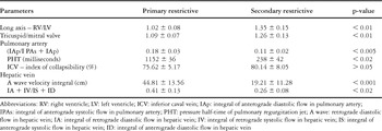
The average follow-up period of all subjects was 5.50 years. The follow-up period, at 5.8 years, was the longest in the subgroup with secondary restriction, albeit in absence of statistically significant difference among the subgroups. A restrictive pattern of flow in the right ventricle was noted in the early postoperative period in 15 out of 36 patients (41.3%). In 5 out of the 15 patients (33%), the restriction was transitory. At midterm follow-up, 18 of 60 patients (30%) were found to exhibit restrictive physiology, this number now including 4 patients who had failed to meet the criterions for restriction over the immediate postoperative period. The percentage of contribution of diastolic anterograde pulmonary arterial flow to total forward pulmonary arterial flow ranged from 8 to 22%. The contribution was significantly higher in the patients with primary as opposed to secondary restriction (p less than 0.005). Echocardiographic data obtained by examination of the patients with restrictive and non-restrictive physiology are presented in Tables 1 and 2.
Table 1. Echocardiographic and Doppler data in patients undergoing surgical correction of tetralogy of Fallot and their subsequent diastolic right ventricle physiology.

Table 2. Echocardiographic and Doppler data in patients undergoing surgical correction of tetralogy of Fallot with subsequent primary and secondary restrictive right ventricular physiology.

Analysis of cardiac size
We found a significant positive correlation between cardiothoracic ratio and echocardiographic parameters of the size of the right heart, with a statistically significant difference noted in the cardiothoracic ratio of those with restrictive as opposed to non-restrictive physiology (Fig. 1). The average values of the quotients of the long axis of the right and left ventricles, and the diameters of the tricuspid and mitral valves, were significantly smaller in those with primary restriction when compared to the patients with non-restrictive physiology and a small heart (p less than 0.001), as well as in those with non-restrictive physiology and a large heart when compared to the patients with secondary restriction (p less than 0.05). Of five patients with transitory restrictive physiology, 2 had cardiothoracic ratios of more than 0.55 in the midterm period of follow-up.

Figure 1. Cardiothoracic ratio and diastolic right ventricular physiology.
The average value of the diameter of the right ventricular outflow tract measured in early diastole 1 to 2 centimetres below the pulmonary valve in all the patients with a large heart was 27.63 centimetres, with standard deviation of 5.42 centimetres, while in those with small hearts, it was 21.86 centimetres, with standard deviation of 3.58 centimetres, this difference reaching high statistical significance. Although the average value of the diameter of the right ventricular outflow tract was higher in those with secondary restriction in comparison to the patients with non-restrictive physiology and a large heart, the difference was not statistically significant, the numbers being 28 centimetres with standard deviation of 5.39 centimetres versus 25.42 centimetres with standard deviation of 4.38 centimetres.
Systolic function of the left and right ventricles
Systolic function of the both ventricles was grossly maintained in our patients. The average value of the right and left ventricular ejection fractions was 48.45% and 64.5%, respectively. Statistical analysis failed to demonstrate any statistically significant differences in the average values of these parameters between the various groups.
Analysis of flow through the hepatic veins.
The collapsibility index was significantly lower in those with restrictive in comparison to those with non-restrictive physiology, at 74.94% with standard deviation of 6.24% versus 81.29% with standard deviation of 4.75%, this difference having a p value of less than 0.01. The index was also lower in those with primary as opposed to secondary restriction. The velocity of anterograde systolic flow through the hepatic veins was significantly higher in those without restrictive physiology, at 68.19 centimetres per second with standard deviation of 6.5 centimetres per second, compared to 55.51 centimetres per second with standard deviation of 8.3 centimetres per second in those having restrictive physiology, this being significant at a p value of less than 0.01, as well as in the group of non-restrictive patients with a small heart when compared to those with primary restriction. The velocity integral of the diastolic anterograde flow was also significantly higher in the group with primary restriction, with a highly statistically significant difference demonstrated in the average values of the integrals of velocity of retrograde diastolic flow between the patients having primary as opposed to secondary restriction (p less than 0.001). The integral of retrograde systolic flow in the hepatic veins was significantly greater in those with restriction, at 35.22 centimetres with standard deviation of 8.59 centimetres, when compared to values of 25.25 centimetres, with standard deviation of 11.40 centimetres, in those lacking restrictive physiology (p less than 0.02).
Analysis of pulmonary regurgitation
We evaluated pulmonary regurgitation in the patients with non-restrictive physiology using pulsed and colour Doppler.8, 9 Semi-quantification of pulmonary regurgitation was performed based on determination of the ratio between the width of regurgitant flow and the diameter of the pulmonary trunk at the level of the pulmonary valve, presence of retrograde colour flow from the pulmonary trunk or its branches, and the pressure half-time of the regurgitant jet. The pressure half-time of longer than 100 milliseconds was found to be a good indicator of haemodynamically significant regurgitation.10 The criterions listed above were taken as indicators of severe, or “free”, pulmonary regurgitation when the ratio between the width of the regurgitant jet and the diameter of the pulmonary trunk at the level of the valve exceeded 0.5, there was retrograde flow, and the pulmonary pressure half-time was longer than 100 milliseconds, with severe pulmonary regurgitation diagnosed in presence of two of these markers. This permitted classification of the patients into those with mild as opposed to severe pulmonary regurgitation. Of those with non-restrictive physiology, 88.6% with small hearts had mild pulmonary regurgitation, while 77% of those with large hearts had severe regurgitation. The mean value of the pressure half-time jet of the pulmonary regurgitation was higher in those with secondary restriction compared to those with non-restrictive physiology and large hearts, specific values being 152 milliseconds with standard deviation of 36 milliseconds as opposed to 85 milliseconds with standard deviation of 11 milliseconds, giving a p value of less than 0.05. The values were significantly lower, however, when compared to those obtained in the patients with primary restriction, specifically 238 milliseconds with standard deviation of 42 milliseconds (p less than 0.02).
Analysis of flow across the tricuspid valve
Semi-quantification of the tricuspid regurgitation was performed using colour flow mapping by determining the ratio between the area of the regurgitant jet and that of the right atrium. A ratio of greater than 0.5 is taken as our criterion for severe tricuspid regurgitation, permitting division of the patients into those with mild as opposed to severe regurgitation. In those with secondary restriction, 75% had severe tricuspid regurgitation, as did 78% of the patients from the non-restrictive subgroup with large hearts. The 2 patients with transitory restriction had severe tricuspid regurgitation at the time of midterm follow-up. The average velocity and duration of the E wave, and the ratio between the E and A waves, were statistically significantly higher in the patients with secondary restriction, and those with non-restrictive physiology, when compared to the patients with primary restriction.
Analysis of interdependence of the various parameters
Analysis of the degree of dependence showed statistically significant differences between pulmonary regurgitation and cardiothoracic ratio (r = 0.359; p less than 0.01), the diameter of the right ventricular outflow tract (r = 0.423; p less than 0.05), the proportional contribution of diastolic to total anterograde flow through the pulmonary arteries (r = − 0.557, p less than 0.01), the velocity integral of the retrograde diastolic flow in the hepatic veins (r = −0.295, p less than 0.05), and the quotient of the total retrograde and total anterograde flow in the hepatic veins (r = −0.385; p less than 0.008). No correlation was found between the degree of pulmonary regurgitation and presence of a transannular patch. None of the patients with transitory right ventricular restriction had severe pulmonary regurgitation in either the early or midterm periods of follow-up.
Discussion
A restrictive pattern of right ventricular filling was demonstrated in the immediate post-operative period of just over two-fifths of our patients undergoing surgical repair of tetralogy of Fallot. In one-third of these patients, however, the restriction was transitory. According to Norgård and co-workers,11 normalization of diastolic function may be detected in the early postoperative period in a small number of patients. In our series, the transitory restriction persisted from the second postoperative week to the second postoperative month. Severe tricuspid regurgitation seen in two of these five patients was responsible for determining the size of the heart in this variant of restrictive right ventricular physiology. Of the five patients with transitory restriction, 3 had received a transannular patch.
Our findings showed that the anterograde diastolic flow in those with restrictive physiology accounts for up to nearly one-quarter of the total anterograde flow in the pulmonary arteries. Gatzoulis and co-workers also reported values in the range from 3.5 and 25%.1 According to Meyer et al.,12 both retrograde diastolic and systolic flows are recorded in one-quarter of all healthy children. Although all four waves were recorded in the hepatic veins in all the patients evaluated from our series, the profile and velocity of the flow differed between the groups. The fraction of the venous retrograde flow in the hepatic veins, that is, the integral of A wave velocity greater or equal to 30 centimetres, distinguishes between the patients with primary restriction as compared to the remainder of the surgically treated patients in the early postoperative course and over midterm period of follow-up. The lower ratio of E and A waves, and the duration of the E wave of tricuspid flow, also serve to distinguish those with primary restriction from the remaining patients.
Restrictive and non-restrictive physiology
In those with restrictive physiology, right atrial filling predominantly takes place in the phase of the right ventricular diastole, as opposed to those with non-restrictive physiology, in which systolic filling is dominant. In the non-restrictive right ventricle, it is anterograde systolic filling, which develops in the phase of the right atrial relaxation and lowering of the tricuspid valvar annulus during right ventricular systole, which is greatest. The ratio between velocity and duration of E and A waves is significantly lower in those with restriction as compared to non-restriction. This variability of the ratio between the E and A waves may reflect a different degree of right ventricular restriction, as demonstrated by Nishimura et al.13 in the case of disorders of right ventricular relaxation. There are certain data indicating that the ratio of E and A wave of the tricuspid flow in surgically treated patients with tetralogy of Fallot could be normal in spite of the reduced compliance of the right ventricle.1 According to Cullen and colleagues,14 the restrictive pattern of right ventricular filling is more frequently shown in the early postoperative course in patients in whom placement of a transannular patch was inevitable. Norgård, 15 Eroglu,16 et al., showed that the presence of transannular patching is the only variable significant for development of restrictive right ventricular physiology at midterm follow-up of surgically treated patients with tetralogy of Fallot. This was not confirmed in our study. In this respect, it should be kept in mind that, in patients in whom a transannular patch was necessary, the pathoanatomical characteristics of myocardial hypertrophy and fibrosis may, in themselves be sufficient to explain the disorders of right ventricular filling.
Primary and secondary restriction.
We have shown that the proportion of total anterograde flow in the pulmonary arteries occurring during diastole is greater in patients with primary as opposed to secondary restriction. The duration and velocity of pulmonary regurgitation are both smaller in those with primary restriction as opposed to secondary restriction, whereas the pressure half-time was shorter in those with secondary restriction. Patients with primary restriction have smaller cardiothoracic ratios, shorter long axes of the right ventricle, smaller tricuspid valvar annular diameters, and smaller diameters of the right ventricular outflow tract. In spite of the fact that some authors11 found that early restriction was the strongest predictor of subsequent restrictive right ventricular physiology at mid-term follow up, an A wave in the pulmonary artery greater than 30 centimetres per second was recorded in 4 of our patients after an average period of follow-up of 2.4 years, with a range from 1.5 to 3.5 years, subsequent to the complete surgical repair. Echocardiographic indicators of right ventricular volume overload, and a cardiothoracic ratio greater than 0.57, were found in all four patients. The significant pulmonary and tricuspid regurgitation resulting from increased end-diastolic pressure determines the restrictive characteristics of the right ventricle in those patients with secondary or acquired restriction. On the other hand, we showed that the size of the heart was unchanged in those with secondary restriction throughout the period of follow-up. Retrograde flow in the hepatic veins in late diastole was marked in those with primary restriction. Total retrograde flow in the hepatic veins in our patients was significantly higher in those with primary as opposed to secondary restriction. Retrograde diastolic flow was more prominent in the patients with restrictive physiology but milder tricuspid regurgitation in comparison to the number of children with more severe tricuspid regurgitation. We have also showed a statistically significant difference between the size of the heart and the velocity integral of retrograde diastolic flow, with those having smaller hearts having also higher retrograde diastolic flow in the hepatic veins. Although we found that the integral of the retrograde systolic flow in the hepatic veins was significantly greater in those with restrictive compared to nonrestrictive physiology, we are unable to determine whether prominent retrograde systolic flow in the hepatic veins indicates restriction of the right atrium in the settings with the same or similar pathomorphological substrates or pathophysiological mechanisms.
Secondary restriction and non-restrictive physiology in the setting of a large heart.
Despite the shortcomings and limitations of echocardiographic quantification of valvar insufficiency, particularly pulmonary regurgitation, it is usually accepted that the size of the right ventricle is conditioned by the presence of significant pulmonary or tricuspid regurgitation. We have shown a correlation between the size of the heart and the criterions used for assessment of the severity of pulmonary or tricuspid regurgitation in the patients with non-restrictive physiology. Systolic retrograde flow in the hepatic veins was significantly higher in those with secondary restriction, while the same relation has not been confirmed with respect to differences in mean values of diastolic retrograde hepatic venous flow between those having secondary restriction as opposed to those lacking restrictive physiology but having large hearts. The patients from these subgroups are candidates for reintervention, owing to the well-known fact that chronic pulmonary regurgitation, as well as tricuspid regurgitation, have detrimental effects on right ventricular function.17, 18 The consensus on the optimal time for reintervention in such patients has yet to be determined. Recovery of the right ventricle may be compromised in adults following replacement of the chronic and significantly regurgitant pulmonary valve subsequent to repair of tetralogy of Fallot.19 Echocardiographic assessment of the size of the right ventricular outflow tract, the recording of anterograde diastolic flow in the pulmonary arteries of patients with large hearts, as well as marked retrograde systolic flow in the hepatic veins, may all play significant roles in the timing and selection of patients for additional investigations and reintervention, particularly bearing in mind that it is now possible to insert new pulmonary valves percutaneously.20
Limitations of our study
The average age at repair of 4.5 years, the small number of patients with secondary restrictive physiology, and the relatively short time of follow-up all represent limitation in the generalisability of our experience.
Conclusions
We suggest that echocardiographic analysis offers great possibilities for assessment of right ventricular diastolic function over the early and mid-term periods of follow-up of patients undergoing surgical correction of tetralogy of Fallot. It permits identification of the presence and type of restrictive physiology, its interrelation with pulmonary and tricuspid regurgitation, and the timing and selection of patients for additional investigations and reinterventions. We assume that the appearance of antegrade diastolic flow in the pulmonary arteries of the patients with large hearts represents one of the adaptive responses of the right ventricle to the volume overload resulting from pulmonary and tricuspid regurgitation.