
Introduction
Calcium phosphate cements (CPCs) are widely used to fill bone voids for fracture fixation.[Reference Chen, Zhang, Kang, Xu, Wang, Cui and Guo1,Reference Larsson and Bauer2] In 1986, Brow and Chow introduced an injectable CPC composed of tetracalcium phosphate (TTCP) and dicalcium phosphate anhydrous. This powder mixture has the ability to set in situ when combined with an aqueous liquid and then precipitate into hydroxyapatite (HA) as the final product.[Reference Chen, Zhang, Kang, Xu, Wang, Cui and Guo1,Reference Brown and Chow3–Reference Sharifi, Imani, Mirzadeh, Atai, Ziaee and Bakhshi5] This precipitated HA has a more similar crystal structure to biological HA than sintered HA with finer grains.[Reference Thai and Lee6] Hence, they are used as injectable bone substitutes (IBS) with their unique properties such as self-setting ability, good injectability, easy moldability, high bioactivity, and feasibility.[Reference Chen, Zhang, Kang, Xu, Wang, Cui and Guo1,Reference Ginebra, Traykova and Planell7] However, the injectable CPCs have some drawbacks, including the possibility of a collapse in body fluids, poor degradability, and weak mechanical properties.[Reference Liu, Li, Cheng, Yang, Zhu and Zhou8] These physical properties should be improved to make them more applicable in practice. In literature, many methods are presented to overcome drawbacks of CPCs, such as adjusting powder-to-liquid ratio, using pore-making agents like acetic acid or citric acid. The setting time was modified by sodium phosphate or calcium sulfate. The mechanical properties were improved by combining with poly(methyl methacrylate) (PMMA). Moreover, methods have been developed to enhance their bone repair ability such as adding calcium silicate, HA, zinc, magnesium, or fluoride ions.[Reference Chen, Zhang, Kang, Xu, Wang, Cui and Guo1]
To adjust the rheological properties of CPCs, the aqueous liquid could be chosen as a polymeric material.[Reference Priya, Sivshanmugam, Boccaccini, Goudouri, Sun, Hwang, Deepthi, Nair and Jayakumar9,Reference O'Neill, McCarthy, Montufar, Ginebra, Wilson, Lennon and Dunne10] In this study, the polymeric phase of the bone filler was prepared by using methylcellulose (MC) and gelatin. MC/gelatin mixture is a thermosensitive formulation which undergoes sol–gel transition at the body temperature. Studies state that the cellulose provides the optimal mechanical properties for orthopedic surgeries, augments the osteoconductivity, and also leads to a longer implant lifetime by causing slow resorption of CPC.[Reference Ghanaati, Barbeck, Hilbig, Hoffmann, Unger, Sader, Peters and Kirkpatrick11,Reference Krause, Oheim, Catala-Lehnen, Pestka, Hoffmann, Huebner, Peters, Barvencik and Amling12] Gelatin, which is a chemical derivative of collagen, has certain biological characteristics comparable with collagen, like easy processability into microspheres, fine gelling behavior, controllable degradation characteristics, and plenty of functional groups for further functionalization and modification.[Reference Wang, Leeuwenburgh, Li and Jansen13] Due to the presence of its arginine-glycine-aspartic acid (RGD) groups, integration of gelatin with CPC supports cell-surface recognition, cell physiology, and cellular affinity of the IBS.[Reference Dessi, Alvarez-Perez, De Santis, Ginebra, Planell and Ambrosio14,Reference Bongio, Nejadnik, Kasper, Mikos, Jansen, Leeuwenburgh and van den Beucken15]
Graphene-based nanocomposites have also attracted great interest in biomedical applications due to their extraordinary physical, electrical, and chemical properties, large surface area, and chemical purity.[Reference Ege, Kamali and Boccaccini16] Accordingly, the use of graphene and its derivatives with CPC may be an efficient way to improve CPCs' mechanical properties, stability, workability, osteoconductivity, cell adhesion, proliferation, and differentiation.[Reference Ege, Kamali and Boccaccini16–Reference Wu, Xia, Han, Xu, Fang, Wang, Chang and Xiao19] However, structural damage of graphene during the sintering process of ceramics and uneven dispersion of graphene in the matrix because of the strong van der Waals force between graphene layers are the main problems of its use.[Reference Gao, Feng, Peng and Shuai20] Therefore, in this study, graphene oxide (GO) was incorporated into MC/gelatin-based polymeric matrix to achieve homogenous and stable dispersion of GO in CPC.
Here, different wt% of GO was added to CPC/MC-based IBS samples. In this study, X-ray diffraction (XRD), scanning electron microscopy (SEM), rheological measurement techniques, and compressive modulus of the samples were used to study physical properties. In addition, the produced IBS samples were analyzed by means of setting time, injectability, degradation, swelling, and biocompatibility studies.
Materials and methods
Preparation of GO-incorporated IBS materials
GO added IBS are prepared from a liquid and powder phase. The desired amount of liquid component was prepared as described previously[Reference Oğuz and Ege21] by using 2.5% w/v Na2HPO4 solution instead of distilled water. Then, 0.5, 1.0, 1.5, and 2 wt% of GO (SigmaAldrich, Taufkirchen, Germany) in the mass of MC were dispersed in Na2HPO4 solution by using a homogenizer (Sonopuls, Bandelin, Berlin, Germany)) for 1 h. After adding this homogenized GO solution to the liquid phase, the final liquid component was sonicated again for 1 h to obtain a homogeneous well-dispersed liquid phase. The powder phase was prepared by using an equal molar ratio of TTCP (Hitemco Medical, Old Bethpage, Newyork, USA) and dicalcium phosphate dihydrate (DCPD; Sigma-Aldrich, Taufkirchen, Germany), and this ceramic mixture was added to calcium sulfate dihydrate (CSD; Sigma-Aldrich, Taufkirchen, Germany) with a 75/25 TTCP–DCPD/CSD mass ratio. Then, the liquid and powder phases with a 1:1 ratio (wt:wt) were mixed manually with a spatula for approximately 45 s at ambient conditions to form IBS samples. Thus, five groups of IBS samples were prepared in this study: (i) 0GO with 0 wt% GO added liquid phase; (ii) 0.5GO with 0.5 wt% GO added liquid phase; (iii) 1GO with 1 wt% GO added liquid phase; (iv) 1.5GO with 1.5 wt% GO added liquid phase; and (v) 2GO with 2 wt% GO added liquid phase.
Characterization of GO-incorporated injectable bone fillers
Setting time and injectability measurements
The setting time of GO-reinforced injectable bone filler samples was tested by Gilmore needles according to ASTM C266-04 standard.[Reference Wang, Zhang, Wang, Sun and Sun22] Samples were molded to form cylindrical tubes with dimensions of 10 mm of diameter and 5 mm of height, and then samples were kept at 37 °C and 100% relative humidity. The initial setting time was tested by the initial needle of Gillmore apparatus (m = 113.4 ± 0.5 g, d = 2.12 ± 0.05 mm), and the final setting time was tested by the final needle Gillmore apparatus (m = 453.6 ± 0.5 g, d = 1.06 ± 0.05 mm). The time was recorded until the needle could not create a 1 mm deep indentation in three separate areas.
The workability of samples is defined as the degree of injectability when a homogeneous paste was formed after mixing the powder and the liquid phase and also the ability to give shape before setting of the samples.[Reference Campana, Milano, Pagano, Barba, Cicione, Salonna, Lattanzi and Logroscino23] Therefore, injectability was qualitatively assessed by considering the percentage mass of the samples extruded from the syringe divided by the original mass of the samples filling the syringe.[Reference Lian, Xiao, Bian, Xia, Guo, Wang and Lang24] The 18-gauge needle was used to extrude the samples, then samples extruded from the syringe manually within a few seconds at a relatively constant speed.
Phase composition and morphology
GO-reinforced injectable bone filler samples were molded into columns of 10.0 mm of diameter and 5.0 mm of length and incubated at 37 °C with a 100% relative humidity for 1 day. Then, samples were removed from the molds and lyophilized. The surface of the samples was coated with gold and examined with scanning electron microscopy (SEM; Philips-FEI XL30). Phase analysis of the samples was performed by using the X-ray diffractometry (XRD; Rigaku D/MAX-Ultima+/PC) operated at 30 kV and 30 mA at a scanning speed of 1°/min. Fourier transform infrared spectroscopy (FTIR) measurements were performed to detect the functional groups and to determine the interaction of GO and other phases (Nicolet FTIR Instruments, Thermofischer).
Mechanical analysis
Rheological measurements of GO-reinforced IBS samples were performed by using a stress-controlled rheometer with a parallel plate geometry (15 mm) (Anton Paar, MCR302). The viscoelastic properties of the samples were obtained at 0.1% strain and between 0.1 and 10 Hz frequency within the linear viscoelastic region. Measurements were carried out at 25 and 37 °C.[Reference Dessi, Alvarez-Perez, De Santis, Ginebra, Planell and Ambrosio14]
Samples were injected into cylindric Teflon molds (10 mm of diameter, 20 mm of height) and allowed to set at 37 °C, 100% relative humidity overnight. After setting, IBS samples (n = 4) were dried at 70 °C, and then compressive modulus of IBS samples was measured at a loading rate of 1 mm/min with Lloyd Instruments (LF Plus, Ametek Inc.).[Reference Nasrollahi, Dehkordi, Jamshidizad and Chehelgerdi25]
In vitro degradation and swelling studies
For the in vitro degradation and swelling studies, preset samples were incubated in 5 mL of PBS at 37 °C, and the measurements were done at predetermined time intervals.[Reference Liu and Yao26] The weight was measured both before and after soaking for degradation studies using the formula for mass loss (%, t) = (W 0 − W t)/W 0 × 100%, where W 0 and W t are the dry weights of the samples after 0 and t days of the incubation. The swelling studies were carried out for 1 and 24 h. The swelling ratio was evaluated using the formula for swelling ratio (%, t) = (W t − W i)/W i × 100%, where W i is the initial weight of the sample, and W t is the wet weight of the sample after t hour of the immersion.
In vitro biocompatibility
The human bone marrow mesenchymal stem cells (hBMMSCs; ATCC) were used to evaluate the biocompatibility of the prepared IBS samples. Cells were cultured using low Dulbecco's Modified Eagle's Medium-low glucose (DMEM-low glucose; Sigma) supplemented with 10% fetal bovine serum (Biowest) and 1% gentamicin sulfate (50 mg/mL) solution (Biowest). To harden the samples (n = 6), they were placed in a 24-well plate and stored in an incubator at 100% relative humidity and 37 °C for 1 day. Then, before the cell incubation, the hardened samples were sterilized by soaking in a 70% ethanol solution for 1 h and then by exposing to ultraviolet (UV) light for 30 min for both sides of the prepared discs. hBMMSC suspensions at densities of 3000 cell/cm2 were directly seeded over sterilized samples and incubated at 37 °C in a 5% CO2 atmosphere. After 1, 4, 7, and 14 days, cell viability was examined by using the alamarBlue assay (Bio-rad, UK). Briefly, the present medium was discarded, and then each sample group was incubated in fresh medium containing 4% alamarBlue reagent. After 3 h of incubation at 37 °C in a 5% CO2 atmosphere, the color change was measured at 570 and 595 nm with a microplate reader (Bio-rad, iMark™).
Statistical analysis
One-way analysis of variance (ANOVA) was used to evaluate significant differences between the means of the groups. Then, Dunnett's multiple comparison test was used to determine the significance of standard deviations in the measured data from each specimen under different experimental conditions. All results were considered statistically significant with a P-value which is less than 0.05.
Results and discussion
Characterization of the GO added IBS samples
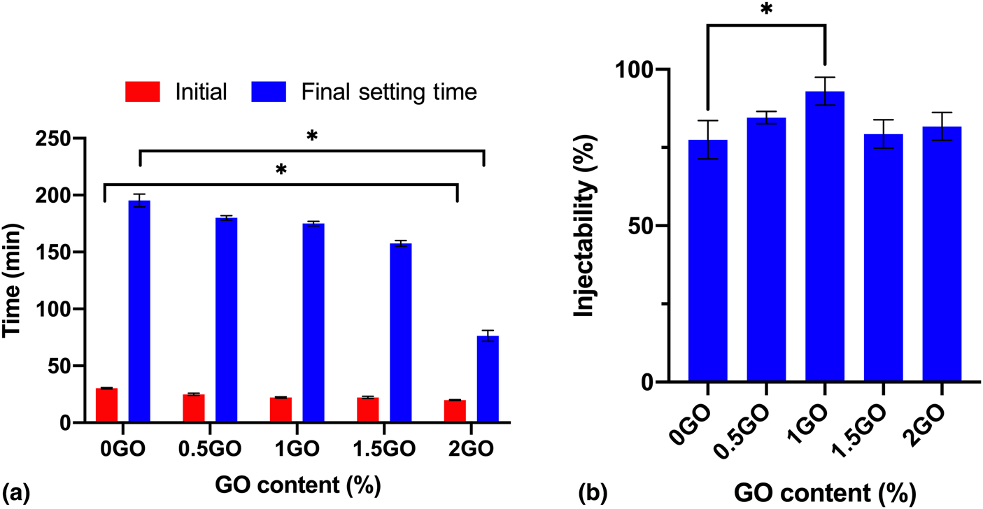
The setting time and injectability measurements of the samples are presented in Fig. 1. The initial and the final setting time was recorded when the specimen could bear the initial and final needles without appreciable indentation. Depending on the amount of GO, the setting time of the cement phase varied. There are no statistically significant differences between the initial and final setting time of 0.5GO, 1GO, and 1.5GO samples compared to 0GO. However, the setting time of 2GO was significantly lower than 0GO samples with P < 0.05. In literature, it is stated that the setting time is the critical factor during clinical application, and it may be approximately 20 min for IBS.[Reference O'Neill, McCarthy, Montufar, Ginebra, Wilson, Lennon and Dunne10,Reference Wang, Zhang, Wang, Sun and Sun22,Reference Chiang, Ho, Chen, Lai and Ding27] This revealed that the prepared injectable bone fillers should be handled according to the initial and the final setting time which are 19.93 and 76.33 min for 2GO samples, respectively.
(a) Setting time and (b) injectability results (n = 4). Values are mean ± standard deviation. Asterisk means that there is statistically significant difference between the groups (P < 0.05), and ns means that there is no statistically significant difference according to ANOVA and Dunnett's multiple comparison test.
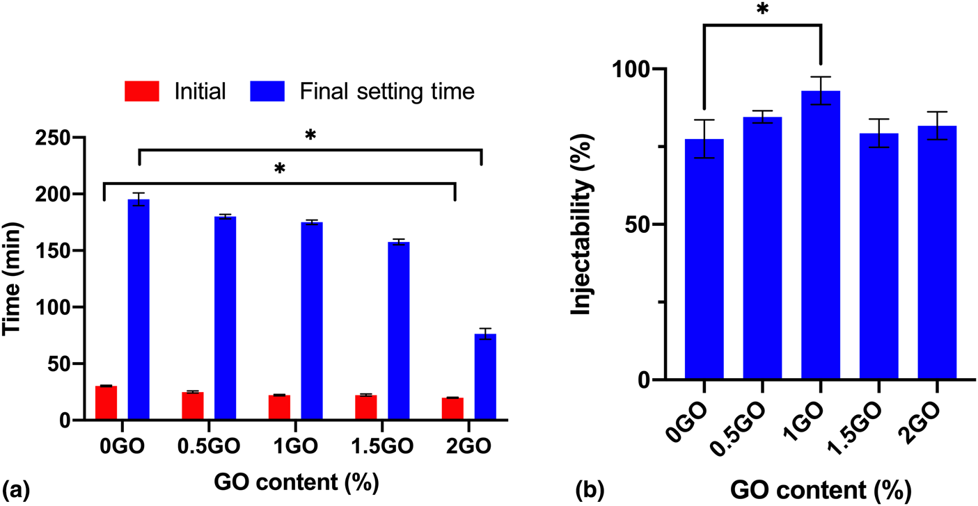
According to Fig. 1(b), all samples showed a high degree of injectability. 1GO samples had significantly higher % injectability than 0GO samples. There is no statistically significant difference between 0GO and the other GO added IBS samples. The incorporation of GO up to 1 wt% increased the injectability by approximately 15%. Visual observation of the injected samples showed that the addition of GO did not cause any segregation or phase separation during the injection. Overall, the setting time of GO-reinforced IBS samples was significantly shortened for 2GO and the fluidity of samples decreased for 1.5GO and 2GO.
Supplementary Figure S1 shows the XRD pattern of 0GO and 2GO samples after 24 h of setting. Samples possessed the same XRD pattern independent of incorporation of GO because of the small content of GO. Depending on the qualitative analysis, no new crystal structure was obtained after setting of the samples, only the added raw powders, namely TTCP, DCPD, and CSD, were detected which matched with JCPDS files no. 72-1240, 70-1379, and 74-1905, respectively. Supplementary Figure S2 shows the FTIR spectrum of GO, MC, polymer phase with 2 wt% GO, 0GO, and 2GO. Details of the analysis were given in Supplementary Material. According to the results, there were only small shifts since the materials in the samples had noncovalent interactions.
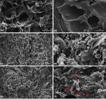
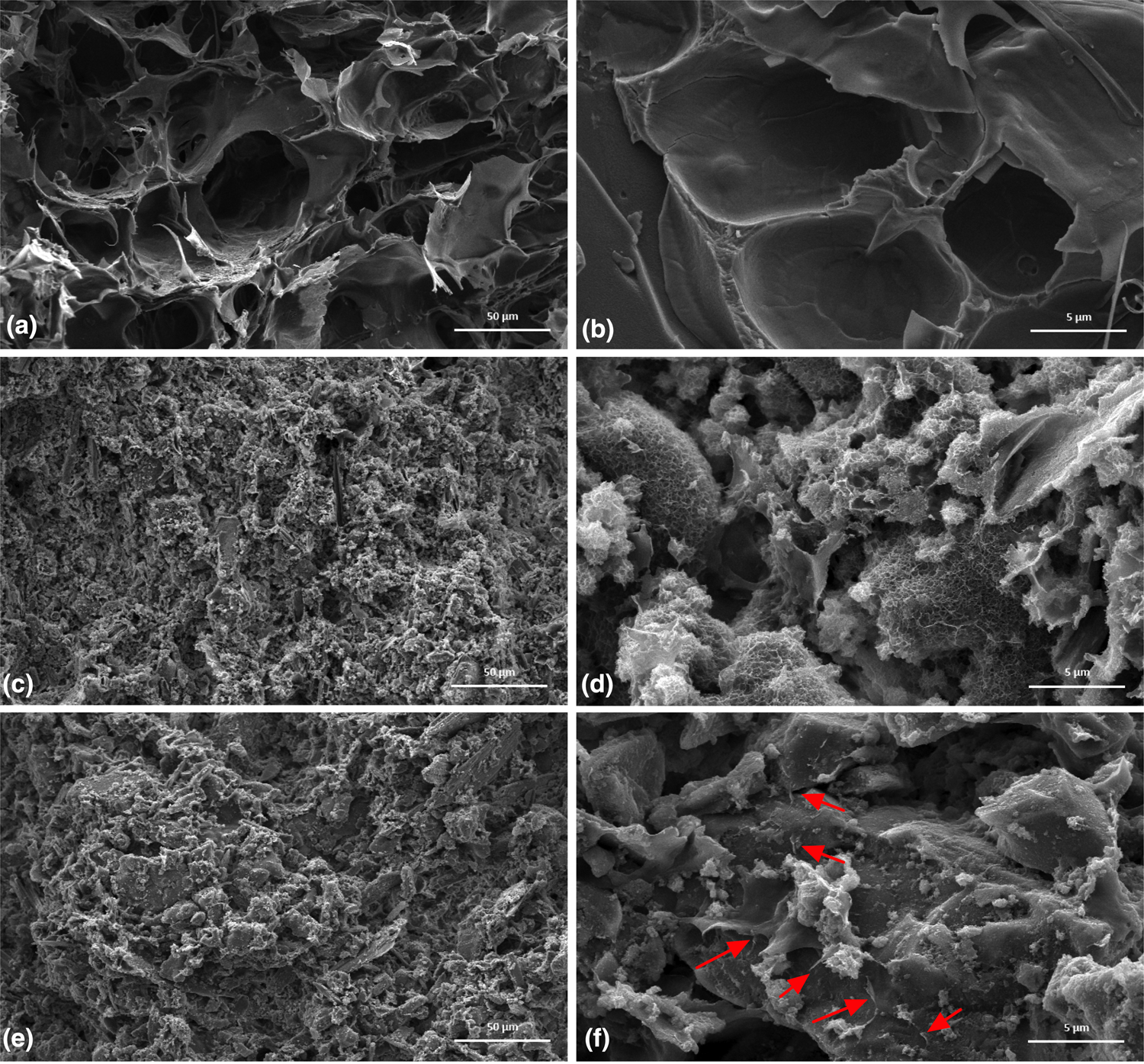
Figures 2(a) and 2(b) show the cross-sectional morphology of lyophilized liquid phase of IBS samples. It was observed that these samples had a rough microporous structure. In Figs. 2(c) and 2(d), 0GO samples had a flower-like morphology which disappeared with the incorporation of GO. With the addition of GO, the samples appeared less porous and denser when Figs. 2(d) and 2(f) were compared. GO particles were indicated with a red arrow in the SEM images; however, most of the GO particles were possibly covered inside the powder phase and fill the pores between the powder/liquid matrix. As Liu et al.[Reference Liu, Li and Xu28] indicated, incorporated GO was cross-linked by the hydration product; therefore, it was hard to detect GO particles/flakes in the samples.
(a–f) Cross-sectional SEM images of without GO and GO added lyophilized samples. (a and b) Liquid phase of IBS samples without GO; (c and d) 0GO samples; (e and f) 2GO samples; (a, c, and e) with 1000× magnification; and (b, d, and f) 10,000× magnification.
Mechanical analysis
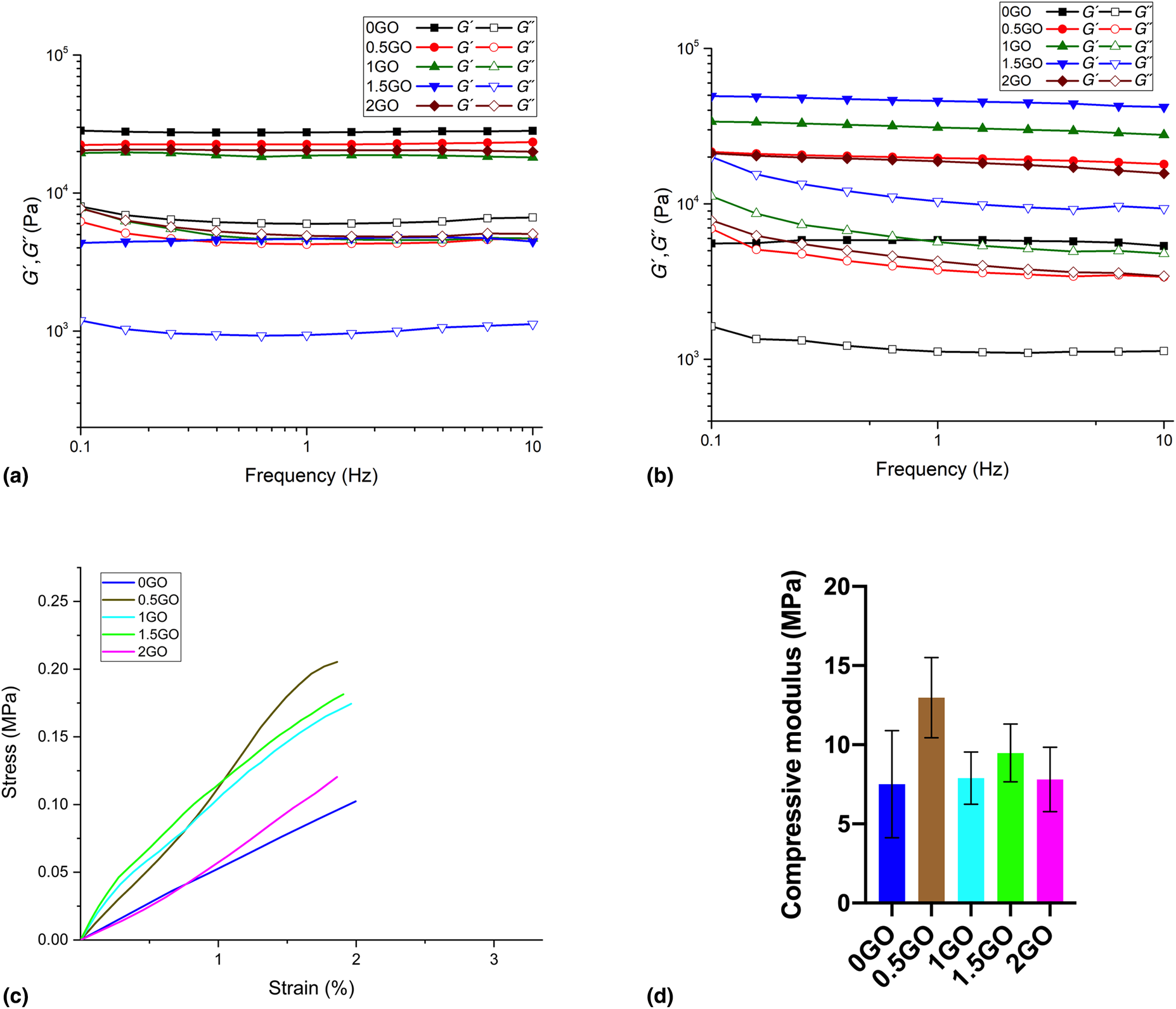
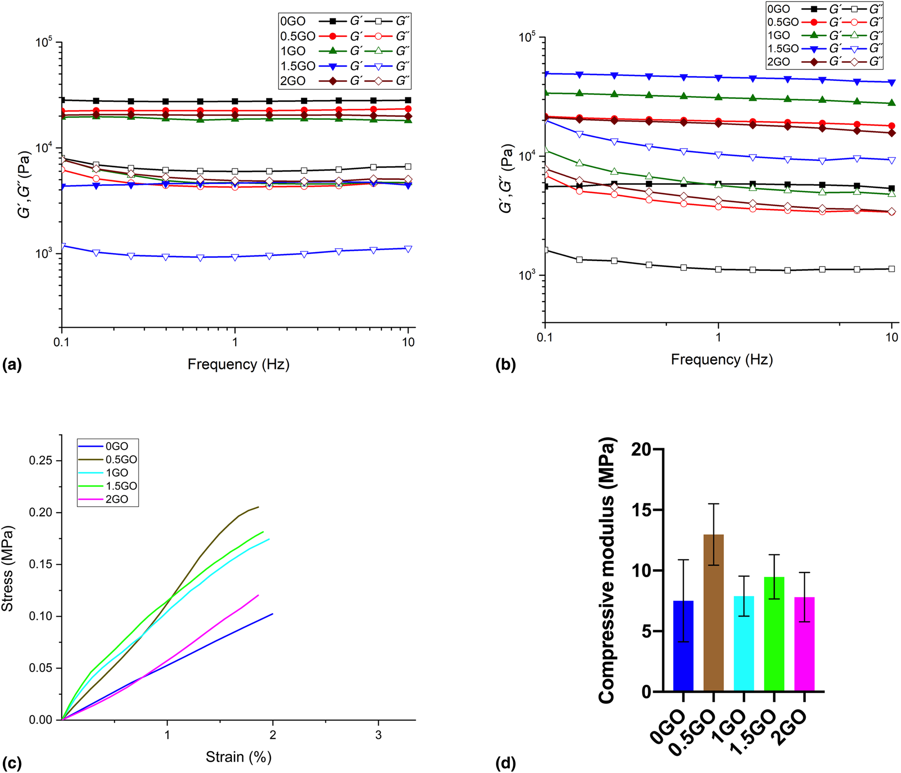
Figures 3(a) and 3(b) shows the frequency-dependent loss (G″) and storage (G′) modulus of GO-reinforced IBS samples at 25 and 37 °C using 1% strain. All samples revealed linear viscoelastic properties both at 25 and 37 °C. Also, they preserved their linear viscoelastic properties between 0.1 and 10 Hz frequency. IBS samples without GO incorporation had the highest storage modulus (G′) value at 25 °C and the lowest storage modulus (G′) value at 37 °C. All samples revealed gel-like behavior with their G′ being rather constant and much higher than their G″.[Reference von Lospichl, Hemmati-Sadeghi, Dey, Dehne, Haag, Sittinger, Ringe and Gradzielski29] 1.5GO samples showed a higher G′ value than the other samples at 37 °C, while it had the lowest G′ value at 25 °C. G′ of 1.5GO was dramatically increased with the increase of temperature from 25 to 37 °C. However, 2GO samples had a similar G′ value with 0.5GO samples. Overall, the results demonstrated that the addition of GO up to 1.5 wt% improved G′ at 37 °C. Figure 3(c) shows the stress–strain curve, and Fig. 3(d) shows the compressive modulus of the prepared samples. There was no statistically significant difference between the compressive modulus of IBS samples. Although there was no statistical significance, 0.5GO samples had a higher mean compressive modulus. These values were in the range of compressive modulus of trabecular bone which is between 3.5 and 125 MPa. Therefore, these bone cements could be used for bone replacement applications in terms of their mechanical properties.[Reference Misch, Qu and Bidez30]
Frequency-dependent loss (G″) and storage (G′) modulus of all samples group using 1% strain at (a) 25 °C and (b) 37 °C. (c) Stress–strain curve of samples. (d) Compressive modulus graph of samples.
In vitro degradation, swelling, and biocompatibility
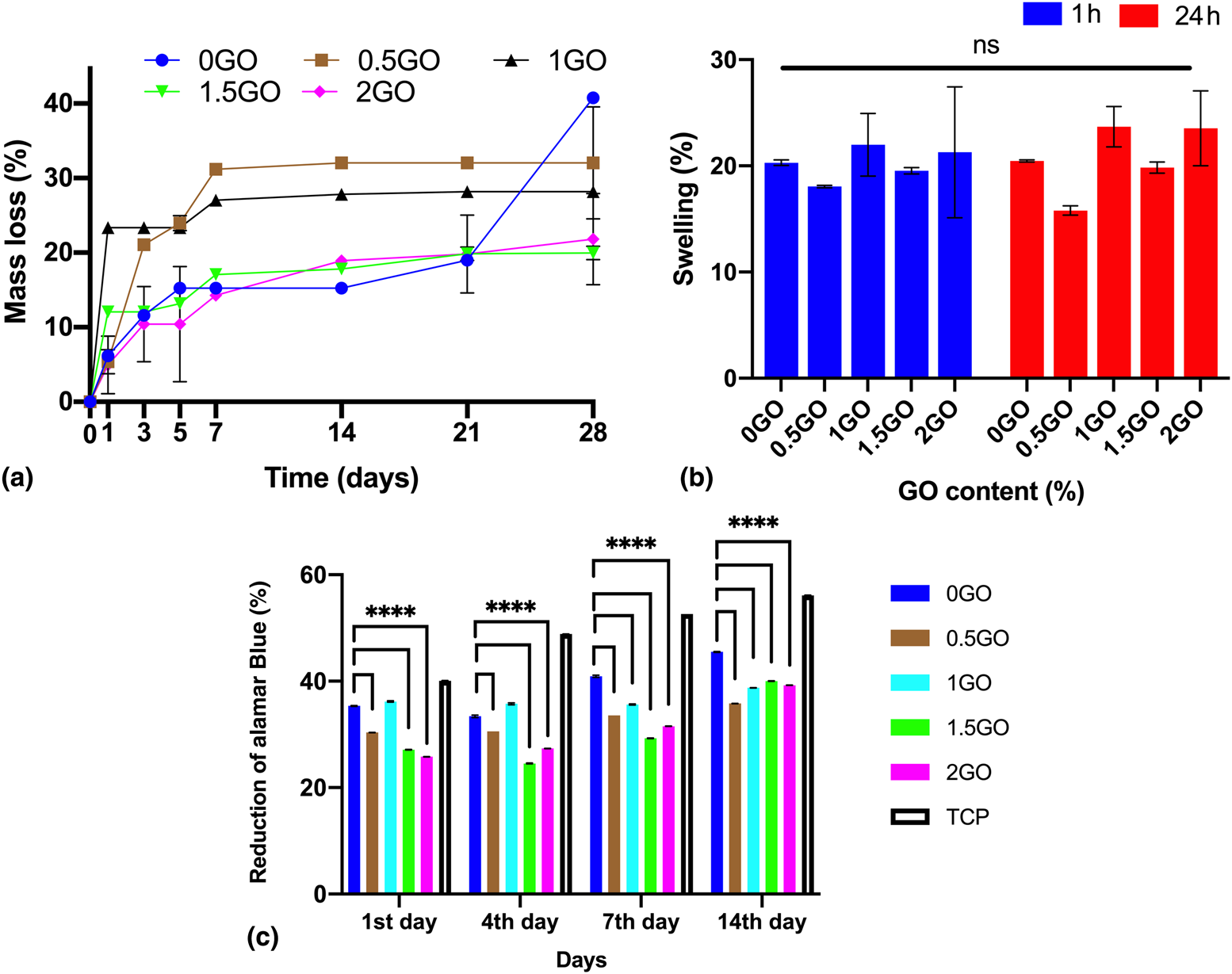
Figure 4(a) shows in vitro degradation behavior of GO added IBS samples by the measurement of mass loss (%) in PBS at 37 °C after setting of the cement phase. Until day 7, 0GO, 1.5GO, and 2GO samples lost ~20% of their weight, whereas 0.5GO and 1GO samples lost ~30% of their weight. 2GO and 1.5GO revealed nearly the same degradation pathway during the incubation period. 0GO samples also showed similar degradation profiles with 1.5GO and 2GO samples until day 14. After 28 days, 0GO samples lost 50% of their weight. Overall, it could be concluded from these results that the GO incorporation reduced the % mass loss of the samples on day 28. Figure 4(b) shows % of swelling of the GO added IBS samples which were incubated in PBS for 1 and 24 h at 37 °C. Their dry weights and wet weights were recorded, and % of swelling was calculated. The results indicated that GO incorporation did not significantly affect the swelling of the samples.
(a) In vitro degradation (n = 4), (b) swelling (%) of the all samples groups in the PBS solution at 37 °C (n = 4), and (c) alamarBlue assay results. TCP is the tissue culture plate used as a control for this study (n = 6). Values are mean ± standard deviation. ****P < 0.0001 means that there is statistically significant difference between the groups, and ns means that there is no statistically significant difference according to ANOVA and Dunnett's multiple comparison test.
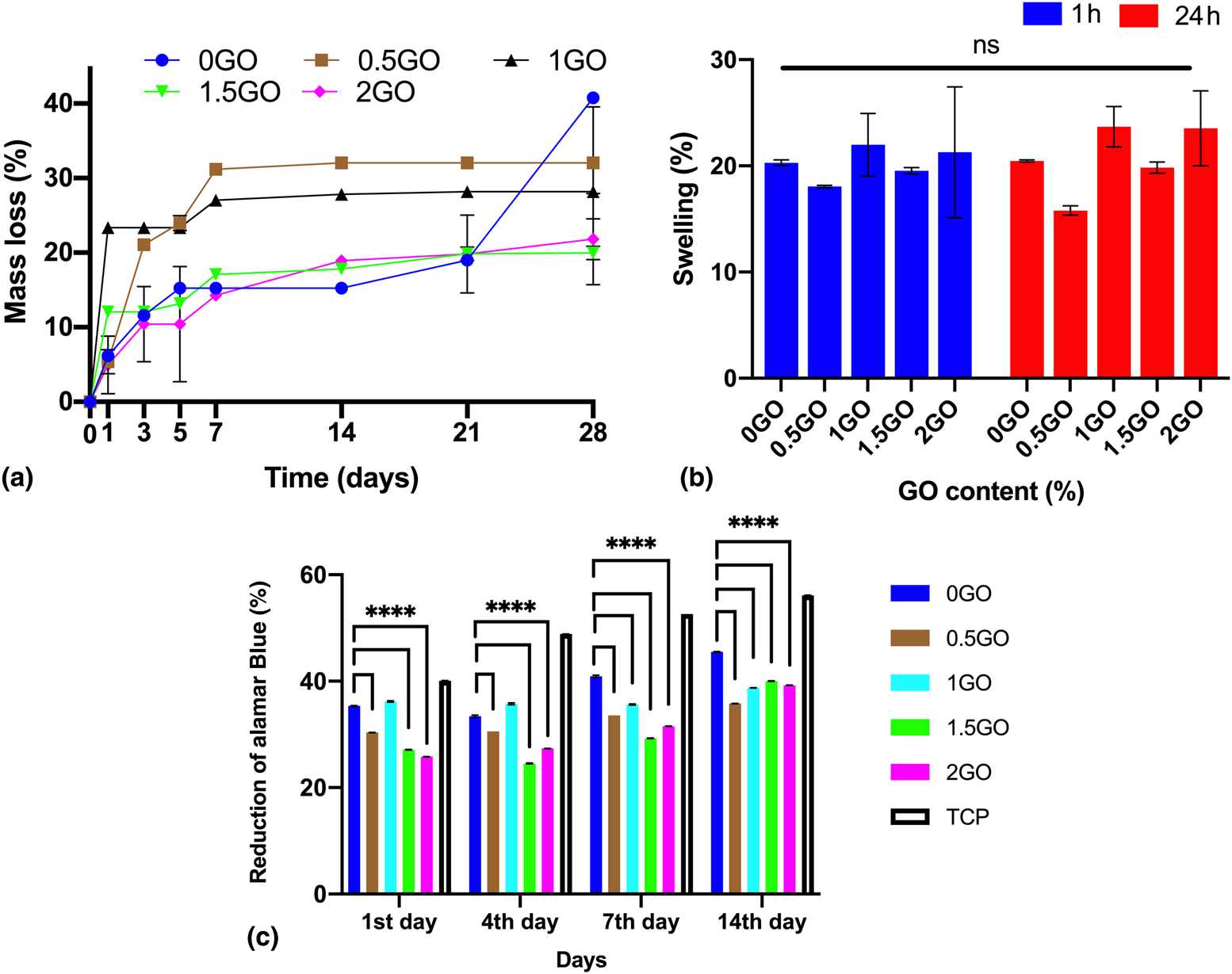
In literature, it is stated that GO shows dose-dependent toxicity to various types of cells. Therefore, bone marrow mesenchymal stem cells (BMMSCs) were seeded on IBS samples, and the cytotoxicity of prepared GO added IBS samples were tested by using the alamarBlue viability assay. The results of cell proliferation during the incubation period are shown in Fig. 4(c). Statistical analysis was conducted between the group by using Dunnett's multiple comparison test. On days 1 and 4, 0GO and 1GO samples showed significantly higher cell viability compared to the other study groups, except tissue culture plate (TCP). On days 7 and 14, the proliferation rate of 0GO was increased, and it became significantly higher than 1GO and the other GO added IBS samples. On day 7, the viability of 1GO was significantly higher than the other GO added IBS samples. When wt% of GO was increased above 1, the % cell viability did not significantly increase or decrease on day 14. There were no direct dose-dependent viability results of GO added IBS samples; however, there was an upward trend of cell viability with the time of incubation supporting that cells were growing on or in the samples.
The presence of GO in the IBS samples, affect the interaction of mesenchymal stem cells with the sample surfaces. The negative charge and polarity of GO lead to its interaction with the functional groups of cellular proteins which changes the degree of biocompatibility of the samples.[Reference Prasadh, Suresh and Wong31] Moreover, the addition of GO might have changed the dissolution and the re-precipitation rate of calcium phosphate phase. This might have also led to a change of the % viability of cells after GO incorporation.[Reference Gurunathan, Kang, Jeyaraj and Kim32] The % cell viability did not significantly increase or decrease on day 14 when GO concentration was above 1 wt%. Therefore, probably further increase in GO concentration did not have a significant impact on the mechanism of interaction between the cells and the sample surface.
Conclusion
In this study, samples were prepared by incorporating different wt% of GO to enhance the physical and biological properties of IBS. GO addition significantly reduced the setting time of the cements, improved in vitro degradation and rheological properties at 37 °C. Although the compressive modulus of different study groups did not have any significant difference, the found values were within the range of trabecular bone.
Interestingly, 1GO and 1.5GO showed higher stability, workability, rheological properties, and biocompatibility than the other GO added IBS samples. Therefore, the optimum amount of GO that our powder and liquid phase can accommodate was found to be between 1 and 1.5 wt%. Further improvement may be achieved by the surface modification of GO to improve mechanical properties. Still, more research is needed to apply such an injectable material for bone regeneration by investigating them with other cell lines and in vivo animal tests in the future.
Supplementary material
The supplementary material for this article can be found at https://doi.org/10.1557/mrc.2019.125.
Acknowledgment
This work was supported by Boğaziçi University Research fund (No. 12240) and the Scientific and Technological Research Council of Turkey (TUBITAK) (No. 117M231).