Introduction
Of the neglected tropical diseases (NTDs) prioritised by the World Health Organization, parasitic infections account for the majority, and human enterobiasis caused by Enterobius vermicularis is among them (Molyneux et al Reference Molyneux, Savioli and Engels2017). Pinworm is one of the most common nematode infections of humans; more than 200 million people, and over 20% of children in some settings, are estimated to be infected, with humans as the only known host (Lala and Upadhyay Reference Lala and Upadhyay2016). Gravid female worms migrate to the peri-anal folds at night to deposit thousands of eggs, provoking peri-anal and perineal pruritus. Eggs are transmitted by the faecal–oral route through contaminated fingers, dust, bedding, clothing and food, and embryonate within about six hours, allowing frequent retro-infection and household transmission (Song et al Reference Song, Cho, Kim, Choi and Hong2003).
Although often regarded as a nuisance infection, enterobiasis in young children can cause peri-anal itching, restlessness, bruxism, disturbed sleep, and poor appetite and, less commonly, abdominal pain, and nausea. Ectopic migration of female worms into the female genital tract, appendix, peritoneal cavity, or urinary tract has been associated with salpingitis, appendicitis, and other inflammatory lesions in numerous case reports (Erhan et al Reference Erhan, Zekioğlu, Ozdemir and Sen2000; Fleming et al Reference Fleming, Kearney, Moriarty, Redmond and Andrews2015; Kumar et al Reference Kumar, Sharma, Sachdeva and Jain2003; Macedo and MacCarty Reference Macedo and MacCarty2000; Sizer et al Reference Sizer, Nirmal, Shannon and Davies2004; Tandan et al Reference Tandan, Pollard, Money and Scheifele2002). Detection of pinworm eggs in faeces has a low yield (typically <5%) and is not the diagnostic method of choice; both classic survey work and family-medicine guidance recommend the peri-anal cellophane/adhesive-tape swab, performed in the morning before defaecation, as the most accurate and practical way to demonstrate eggs on the peri-anal skin (Chang et al Reference Chang, Liao, Huang, Chang, Chou, Tsay, Huang, Guu, Kao and Fan2009; Kucik et al Reference Kucik, Martin and Sortor2004).
Beyond enterobiasis, soil-transmitted helminths (Ascaris lumbricoides, hookworms, and Trichuris trichiura) and intestinal protozoa remain major contributors to childhood morbidity in low- and middle-income countries (Jourdan et al Reference Jourdan, Lamberton, Fenwick and Addiss2018). In young children, chronic intestinal parasitic infection can cause diarrhoea, abdominal pain, malnutrition, weight loss, intestinal blood loss, and anaemia, rectal prolapse, and impaired growth and learning, and in severe cases can be life-threatening; controlling these infections is therefore a priority public-health objective for the World Health Organization and national health ministry’s alike (Fletcher et al Reference Fletcher, Stark, Harkness and Ellis2012; WHO 2017).
Urogenital schistosomiasis due to Schistosoma haematobium is endemic in Eswatini, particularly in the low-lying, water-rich eastern (Lubombo) and south-western (Shiselweni) regions and in parts of the north, and remains a major cause of chronic morbidity in sub-Saharan Africa (Colley et al Reference Colley, Bustinduy, Secor and King2014; WHO 2022). Infection follows contact with cercaria-infested fresh water during bathing, washing, water collection, and play. In an earlier survey by our team (2018), primary schoolchildren in the Piggs Peak area showed the highest local S. haematobium prevalence (17.8%, 21/118), and the intestinal-parasite prevalence among Eswatini primary schoolchildren was 37.6% (97/258), with no helminth infection detected.
In Eswatini, the National Malaria and Neglected Tropical Diseases Programme (NMNTDP) delivers annual mass deworming to primary, junior and senior schoolchildren, but kindergarten and community-care children – those below school-entry age – are not included and had never been surveyed. At the request of the Programme, and in the absence of any baseline data for this age group, we set out to establish the first prevalence estimates of E. vermicularis, S. haematobium and intestinal parasites among Eswatini preschool children, using three simple, field-deployable methods (the cellophane peri-anal swab, the CTMS urine-sedimentation method and MIF stool concentration), and the work supports Sustainable Development Goals 3 and 17.16 (Molyneux et al Reference Molyneux, Savioli and Engels2017).
Materials and methods
Study design, setting, and participants
This cross-sectional survey was conducted during June 2019 in four administrative provinces of the Kingdom of Eswatini – Hhohho (Mbabane and Piggs Peak areas), Manzini, Lubombo (Siteki area), and Shiselweni (Nhlangano/Hlatikulu areas) (Figure 1). Sixteen kindergartens and community care points were selected in cooperation with the responsible provincial education authorities and the NMNTDP. Participating institutions enrolled voluntarily; because rural centres frequently cared for fewer children than urban ones, the number sampled per centre varied. In total, 261 preschool children were examined for pinworm; urine and stool specimens were obtained from those able to provide them (232 and 175 children, respectively).
Map of the Kingdom of Eswatini showing the four administrative provinces (Hhohho, Manzini, Lubombo, and Shiselweni) and the preschool survey sites (red circles) – Mbabane and Piggs Peak (Hhohho), Manzini (Manzini), Siteki (Lubombo), and Nhlangano and Hlatikulu (Shiselweni) – sampled during the June 2019 cross-sectional survey. Inset: Location of Eswatini within southern Africa. Provincial boundaries were digitised from a published administrative map of Eswatini and are shown schematically.

Figure 1. Long description
A geographical map plotted on a coordinate system with Latitude degrees South on the Y axis and Longitude degrees East on the X axis.
* In the North, the Hhohho province is shaded light blue and contains two survey sites: Piggs Peak and Mbabane.
* In the center, the Manzini province is shaded light green and contains the Manzini survey site.
* In the East, the Lubombo province is shaded light orange and contains the Siteki survey site.
* In the South, the Shiselweni province is shaded light pink and contains two survey sites: Hlatikulu and Nhlangano.
An inset map in the top-right corner shows Eswatini as a red dot on the eastern border of South Africa, south of Mozambique. A North arrow is located in the top-left, and a 20 kilometer scale bar is at the bottom-right. A legend at the bottom identifies the four provinces by color and the red circle as a Survey site town or area.
Peri-anal cellophane/Scotch-tape swab for E. vermicularis
Children were asked, where possible, to refrain from defaecation on the morning of collection. A transparent adhesive (cellophane/Scotch) tape mounted on a glass slide was pressed against the peri-anal folds to recover eggs, following the classic peri-anal swab technique. Slides were transported to the laboratory of the Mbabane hospital and examined by light microscopy for the characteristic asymmetrical, flattened, embryonated eggs of E. vermicularis.
Urine examination for S. haematobium (CTMS method)
A single urine specimen (about 10 mL) was collected from each participating child. Specimens were centrifuged at 1000–2000 rpm for 2 min (or left to sediment for 30 min); the supernatant was gently discarded, leaving approximately 0.6 mL of sediment, which was transferred with a 0.6-mL dropper into one chamber tube microscope-slide (CTMS) system and examined microscopically for the terminal-spined ova of S. haematobium.
Stool examination for intestinal parasites (MIF method)
A peanut-sized portion of stool was placed in a collection container using a stir bar and mixed thoroughly with 5 mL of merthiolate–iodine–formaldehyde (MIF) solution. After releasing the clamp and homogenising, the suspension was eluted through a mesh into a collection tube. Preparations were stained for at least 4 h (or overnight); the sediment was then aspirated onto a slide and examined by light microscopy for helminth ova and protozoan cysts and trophozoites. A child was classified as positive if any parasite stage was identified; ‘single infection’ denoted one species and ‘multiple infection’ two or more species.
Data handling and statistical analysis
Results were recorded per child with age, sex, province, and institution. Prevalence was expressed as the number of positive children over the number examined. Differences between sexes and among provinces, and the comparison of preschool with previously reported primary-schoolchildren data, were assessed using Fisher’s exact test or the χ2 test, as appropriate; a two-tailed p < 0.05 was considered statistically significant. Analyses were undertaken using IBM SPSS Statistics version 20.0 (IBM Corp., Armonk, NY, USA). All positive kids were reported to the NMNTDP to enable follow-up and treatment.
Results
Participants
A total of 261 preschool children were screened for pinworm (141 boys, 120 girls; mean age 4.8 ± 0.75 years). Urine specimens suitable for schistosome examination were available from 232 children (136 boys, 96 girls), and stool specimens suitable for MIF examination from 175 children (105 boys, 70 girls).
Enterobius vermicularis infection
The overall peri-anal swab prevalence of E. vermicularis was 3.8% (10/261) (Table 1). Prevalence was marginally higher in boys (4.3%, 6/141) than girls (3.3%, 4/120), a difference that was not statistically significant (p = 0.76). Province-level prevalence was highest in Manzini (6.7%, 2/30), followed by Siteki (4.8%, 2/42), Mbabane (4.0%, 4/100), Nhlangano (3.3%, 1/30), and Piggs Peak (1.7%, 1/59); regional variation was not significant (χ2 = 1.5, p = 0.83). Positive children were identified at nine institutions, and the capital, Mbabane, contributed the largest number of positive cases, consistent with its larger sampled population.
Prevalence of Enterobius vermicularis infection among preschool children by province and sex, Eswatini, 2019 (peri-anal cellophane/Scotch-tape swab)

Table 1. Long description
The table consists of four columns: Province and sex, No. examined, Age (mean plus or minus S D, years), and E. vermicularis no. (%).
* Mbabane: 100 examined, age 5.0 plus or minus 0.82, 4 (4.0%) positive. Males: 50 examined, 1 (2.0%) positive. Females: 50 examined, 3 (6.0%) positive.
* Manzini: 30 examined, age 5.1 plus or minus 0.96, 2 (6.7%) positive. Males: 17 examined, 1 (5.9%) positive. Females: 13 examined, 1 (7.7%) positive.
* Nhlangano: 30 examined, age 4.8 plus or minus 0.50, 1 (3.3%) positive. Males: 15 examined, 1 (6.7%) positive. Females: 15 examined, 0 (0.0%) positive.
* Piggs Peak: 59 examined, age 4.5 plus or minus 0.60, 1 (1.7%) positive. Males: 35 examined, 1 (2.9%) positive. Females: 24 examined, 0 (0.0%) positive.
* Siteki: 42 examined, age 4.7 plus or minus 0.55, 2 (4.8%) positive. Males: 24 examined, 2 (8.3%) positive. Females: 18 examined, 0 (0.0%) positive.
* Total: 261 examined, age 4.8 plus or minus 0.75, 10 (3.8%) positive. Total Males: 141 examined, 6 (4.3%) positive. Total Females: 120 examined, 4 (3.3%) positive.
Footnote: S D stands for Standard deviation. Statistical analysis shows p equals 0.76 for male versus female and chi-squared equals 1.5, p equals 0.83 among provinces.
SD, Standard deviation. Male versus female, p = 0.76 (Fisher’s exact test); among provinces, χ2 = 1.5, p = 0.83.
Schistosoma haematobium infection
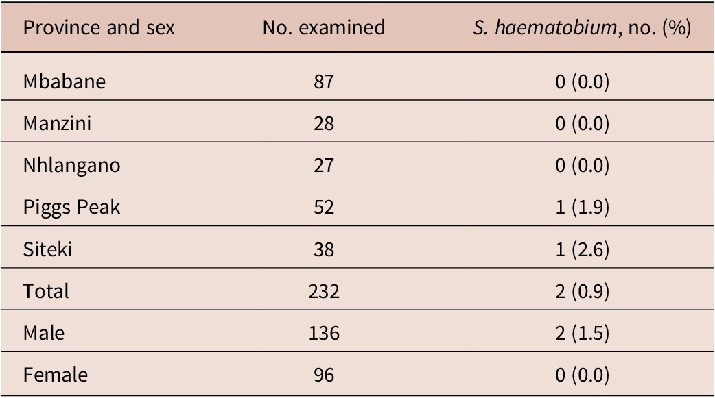
S. haematobium ova were detected by CTMS in 0.9% (2/232) of children (Table 2). Both positive children were boys (2/136, 1.5%); no girl was positive (0/96). The two cases originated from Maland Care Point in the Piggs Peak area (north) and Ward 6 Preschool in the Siteki area (east) – both within recognised endemic foci – consistent with the known endemicity of the low-lying eastern (Lubombo) and south-western regions, where surface water bodies are abundant.
Prevalence of Schistosoma haematobium infection among preschool children by province and sex, Eswatini, 2019 (urine, CTMS sedimentation)

Table 2. Long description
The table consists of three columns: Province and sex, Number examined, and S. haematobium number (percent).
Data by province:
* Mbabane: 87 examined, 0 (0.0 percent) positive.
* Manzini: 28 examined, 0 (0.0 percent) positive.
* Nhlangano: 27 examined, 0 (0.0 percent) positive.
* Piggs Peak: 52 examined, 1 (1.9 percent) positive.
* Siteki: 38 examined, 1 (2.6 percent) positive.
Summary data:
* Total: 232 examined, 2 (0.9 percent) positive.
* Male: 136 examined, 2 (1.5 percent) positive.
* Female: 96 examined, 0 (0.0 percent) positive.
Both positive cases were boys, from the Piggs Peak (Maland Care Point) and Siteki (Ward 6 Preschool) endemic foci. Male versus female, p = 0.51 (Fisher’s exact test).
Intestinal parasites: overall prevalence
Of the 175 children examined by MIF, 41.1% (72/175) were infected with at least one intestinal parasite (Table 3). Prevalence did not differ significantly between boys (42.9%, 45/105) and girls (38.6%, 27/70; p = 0.64). Province-level prevalence exceeded 40% in four of five areas – Mbabane 44.6% (25/56), Manzini 44.4% (8/18), Siteki 42.4% (14/33), and Piggs Peak 41.9% (18/43) – and was lowest in Nhlangano (28.0%, 7/25); the regional differences were not significant (χ2 = 2.2, p = 0.70).
Prevalence of intestinal parasitic infection among preschool children by province and sex, Eswatini, 2019 (MIF stool concentration)

Table 3. Long description
The table contains four columns: Province and sex, Number examined, Age (mean plus or minus S D, years), and Any parasite, number (percent).
* Mbabane: 56 examined, age 5.1 plus or minus 0.70. Total 25 (44.6 percent). Male: 28 examined, 14 (50.0 percent). Female: 28 examined, 11 (39.3 percent).
* Manzini: 18 examined, age 4.9 plus or minus 0.94. Total 8 (44.4 percent). Male: 13 examined, 5 (38.5 percent). Female: 5 examined, 3 (60.0 percent).
* Nhlangano: 25 examined, age 4.7 plus or minus 0.54. Total 7 (28.0 percent). Male: 12 examined, 3 (25.0 percent). Female: 13 examined, 4 (30.8 percent).
* Piggs Peak: 43 examined, age 4.6 plus or minus 0.59. Total 18 (41.9 percent). Male: 30 examined, 13 (43.3 percent). Female: 13 examined, 5 (38.5 percent).
* Siteki: 33 examined, age 4.8 plus or minus 0.56. Total 14 (42.4 percent). Male: 22 examined, 10 (45.5 percent). Female: 11 examined, 4 (36.4 percent).
* Total: 175 examined, age 4.8 plus or minus 0.69. Total 72 (41.1 percent). Total Male: 105 examined, 45 (42.9 percent). Total Female: 70 examined, 27 (38.6 percent).
SD, Standard deviation. Male versus female, p = 0.64 (Fisher’s exact test); among provinces, χ2 = 2.2, p = 0.70.
Intestinal parasites: species composition
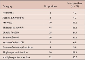
Among the 72 positive children, intestinal protozoa overwhelmingly predominated (97.2%, 70/72) over helminths (4.2%, 3/72) (Table 4). All three helminth infections were A. lumbricoides; no hookworm or T. trichiura was detected. Blastocystis hominis was the most frequent species (61.1%, 44/72), followed by Giardia lamblia (34.7%, 25/72), Entamoeba coli (22.2%, 16/72), Iodamoeba butschlii (6.9%, 5/72), and Entamoeba histolytica/dispar (5.6%, 4/72). Single-species infection accounted for 69.4% (50/72) of positive children, and multiple infection for 30.6% (22/72). The three A. lumbricoides infections occurred at Mangwaneni Social Centre (Mbabane) and Town Ward 5 (Nhlangano).
Species composition and infection multiplicity among the 72 intestinal-parasite-positive preschool children, Eswatini, 2019

Table 4. Long description
The table consists of three columns: Category, Number positive, and Percentage of positives (n = 72).
• Helminths: 3 positive cases (4.2 percent). All helminth infections were Ascaris lumbricoides.
• Protozoa: 70 positive cases (97.2 percent).
• Blastocystis hominis: 44 positive cases (61.1 percent).
• Giardia lamblia: 25 positive cases (34.7 percent).
• Entamoeba coli: 16 positive cases (22.2 percent).
• Iodamoeba butschlii: 5 positive cases (6.9 percent).
• Entamoeba histolytica or dispar: 4 positive cases (5.6 percent).
• Single-species infection: 50 positive cases (69.4 percent).
• Multiple-species infection: 22 positive cases (30.6 percent).
Note: Percentages sum to more than 100 percent due to multiple infections. No hookworm or Trichuris trichiura was detected.
Percentages are of the 72 positive children and sum to more than 100% because of multiple infections. All helminth infections were Ascaris lumbricoides; no hookworm or Trichuris trichiura was detected.
Discussion
To our knowledge, this is the first survey to establish baseline prevalence of E. vermicularis, S. haematobium and intestinal parasites among preschool children in the Kingdom of Eswatini. The three simple methods used – the cellophane/Scotch-tape swab, CTMS urine sedimentation and MIF stool concentration – proved readily transferable to local laboratory staff and, together, gave a rounded picture of the parasitic burden in a previously unmonitored age group.
The pinworm prevalence of 3.8% is modest but not negligible for a group in which no prior data existed. The slightly higher prevalence in boys and the absence of significant regional variation are consistent with the crowding- and hygiene-driven, largely age-dependent epidemiology of enterobiasis, in which household and classroom transmission tends to homogenise risk across settings. Because pinworm eggs are notoriously under-detected in faeces, the peri-anal swab is essential; the low faecal yield (<5%) reported in the literature underlines why routine faecal screening would have substantially underestimated the true burden (Chang et al Reference Chang, Liao, Huang, Chang, Chou, Tsay, Huang, Guu, Kao and Fan2009).
The two S. haematobium infections were both in boys and both occurred in the northern (Piggs Peak) and eastern (Siteki) endemic foci, mirroring the recognised endemicity of the low-lying, water-rich regions of Eswatini (Colley et al Reference Colley, Bustinduy, Secor and King2014). The exclusively male distribution is compatible with greater freshwater contact among boys, although the small number of cases precludes firm inference. Even a low measured prevalence in preschoolers is programmatically important, because early and repeated exposure sustains transmission and because this age group is not currently reached by school-based interventions (WHO 2022). More recent data corroborate this continuing risk: a 2023 cross-sectional survey of senior primary-school pupils in the Siphofaneni area of the Eswatini Lowveld – an older age group and a considerably smaller sample (n = 200) than the present study – found a S. haematobium prevalence of 16.0% (32/200), confirming that urogenital schistosomiasis remains actively transmitted in Eswatini’s low-lying endemic foci in the years since our 2019 survey and reinforcing the case for extending screening to preschool children in these same catchment areas (Maseko et al Reference Maseko, Masuku, Dlamini and Fan2023).
The most striking finding was the high burden of intestinal parasites (41.1%), which was dominated by intestinal protozoa (97.2% of positives), led by Blastocystis hominis and Giardia lamblia – a pattern typical of settings where waterborne and faecal–oral protozoan transmission outweighs soil-transmitted helminthiasis, and consistent with the central role of water, sanitation and hygiene in protozoan control (Ryan and Cacciò Reference Ryan and Cacciò2013; Speich et al Reference Speich, Croll, Fürst, Utzinger and Keiser2016; Stensvold and Clark Reference Stensvold and Clark2016). This predominance also has therapeutic implications, since standard albendazole/mebendazole deworming does not treat giardiasis or other protozoan infections. The preschool prevalence numerically exceeded the 32.2% and 40.5% we recorded among Eswatini primary schoolchildren in 2010 and 2019, respectively (Fan et al Reference Fan, Liao, Lyu, Sukati, Ji, Cho, Jien, Huang, Chang, Chiu, Nara, Tsubouchi, Huang, Tu, Lan and Chao2012; Yin et al Reference Yin, Lee, Dlamini, Maphalala, Liao and Fan2022), although the difference was not statistically significant (p > 0.5), indicating that the youngest, untreated children carry a burden at least as high as that of older, treated children. A further contrast with 2010 and 2019 was the reappearance of A. lumbricoides in three preschool children, whereas no helminth was found in the earlier schoolchildren survey in 2010; although small, this signals that soil-transmitted helminthiasis and its sequelae in early childhood warrant continued vigilance alongside chemotherapy (Jourdan et al Reference Jourdan, Lamberton, Fenwick and Addiss2018).
Several limitations should be noted. Prevalence estimates rest on a single specimen per child for each method; because pinworm egg deposition, schistosome egg excretion and protozoan cyst shedding all fluctuate, single-sample screening tends to underestimate infection, so our figures are best regarded as minimum estimates. Participation by institution and child was voluntary, and centre sizes varied, which may limit representativeness, and the number of positive cases for pinworm and schistosomiasis was small, so subgroup comparisons are exploratory. Speciation of the E. histolytica/dispar complex by microscopy alone cannot distinguish pathogenic E. histolytica from non-pathogenic E. dispar.
The public-health implications are clear. Eswatini’s annual school-based deworming reaches primary, junior and senior pupils but not kindergarten or community-care children; our data indicate that preschoolers carry a high burden of intestinal protozoa and a measurable burden of pinworm and urogenital schistosomiasis, supporting their inclusion in screening and preventive-chemotherapy planning by the Ministries of Health and Education, together with attention to water, sanitation and hygiene for protozoan control. By building local capacity in the cellophane-swab, CTMS and MIF methods and by feeding results back promptly to the NMNTDP, the future work should help establish a sustainable sample–diagnose–treat–follow-up pathway for childhood parasitic infection, in keeping with Sustainable Development Goals 3 and 17.16.
Acknowledgements
We thank the Ministry of Health of the Kingdom of Eswatini; the Taipei Medical University Medical Mission in Eswatini; Mrs Fortunate Lushaba and Mr Phumlani Motsa for field and logistical assistance; and the participating kindergartens, community care points, children, and their guardians.
Financial support
This work was supported by Taipei Medical University Hospital [107-1604-008-400].
Competing interests
The author(s) declare none.
Ethical standard
Informed/verbal consent was obtained from the parents/guardians and carers of participating children. Formal ethical approval was granted by the Eswatini Health and Human Research Review Board (SHR172/2019).