Introduction
Trichinellosis is a cosmopolitan zoonosis caused by the ingestion of raw meat infected by roundworms of the genus Trichinella. Currently, the Trichinella genus consists of two clades: the non-encapsulated clade with three species infecting mammals and birds (T. pseudospiralis), or mammals and reptiles (T. papuae and T. zimbabwensis), and the encapsulated clade with six species (Trichinella spiralis, T. nativa, T. britovi, T. murrelli, T. nelsoni and T. patagoniensis) and three genotypes (Trichinella T6, T8 and T9) infecting only mammals (Pozio et al., Reference Pozio, Hoberg, La Rosa and Zarlenga2009; Krivokapich et al., Reference Krivokapich, Pozio, Gatti, Prous, Ribicich, Marucci, La Rosa and Confalonieri2012). The differentiation of Trichinella taxa is of importance for biological and epidemiological studies. The tools for the identification of Trichinella species were first based on biological (e.g. host specificity, geographical distribution, cross-breeding, nurse cell development and resistance to freezing), biochemical (e.g. isoenzyme analysis) and molecular methods (Murrell et al., Reference Murrell, Lichtenfels, Zarlenga and Pozio2000). A plethora of molecular diagnostic methods based on the conventional polymerase chain reaction (PCR) (Dick et al., Reference Dick, Lu, deVos and Ma1992; Bandi et al., Reference Bandi, La Rosa, Bardin, Damiani, Comincini, Tasciotti and Pozio1995), PCR-restriction fragment length polymorphism (RFLP) (Wu et al., Reference Wu, Nagano, Pozio and Takahashi1999), PCR-based single strand conformation polymorphism analysis (Gasser et al., Reference Gasser, Zhu, Monti, Dou, Cai and Pozio1998), multiplex PCR (Zarlenga et al., Reference Zarlenga, Chute, Martin and Kapel1999) and real-time PCR (Guenther et al., Reference Guenther, Nockler, von Nickisch-Rosenegk, Landgraf, Ewers, Wieler and Schierack2008; Tantrawatpan et al., Reference Tantrawatpan, Intapan, Thanchomnang, Lulitanond, Boonmars, Wu, Morakote and Maleewong2012) are available to distinguish Trichinella taxa.
Recently, the pyrosequencing technique has facilitated direct sequencing by the synthesis of short nucleotide fragments using an enzymatic-cascade system (Ahmadian et al., Reference Ahmadian, Ehn and Hober2006). This method has been used for high-throughput genotyping of protozoan parasites (Sreekumar et al., Reference Sreekumar, Hill, Miska, Vianna, Yan, Myers and Dubey2005; Stensvold et al., Reference Stensvold, Traub, von Samson-Himmelstjerna, Jespersgaard, Nielsen and Thompson2007, Reference Stensvold, Lebbad, Verweij, Jespersgaard, von Samson-Himmelstjerna, Nielsen and Nielsen2010; Lulitanond et al., Reference Lulitanond, Intapan, Tantrawatpan, Sankuntaw, Sanpool, Janwan and Maleewong2012) and allows the detection of nucleotide polymorphisms that can be used for parasite differentiation at the species level. The aim of the present work was to develop a tool to identify, by PCR and pyrosequencing, four Trichinella species, three of which are circulating in humans and animals of South-East Asia.
Materials and methods
Sources of Trichinella larvae
Trichinella sp. larvae belonging to four species, i.e. T. spiralis, T. papuae, T. pseudospiralis and T. zimbabwensis, were used in this study. The T. spiralis strain (code ISS62), maintained in laboratory mice by serial passages, originated from a domestic pig that was the source of infection for a human outbreak in the Mae Hong Son Province, Thailand, in 1986 (Pozio & Khamboonruang, Reference Pozio and Khamboonruang1989). The T. pseudospiralis strain (code ISS13) was the original strain isolated from Russia in 1972 (Garkavi, Reference Garkavi1972). The T. zimbabwensis (code ISS1029) strain was isolated from a Nile crocodile of Zimbabwe in 1996 (Pozio et al., Reference Pozio, Foggin, Marucci, La Rosa, Sacchi, Corona, Rossi and Mukaratirwa2002). The T. papuae (code ISS4120) strain was isolated from a muscle biopsy of a patient who had worked in Malaysia and had a history of eating raw pork from a wild boar in 2005 (Intapan et al., Reference Intapan, Chotmongkol, Tantrawatpan, Sanpool, Morakote and Maleewong2011). Trichinella larvae were isolated from mouse muscles by digestion and stored in 70% ethanol at − 20°C. This study was approved by the Animal Ethics Committee of the Khon Kaen University, based on the Ethics of Animal Experimentation of the National Research Council of Thailand (Reference No. 0514.1.12.2/70).
Molecular analysis
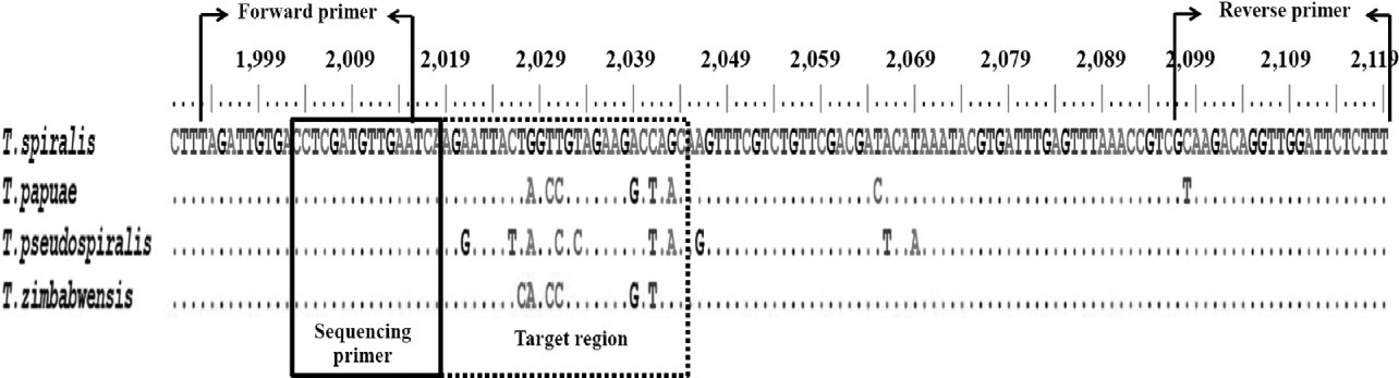
The mitochondrial large subunit ribosomal RNA (lsu-RNA) of T. spiralis (GU339148), T. papuae (AY851286), T. pseudospiralis (DQ159091) and T. zimbabwensis (EF517131), available in GenBank were selected to find a suitable region to distinguish the four species. Trichinella genus specific PCR primers (TriPyr_F, 5′-TAGATTGTGACCTCGATGTTGAA-3′ and biotinylated TriPyr_R, biotin-5′-AAAGAGAATCCAACCTGTCTTGC-3′) and sequencing primer (TriPyr_S, 5′-CCTCGATGTTGAATCA-3′) were designed by using pyrosequencing assay design software (PyroMark™ Q96 ID software version 1.0; Biotage, Uppsala, Sweden) (fig. 1).
Alignment of the mitochondrial large subunit ribosomal RNA gene of Trichinella spiralis (GU339148), T. papuae (AY851286), T. pseudospiralis (DQ159091) and T. zimbabwensis (EF517131), showing the position of the forward and reverse PCR primers (arrows), of the sequencing primer (solid rectangle) and the position of target regions used for the species differentiation (dotted rectangle).

Muscle larvae were isolated from mouse muscles by an artificial pepsin digestion (Nöckler & Kapel, Reference Nöckler, Kapel, Dupouy-Camet and Murrell2007). Ten larvae of each species mixed with 250 mg of mouse muscles (equivalent to approximately 40 larvae/g), and each species of Trichinella larvae (one or ten larvae), were homogenized with a disposable polypropylene pestle, followed by DNA extraction using a NucleoSpin Tissue kit (Macherey-Nagel GmbH & Co., Duren, Germany) according to the manufacturer's instructions. The DNA was eluted in 50 μ of distilled water, and 5 μl of this solution was used for PCR. The PCR products were obtained from conventional PCRs using the TriPyr_F and TriPyr_R primers. Positive control plasmids containing T. spiralis, T. papuae, T. pseudospiralis and T. zimbabwensis DNA were constructed by cloning the 127 bp PCR product of the lsu-RNA into the pGEM®-T Easy vector (Promega, Madison, Wisconsin, USA) according to the manufacturer's instructions. The recombinant plasmids were propagated in Escherichia coli, and the cloned inserts were sequenced to confirm their identity.
The 127 bp of the lsu-RNA were amplified from genomic DNAs of larvae of the four Trichinella species, of mouse muscle samples spiked with Trichinella larvae, and of positive control plasmids, on a GeneAmp PCR system 9700 thermal cycler (Applied Biosystems, Singapore). The PCR mix consisted of 1 × PCR buffer (Invitrogen, Carlsbad, California, USA) with 0.2 mm of each deoxyribonucleoside triphosphate (dNTP), 1.5 mm MgSO4, 0.2 μ m each of TriPyr_F primer and biotinylated TriPyr_R primer, 0.625 U of Platinum Taq DNA polymerase high fidelity (Invitrogen) and 5 μl of genomic DNA, in a final reaction volume of 25 μl. The amplification procedure was as follows: 5 min at 94°C for initial denaturation followed by 35 cycles of denaturation at 94°C for 30 s, annealing at 56°C for 30 s, and extension at 72°C for 30 s, followed by a final extension at 72°C for 7 min. Electrophoresis on a 1.5% agarose gel was performed to verify the amplification of single products. For the analytical specificity, DNA samples of organisms other than those of the genus Trichinella (helminths: Ascaris lumbricoides, Ancylostoma caninum, Trichuris trichiura, Capillaria philippinensis, Strongyloides stercoralis, Trichostrongylus spp., Taenia spp., Opisthorchis viverrini, Haplorchis taichui, Clonorchis sinensis, Centrocestus spp., Stellantchasmus spp., Paragonimus heterotremus, Schistosoma mekongi, Fasciola gigantica, Echinostoma malayanum and Phaneropsorus boneii; protozoa: Giardia lamblia and Isospora belli; human and mouse) were tested.
Following PCR amplification, biotinylated PCR products were placed in 96-well plates and bound to Streptavidin Sepharose™ beads (GE Healthcare BioSciences AB, Uppsala, Sweden). The PCR products immobilized with beads were denatured and the non-biotinylated fragments were washed from the beads using a PyroMark™ Vacuum Prep Workstation (Biotage). Subsequently, the beads were released and resuspended in 40 μl annealing buffer (20 mm Tris-acetate, 5 mm magnesium acetate, pH 7.6) containing 0.4 μ m TriPyr_S sequencing primer. Samples in duplicate were heated at 80°C for 2 min before performing the pyrosequencing reaction using PyroMark™ Gold Q96 SQA reagents (Biotage) on a PSQ™ 96MA instrument (Biotage). For each assay, two negative control samples were included (sequencing primer and biotinylated primer, only). Following the completion of the pyrosequencing reaction, the PyroMark™ Q96 ID software version 1.0 (Biotage) was run to produce a pyrogram and to analyse the sequencing data. The readouts were interpreted manually in cases where the target sequences contained up to four homopolymers, because of software limitations.
Results and discussion
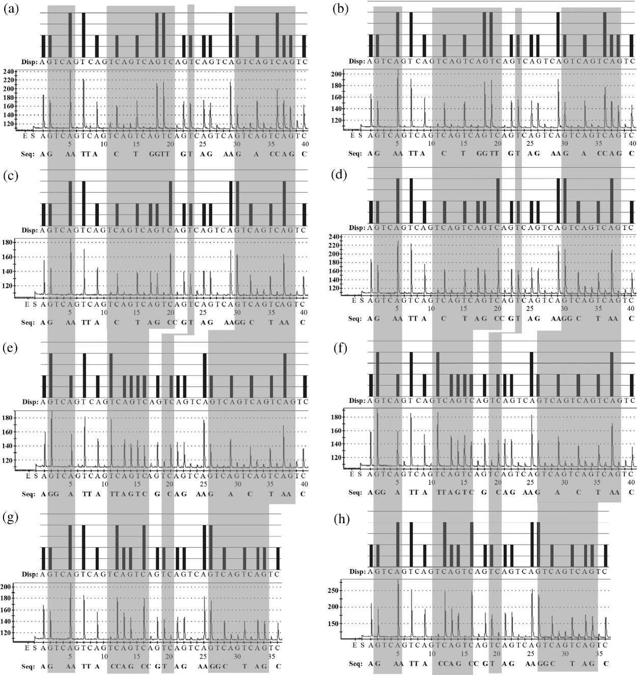
No amplification products were obtained with DNA samples of organisms different from those of the genus Trichinella (see the Materials and methods section for a list). The DNA pyrosequencing of 26 nucleotides of the target region (nucleotide number ranging from 2019 to 2044) (fig. 1) unequivocally identified each of the four Trichinella species (fig. 2). The nucleotides present at positions 2021, 2026–2028, 2030–2031, 2033, 2039, 2041 and 2043, were used for the species identification. For T. spiralis, specific nucleotides were found at positions 2028 (G), 2031 (T) and 2041 (C). For the non-encapsulated clade, specific nucleotides were found at positions 2028 (A), 2031 (C) and 2041 (T) (fig. 2 and table 1). The species T. papuae and T. zimbabwensis were distinguished at positions 2030 (C) and 2039 (G). In the case of T. pseudospiralis, specific nucleotides were found at positions 2021 (G), 2026 (T) and 2033 (C). Finally, in the case of T. zimbabwensis, the presence of a C at position 2027 allows its identification (fig. 2 and table 1). No difference was observed for each species among control plasmids, DNA extracted from one or ten larvae, and DNA extracted from mouse muscles spiked with ten Trichinella larvae. DNA sequences from pyrosequencing of each sample were identical to the sequences produced by Sanger sequencing. The negative controls did not yield pyrograms.
Pyrograms showing the sequence analyses of Trichinella spiralis (a, b), T. papuae (c, d), T. pseudospiralis (e, f) and T. zimbabwensis (g, h) genes. The pyrogram patterns (top of each panel), representative raw data (bottom of each panel) of control plasmids (a, c, e, g), and DNA extracted from each larva of the four Trichinella species spiked in mouse muscles (b, d, f, h), are shown. The letters under the black bars show the dispensation (Disp) order. The sequence (Seq) detected by pyrosequencing is shown below each panel. The y-axis shows the fluorescence emission by the incorporation of a nucleotide base; the x-axis shows the bases added at that point in time of the pyrosequencing. The light grey areas show the pyrogram for the differentiation of each of the four target Trichinella species. E, enzyme; S, substrate; A, G, T, C, four different nucleotides.
Regions of the mitochondrial large subunit ribosomal RNA (lsu-RNA) gene and nucleotide patterns used for differentiation of Trichinella spiralis, T. pseudospiralis, T. papuae and T. zimbabwensis by pyrosequencing; GenBank numbers for T. spiralis (GU339148), T. papuae (AY851286), T. pseudospiralis (DQ159091) and T. zimbabwensis (EF517131).

aSpecific nucleotide for non-encapsulated Trichinella species, bT. pseudospiralis, cT. zimbabwensis, dT. papuae and T. zimbabwensis, and eT. spiralis.
Pyrosequencing is becoming more common for the rapid differentiation and single nucleotide genotyping of protozoan parasites such as Toxoplasma gondii (Sreekumar et al., Reference Sreekumar, Hill, Miska, Vianna, Yan, Myers and Dubey2005), Blastocystis hominis (Stensvold et al., Reference Stensvold, Traub, von Samson-Himmelstjerna, Jespersgaard, Nielsen and Thompson2007), Entamoeba complex (Stensvold et al., Reference Stensvold, Lebbad, Verweij, Jespersgaard, von Samson-Himmelstjerna, Nielsen and Nielsen2010), and Plasmodium vivax and P. falciparum (Lulitanond et al., Reference Lulitanond, Intapan, Tantrawatpan, Sankuntaw, Sanpool, Janwan and Maleewong2012). Laboratory personnel can be easily trained to perform this technique because the pyrosequencing procedure is relatively simple. Furthermore, the cost of pyrosequencing reagents is lower than that of conventional sequencing reagents. After the PCR amplification, the pyrosequencing run time for 96 samples is approximately 1 h.
To the best of our knowledge, this is the first work to identify Trichinella larvae at the species level by pyrosequencing. DNA pyrosequencing coupled with PCR amplification, using a new primer set targeting the highly conserved region of the lsu-RNA sequence, yields species-level differentiation of the three non-encapsulated Trichinella species and the encapsulated T. spiralis, three of which are circulating in South-East Asia (Jongwutiwes et al., Reference Jongwutiwes, Chantachum, Kraivichian, Siriyasatien, Putaporntip, Tamburrini, La Rosa, Sreesunpasirikul, Yingyourd and Pozio1998; Pozio et al., Reference Pozio, Hoberg, La Rosa and Zarlenga2009; Kusolsuk et al., Reference Kusolsuk, Kamonrattanakun, Wesanonthawech, Dekumyoy, Thaenkham, Yoonuan, Nuamtanong, Sa-Nguankiat, Pubampen, Maipanich, Panitchakit, Marucci, Pozio and Waikagul2010; Intapan et al., Reference Intapan, Chotmongkol, Tantrawatpan, Sanpool, Morakote and Maleewong2011; Van De et al., Reference Van De, Trung, Ha, Nga, Ha, Thuy, Duyet le and Chai2012). This new diagnostic tool shows high sensitivity and specificity. In fact, it allows identification of single Trichinella larvae, and no amplification product was detected with DNAs from other parasites, either helminths or protozoa.
In this study, only one isolate has been analysed for each of the four Trichinella species. Therefore, nucleotide variation in the target region needs to be investigated among isolates of these species from different geographical origins. For the bioinformatic analysis, the comparison with the other Trichinella taxa needs further investigations, since the 26-nucleotide sequence of T. spiralis is identical to that of other encapsulated taxa (data not shown). This rapid and specific assay is a promising alternative method that can be used for the differentiation of larvae of Trichinella taxa present in muscle tissues of both domestic and wild animals.
Acknowledgements
This research was funded by grants from the National Science and Technology Development Agency (Discovery Based Development Grant); the Higher Education Research Promotion and National Research University Project of Thailand, Office of the Higher Education Commission, Thailand; the Faculty of Medicine, Khon Kaen University. L.S. was supported by the Khon Kaen University grant. W.M. was supported by TRF Senior Research Scholar Grant, Thailand Research Fund grant number RTA5580004. This work was also partially supported by the Directorate-General for Health and Consumers (DG SANCO) of the European Commission (2011 funds).