Introduction
Natural killer T cell lymphoma of the sinonasal tract is an important, destructive lesion of the nose and midface which runs a progressive and relentless course. Diagnosis can be difficult due to this lesion's nonspecific presentation, large amounts of ischaemic necrosis, and frequent paucity of malignant cells on histological examination. Therefore, clinicians should be aware of the seemingly innocuous presentation of this potentially fatal disease.
Case reports
Case one
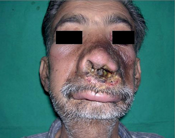
A 45-year-old man presented with swelling over the dorsum of the nose and lips, together with a palatal perforation (Figure 1). The patient had undergone dacryocystorhinostomy four years previously, after which he had begun to suffer symptoms. He had also undergone multiple intranasal biopsies, which had revealed chronic inflammation of inconclusive aetiology. He reported no chest or renal symptoms.
Clinical photograph of case one, showing nasal crusting and necrosis, which developed after dacryocystorhinostomy.

Diagnostic nasal endoscopy revealed complete destruction of the cartilaginous and bony parts of the nasal septum, with profuse, foul-smelling discharge and crusting.
Blood test results were normal, except for a raised erythrocyte sedimentation rate (ESR) of 65 mm/hour (normal range 0–20mm/hr) and a raised C-reactive protein concentration (CRP) of 20 mg/ml. Biochemical test results were normal. Immunological test results for antineutrophilic cytoplasmic antibodies, anti-double-stranded-DNA and serum angiotensin-converting enzyme were within normal limits. Urine analysis was normal.
A computed tomography (CT) scan of the paranasal sinuses suggested bony destruction and soft tissue inflammation, with sclerosis of the maxillary sinus wall. High resolution CT of the chest was normal.
Bone marrow studies did not reveal any evidence of lymphoma infiltration. Histopathological examination of the nasal biopsy suggested acute necrotic inflammation with small vessel vasculitis.
A repeated biopsy was performed. Immunohistochemical analysis showed atypical lymphocytes with angiocentric destruction, with CD3− and CD43+ but negative for CD20 (B cell marker). This confirmed the diagnosis of natural killer T cell lymphoma.
The patient was commenced on chemotherapy, but unfortunately succumbed to intercurrent infection.
Case two
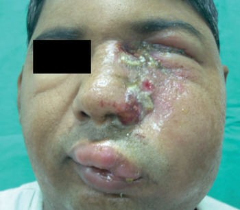
A 35-year-old man presented one month after endoscopic sinus surgery for left-sided nasal polyposis. In the immediate post-operative period, he had developed progressive, left-sided facial swelling with erythema, mimicking cellulitis, followed by ulceration (Figure 2).
Clinical photograph of case two, showing facial swelling and necrosis, which developed after endoscopic sinus surgery.

Diagnostic nasal endoscopy revealed oedematous, necrotic tissue with an intact septum.
As in the previous case, this patient did not complain of any chest or renal symptoms. There was no evidence of lymphadenopathy on general examination.
Blood tests revealed a raised ESR of 50 mm/hour and a raised CRP of 18 mg/ml. Anti-neutrophilic cytoplasmic antibody testing was negative. Urine analysis was normal.
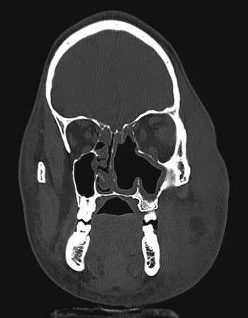
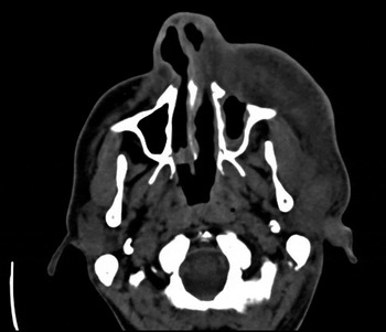
Computed tomography of the paranasal sinuses revealed an enhancing soft tissue swelling on the left side of the face, with periorbital oedema, without any localised collection or bony erosion (Figures 3 and 4).
Coronal computed tomography scan of the paranasal sinuses of case two, showing soft tissue oedema with an intact septum.

Axial computed tomography scan of the paranasal sinuses of case two, showing destruction of the anterior wall of the maxilla.

Histopathological examination of a nasal biopsy showed areas of necrosis with inflammatory granulation tissue, with sheets of histiocytes, lymphocytes and neutrophils. Fungal staining was negative.
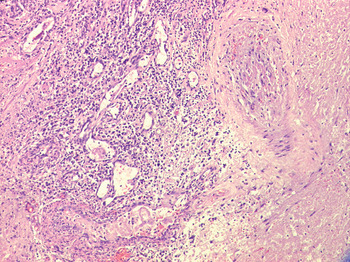
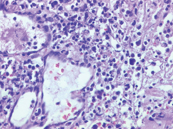
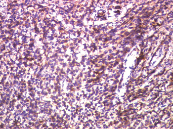
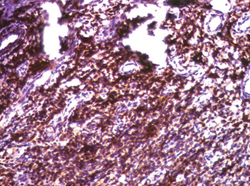
Due to a high index of clinical suspicion, a repeated biopsy was undertaken. Immunohistochemical analysis showed atypical lymphocytes with angiocentric destruction (Figures 5 and 6), with Clusters of Differentiation (CD)3− and CD43+, but negative for CD20 (B cell marker). This confirmed the diagnosis of natural killer T cell lymphoma (Figures 7 and 8).
Photomicrograph of nasal tissue biopsy showing angiocentricity. (H&E; ×10)

Photomicrograph showing angiocentricity and a polymorphous population of atypical lymphocytes. (H&E; ×40)

Photomicrograph showing immunohistochemical staining of case two biopsy, with the lymphoid cell population staining positively for antibodies against cluster of differentiation 43+ glycoprotein. (×40)

Photomicrograph showing immunohistochemical staining of case two, with the lymphoid cell population staining positively for antibodies against cluster of differentiation 3− glycoprotein. (×40)

The patient was commenced on chemotherapy, but unfortunately succumbed to intercurrent infection within two months.
Discussion
Lethal midline granuloma is a rare clinical entity characterised by progressive and fatal destruction of the midline facial structures. In the past, it has been referred to by various other names, such as polymorphic reticulosis, granuloma gangraenescens and angiocentric immunoproliferative lesion, thereby adding confusion to the disease description. Now, it has been universally accepted that most cases can be considered as centrofacial malignant lymphomas of T cell origin, including those cases with an earlier diagnosis of inflammation.Reference Chen, Fong, Su, Ting, Hong and Leung1
Extranodal nasal and nasal-type T cell lymphomas are the most common and well characterised natural killer cell neoplasms.Reference Patel, Mahajan, Kharkar and Khopkar2 Nasal-type lymphomas show the same histological features as nasal lymphomas but arise from extranasal sites, such as the skin, gastrointestinal tract, testes, kidney, upper respiratory tract, and, rarely, the eye and orbit.Reference Patel, Mahajan, Kharkar and Khopkar2
These tumours occur most commonly in males, with a high propensity for those of Oriental racial origin, and a median age group of 50–55 years. Both our patients were younger than this previously reported median age. Common symptoms at presentation are nasal obstruction, nasal discharge and epistaxis; these were not seen in our patients initially. As the disease progressed, our patients developed nasal discharge and crusting.
The classic histological pattern comprises angiocentric and angiodestructive growth with zonal necrosis, as seen in our two cases.Reference Patel, Mahajan, Kharkar and Khopkar2 Despite the malignant clinical course, the histological diagnosis may be difficult because of the extensive tissue necrosis, thus requiring multiple biopsies to confirm the diagnosis.Reference Chim, Ooi, Shek, Liang and Kwong3
Because of this difficulty, the central role of immunophenotyping must be emphasised. Immunohistochemical and flow cytometric analysis show T cell associated markers such as cluster of differentiation 2, 7, 45RO and 43 glycoproteins. In addition, these tumours often express cluster of differentiation 56 glycoprotein, a natural killer cell marker. However, this glycoprotein is not specific to natural killer cells, and is expressed even on Merkel cells and cytotoxic T cell clones.Reference Patel, Mahajan, Kharkar and Khopkar2 In both our cases, the tumour cells expressed cluster of differentiation 3− and 43+ glycoproteins but were negative for cluster of differentiation 20 glycoprotein (a B cell marker). Immunophenotyping improves the diagnostic reliability from 41 to 86 per cent.Reference Rudiger, Weisenburger, Anderson, Armitage, Diebold and MacLennan4 In a study by Rudiger et al., 100 per cent of patients with angiocentric nasal T cell lymphoma were positive for cluster of differentiation 43 glycoprotein.Reference Rudiger, Weisenburger, Anderson, Armitage, Diebold and MacLennan4 This study emphasised the role of immunophenotyping, not only for the diagnosis of peripheral T cell lymphoma but also for its classification.
Trauma and infection are the most common causes of necrotising intranasal lesions, but no such specific predisposing factors are known for nasal natural killer T cell lymphoma. However, both our patients presented with exacerbation of the disease following surgical intervention. The disease is aggressive with a fulminant course, as seen in one of our patients. Although our first patient had an insidious course of disease, our second patient showed rapid progression of disease.
In an animal model study conducted by Gadd et al., lymphocyte dysfunction was shown to develop following controlled murine injury. On inducing controlled injury in mice (either musculoskeletal trauma or a 25 per cent burn injury), marked defects in mitogen-induced lymphocyte maturation were seen, as reflected by surface membrane antigen expression. Stimulation and proliferation of small, resting T lymphocytes has been shown to be accompanied by the appearance of new surface antigens, which are termed T cell activation antigens, like transferrin receptor or insulin receptor Ia. In addition, cell surface co-expression of both ‘helper’ and ‘suppressor’ major histocompatibility complex antigens has been identified as an early step in T cell blastogenesis, thus constituting a useful indicator of post-injury T cell dysfunction.Reference Gadd, Hansbrough, Hoyt and Ozkan5
Little is known about the imaging features of extranodal natural killer T cell lymphoma. The tumour shows a predilection for diffuse invasion of the nasal cavity, often involving both sides, with destruction of midline structures. However, this was not seen on the computed tomography scans of our second patient, which showed an intact nasal septum (Figure 3).
• Nasal natural killer T cell lymphomas are a rare clinical entity with a nonspecific presentation
• They are an important, destructive lesion of the nose and midface, and have a relentlessly progressive course
• A high degree of clinical suspicion is required in cases of atypical inflammation following surgery to the nose and/or sinuses
• The initial biopsy may be negative, due to much ischaemic necrosis and few malignant cells; thus, early immunophenotyping is needed
• Early, correct diagnosis of natural killer T cell lymphoma, with prompt treatment, is important, in view of its potentially aggressive behaviour and poor response to treatment when additional tumour sites become involved
Differential diagnoses should include non-neoplastic conditions such as Wegener's granulomatosis, sarcoidosis and fungal infections, as well as neoplastic conditions such as squamous cell carcinoma and minor salivary gland tumours.Reference Lee, Im, Goo, Kim, Choi and Chang6
Once diagnosed, the treatment of natural killer T cell lymphoma includes radiotherapy and chemotherapy. Patients with localised disease involving the nose and adjacent tissues are treated with radiotherapy alone to the midfacial region, to a total dose of 4500–5000 cGy. The reported overall survival rate is 59.5 per cent at five years and 56.2 per cent at 10 years (Kaplan–Meier analysis).Reference Chen, Fong, Su, Ting, Hong and Leung1 Combination chemotherapy has also been widely used, combining cyclophosphamide, doxorubicin, vincristine and prednisone together with central nervous system prophylaxis. However, despite intensive combination chemotherapy, patients with localised disease have a median disease-free survival time and overall survival time of only nine and 12 months, respectively, and virtually no patients with disseminated disease survive for more than a few years.Reference Chim, Ooi, Shek, Liang and Kwong3
Recently, autologous stem cell transplantation has been shown to be beneficial; however, definitive proof of benefit is lacking.Reference Armitage7
Acknowledgements
We are grateful to Dr Sanjay Oak, Director (ME and MH), Seth G S Medical College and King Edward VII Memorial Hospital, Mumbai, for permitting us to use the hospital records for publication, and to Dr Shubhada V Kane, Professor and Head, Department of Cytopathology, Tata Memorial Hospital, Mumbai, for providing us with the histopathology photomicrographs.