INTRODUCTION
Cystic echinococcosis (CE) is a widespread chronic endemic helminthic disease caused by infection with the metacestode (larval stage) of the tapeworm Echinococcus granulosus. This parasite is responsible for 95% of echinococcosis cases in humans worldwide. In addition to humans, many livestock species are intermediate hosts for E. granulosus. During the larval stage, the parasite forms a cyst comprised of three membrane layers that is filled with hydatid fluid (HF) (McManus et al. Reference McManus, Zhang, Li and Bartley2003; Zhang et al. Reference Zhang, Zhang, Shi, Li, You, Tulson, Dang, Song, Yimiti, Wang, Jones and McManus2006; Moro and Schantz, Reference Moro and Schantz2009). Significant effort has been invested in order to characterize the host immune response during early and chronic hydatid cyst infection (Gottstein et al. Reference Gottstein, Haag, Walker, Matsumoto, Mejri and Hemphill2006; Vuitton et al. Reference Vuitton, Zhang, Yang, Godot, Beurton, Mantion and Bresson-Hadni2006). In the early stages of infection, the host mounts a significant immune response against the tapeworm. However, the parasite adapts highly effective evasive strategies in order to survive, develop and reproduce (Maizels et al. Reference Maizels, Balic, Gomez-Escobar, Nair, Taylor and Allen2004). Early research suggested that parasites play a passive role in immune evasion, while later studies indicate that a variety of adaptive strategies are employed by the tapeworm to actively interfere with the host immune response (Harnett, Reference Harnett2005). However, very little is known about the mechanisms by which the parasites modulate the host immune response.
In this regard, DCs are central players in the activation and subsequent regulation of host and parasite-induced immunity (Jankovic et al. Reference Jankovic, Steinfelder, Kullberg and Sher2006). This rare population of white blood cells is composed of professional antigen presenting cells (APC), which have a crucial role in modulating innate and adaptive immunity. DCs fine-tune the balance between tolerance and the induction of inflammation that is required to destroy pathogenic organisms (Varol et al. Reference Varol, Vallon-Eberhard, Elinav, Aychek, Shapira, Luche, Fehling, Hardt, Shakhar and Jung2009). Immature DCs are specialized for the detection, uptake and processing of invading pathogens into small peptides. Once activated, these cells migrate to secondary lymphoid tissue, such as the lymph nodes, where they present peptides in cell surface major histocompatibility complex (MHC) molecules. Subsequently, T cells recognize these antigen fragments by their T-cell receptor (TCR) (Steinman, Reference Steinman2007; Naik, Reference Naik2008). During this process, activated DCs undergo distinct changes in phenotype and function, in a process termed DC maturation. Mature DCs exhibit decreased endocytosis capacity, upregulation of MHC-II and co-stimulatory molecules, outgrowth of dendrites and modulation of chemokine receptor expression patterns; this facilitates the migration of antigen-loaded DCs to the T-cell zone in local lymph nodes (Banchereau and Steinman, Reference Banchereau and Steinman1998; Moser and Murphy, Reference Moser and Murphy2000; Guermonprez et al. Reference Guermonprez, Valladeau, Zitvogel, Thery and Amigorena2002).
Considerable progress has been made in characterizing the molecular interplay between DC and helminths. DCs do not only via direct interaction, but also indirectly via host-derived cues. Proteins secreted by parasites, as well as those that are expressed on parasite surfaces, are the major immunodominant antigens and likely confer immunomodulatory properties (Rosenzvit et al. Reference Rosenzvit, Camicia, Kamenetzky, Muzulin and Gutierrez2006). Studies by Rigano et al. suggest that E. granulosus antigen B (AgB) interferes with host DC functions by preventing the differentiation of monocyte precursors into iDCs, by modulating sentinel DC maturation, and by priming DCs to polarize lymphocytes into Th2 cells which is to the benefit of the parasite (Rigano et al. Reference Rigano, Buttari, Profumo, Ortona, Delunardo, Margutti, Mattei, Teggi, Sorice and Siracusano2007).
Our previous studies indicated that recombinant E. granulosus ferritin protein has high immunogenicity. The protein is expressed on the protoscolex (PSC) of E. granulosus. Mice immunized with recombinant ferritin protein were significantly more resistant to PSC challenge (Wang et al. Reference Wang, Li, Bo and Zhao2009). However, the mechanism by which recombinant ferritin protein protects against E. granulosus remains unclear. This is a fundamental gap in our knowledge, as an understanding of the interaction of DCs with ferritin and HF from E. granulosus and the downstream consequences for the immune response may provide a novel strategie for development of vaccines against E. granulosus.
MATERIALS AND METHODS
Animals
C57BL/6 mice were from the Laboratory Animal Center of Ningxia Medical University. All animal experiments were approved and performed to comply with international ethical standards.
Echinococcus granulosus antigens
Ferritin from E. granulosus (hereafter, ‘ferritin’) was expressed and purified by Ni2+ affinity chromatography as described previously (Wang et al. Reference Wang, Li, Bo and Zhao2009). Ferritin was localized in inclusion bodies, and was renatured by using protein renaturation buffer (0·4 M arginine; 1 mm EDTA; 0·15 M NaCl; 1·5 M urea; 2 mm GSH; 0·2 mm GSSG; 50 mm Tris-Cl; 10% glycerol) and dialysed by phosphate buffered saline (PBS) (pH 7·2). HF was obtained by aseptically aspirating fluid from liver cysts of patients with hydatid disease. HF was clarified by centrifugation at 10 000 × g at 4 °C for 30 min, and the supernatant was collected. The protein content was determined using a protein assay kit (Bradford, China). Protein lysates were run on 12% sodium dodecyl sulfate (SDS)-polyacrylamide gel electrophoresis (PAGE) under reducing conditions. HF produced a complex protein banding pattern, with bands at positions ranging from 45 kDa to 150 kDa, and ferritin migrated about at 20 kDa. They were filtered through a 0·45 μm membrane filter (MILLEX-HP; Millipore) for subsequent use in cellular cultures. The level of endotoxin contamination in HF and Eg.ferritin was determined by the ToxinSensorTM Chromogenic LAL Endotoxin Assay Kit (GenScript USA Inc). Eg.ferritin was 0·071 EU ml−1, and HF was 0·0106 EU ml−1.
Bone marrow-derived dendritic cell (BMDC) cultures
DCs were generated from bone marrow (BM) as described by Lutz et al. (Lutz et al. Reference Lutz, Kukutsch, Ogilvie, Rossner, Koch, Romani and Schuler1999). C57BL/6 mice were killed humanely by cervical dislocation. Bone marrow (BM) progenitors were flushed from femurs and tibias, and single-cell suspension was obtained by passing through a 25-gauge needle, followed by erythrolysis in Red Blood Cell Lysis Buffer (Solarbio, China). Cells (2 × 10⁶) were counted and cultured in 12-well plates (Corning Incorporated, USA) in 1 ml of RPMI-1640 medium (Gibco, invitrogen) supplemented with 10% fetal calf serum (Hyclone laboratories, Inc.), 2 mm l-glutamine (Sigma-Aldrich), 50 U ml−1 penicillin, 50 μg ml−1 streptomycin (Gibco invitrogen), 50 μm 2-mercaptoethanol (Sigma-Aldrich), 10 ng ml−1 recombinant mouse granulocyte macrophage colony-stimulating factor (GM-CSF) and 5 ng ml−1 mouse interleukin-4 (IL-4) (R&D Systems, Abingdon, United Kingdom). Half of the culture medium was replaced with fresh RPMI1640 medium on day 3 and day 5. On day 7, nonadherent cells were harvested, counted and resuspended to a concentration of 1 × 10⁶ cells per ml. Cells were plated in 24-well plates with supplemented medium in the absence of GM-CSF for further experiments. Ferritin (0·1, 1 and 5 μg ml−1) and HF (10, 20 and 30 μg ml−1) and lipopolysaccharide (LPS) (positive control, 100 ng ml−1, Escherichia coli strain 0111:B4; Sigma-Aldrich) and an appropriate volume of medium alone (untreated control) were added to cells 20 h before the end of culture.
DC maturation
Preliminary dose response experiments we chose 1 μg ml−1 for ferritin and 30 μg ml−1 for HF as the optimal reagent concentrations for DC stimulation. iDCs were stimulated with or without HF (30 μg ml−1), ferritin (1 μg ml−1) and LPS (100 ng ml−1) for 20 h and then collected and washed. To check LPS can influence of E. granulosus antigens on DC. Firstly, DC was stimulated with HF (30 μg ml−1) or ferritin (1 μg ml−1) for 20 h, later plus LPS (100 ng ml−1) for another 20 h. and then collected and washed. Phenotypic DC maturation was assessed by flow cytometric analysis.
Flow cytometric analysis
For phenotypic analyses, stimulated and non-stimulated control BMDCs were stained with phycoerythrin (PE)-conjugated antibodies to CD11c, fluorescein isothiocyanate (FITC)-conjugated antibodies to CD80 and CD86; PE-Cyanin5-conjugated antibodies to CD40 and MHC-II (eBioscience) for 30 min at 4 °C and fixed with 2% paraformaldehyde in PBS, pH 7·4, before analysis by flow cytometry (Guava plus, USA).
Endocytosis assay
DCs (2 × 10⁵ ml−1) stimulated with or without LPS, ferritin, or HF for 20 h were seeded on coverslips. Cells were stained with Hochest33258 (introvgen) for 10 min, washed twice with PBS, and incubated in RPMI 1640 medium with Texas-red-mini-rubby dextran (1 mg ml−1; molecular weight, 10 kDa; introvgen) for 60 min at 37 °C or at 4 °C (for control binding). After incubation, DCs were washed twice with PBS, fixed with 1% paraformaldehyde, and examined using fluorescence microscopy. The uptake of mini-rubby dextran by DCs was counted and the number of phagocytic DCs cultivated under different conditions was determined using a fluorescence microscope. At least 200 cells from ten different fields per sample were analysed.
Scanning electron microscopy
DCs (1 × 10⁵ ml−1) grown on coverslips were stimulated by RPMI-1640, LPS, ferritin and HF for 20 h, fixed in 2·5% glutaraldehyde for 20 min and washed 3 times with PBS, then stored overnight at 4 °C. Cells were then routinely osmicated and dehydrated. Samples were incubated in tert-butyl alcohol, vacuum dried, sputter-coated with gold and examined under a scanning electron microscope (SEM, S-3400N, Hitachi Ltd., Tokyo, Japan).
Transmission electron microscopy
DCs (3 × 10⁶ ml−1) stimulated with or without ferritin, or HF for 20 h were harvested, respectively, and washed with PBS 0·1 mol/L one time, fixed in 2·5% glutaraldehyde in 0·1 mol/L PBS (pH 7·4) for 2 h, and washed 3 times with 0·1 mol/L PBS. then, specimens were post-fixed with 1% osmium tetroxide (OsO4) for 1 h, dehydrated through ascending series of ethanol, and embedded in Epon618. The serial semi-thin sections were stained with 1% toluidine blue and examined with a light microscope to select Epon blocks. The blocks were then used to prepare ultrathin sections, and the thin sections were placed on grids, double stained with uranyl acetate and lead citrate, viewed and photographed with a transmission electron microscope (TEM. 7650B Hitachi Ltd., Tokyo, Japan)
Cytokine production assay
Level of IL-12 p70, IL-6, IL-10 and tumor necrosis factor alpha (TNF-α) in stimulated and unstimulated DC culture supernatants collected at 20 h were determined by enzyme-linked immunosorbent assays (ELISA) (kits; MultiScienceS Biotech Co., China) according to the manufacturer's instructions.
Purification of T cells
Spleen cells of C57BL/6 mice filtered through a 200 mesh nylon filter were collected, and CD4+T cells were purified from spleen lymphocyte cell by negative selection using an untouched CD4+T-cell isolation kit (Miltenyi Biotec) according to the manufacturer's instructions.
Mixed lymphocyte reaction (MLR)
CD4+T cells were incubated with carboxyfluorescein diacetate succinimidylester (CFSE 2·5 µm, Sigma) at 37 °C for 15 min, followed by incubation with 5 ml of ice-cold RPMI 1640 medium with 10% FCS for 5 min, and then washed twice with cold RPMI 1640 medium with 10% FCS. 1 × 10⁶ ml−1 CD4+T cells were used as effector cells in co-cultures with BMDCs (1 × 10⁵ ml−1). The DC: T ratios used on a 96-well plate were 1:10, 1:20 and 1:50. The BMDCs had previously been cultured in medium alone or stimulated with LPS, ferritin, or HF for 20 h and were harvested and washed twice with RPMI-1640 medium at 300×g, for 5 min. Cells were then treated with mitomycin C (25 μg ml−1 Merck) for 45 min, and were washed twice with RPMI-1640 medium. After 5 days of co-culture, and then were analysed by flow cytometry. At least 10 000 cells per sample were analysed.
Cell migration assays
DCs (2 × 10⁵ ml−1) stimulated with ferritin, HF, LPS and medium alone were incubated with CFSE (Sigma) as described previously and were then injected into the footpads of four mice. The mice were examined 48 h later using a Cri Maestro in vivo imaging system.
RESULTS
Surface molecule expression of E. granulosus antigens stimulated DCs
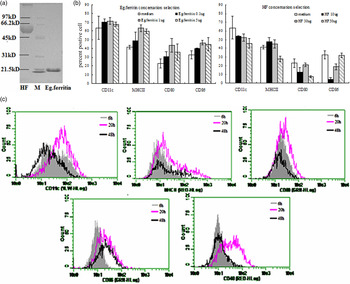
We first confirmed the purity of both HF and ferritin using 12% SDS-PAGE in non-reducing conditions and staining with Coomassie blue. This confirmed that ferritin migrated at ~20 kDa (Fig. 1A). In the absence of parasite antigens, the viability of bone marrow-derived monocytes that were differentiated into immature DC was 90%. During a time course of iDC stimulation, we found that 1 μg ml−1 ferritin was a more effective inducer of iDC maturation than other concentration of the protein. HF (30 μg) did not stimulate iDC maturation and significantly reduced the percentage of CD11c cells compared with unstimulated iDCs (P < 0·05) (Fig. 1B). We found that ferritin treatment for 20 h elicited the greatest increase in mature DCs, and also led to the highest expression of CD80, CD86, MHCII and CD40. Conversely, a 6-h and 48 h post-ferritin treatment did not induce a significant increased on surface marker (Fig. 1C). From these results, we determined that the optimum conditions for DC treatment in subsequent experiments were a 20-h exposure to 1 μg ferritin and 30 μg HF. Although LPS was used as positive control for induction of iDC maturation, we found that 1 μg ferritin was actually more effective. FACS analysis revealed that 30 μg HF significantly inhibited the upregulation of CD80, CD86, MHCII and CD40 compare to medium (P > 0·05). We found that ferritin increased the expression of CD80, CD86 and CD40 (P < 0·002), but had no effect on MHC-II expression. Strikingly, co-treatment of DCs with both ferritin and LPS for 20 h did not significantly increase expression of CD80, CD86 and CD40 beyond that observed with either agent alone. However, HF did reduce the LPS-dependent increase in MHCII and CD86, CD80 (P > 0·08) (Table 1).
Effect of ferritin and HF on the differentiation of monocytes into DCs. (A) HF and ferritin were separated by 12% SDS-PAGE. Left lane, HF. Second lane, molecular weight ladder. Third lane, ferritin. (B) Monocytes were cultured with GM-CSF plus IL-4 with HF (10, 30 or 50 μg ml−1) and ferritin (0·1, 1 or 5 μg ml−1). Culture medium served as a control. Expression of cell surface molecules was assessed by flow cytometry as described in Materials and Methods. The data are the means ± s.d. for the percentages of positive cells from four independent experiments. (C) IDCs were stimulated with ferritin for 6, 20 and 48 h. Fluorescence intensities of CD11c, CD80, CD86 and CD40 are shown. Black lines, 48 h ferritin treatment; grey line, 6 h differentiation; rose red line, differentiation for 20 h.

Phenotypic characterization of DCs following treatment with different stimuli

Monocytes were induced to differentiate to immature DCs on 7 day. The immature DCs were stimulated with ferritin (1 μg ml−1) and HF (30 μg ml−1) alone or with LPS for an additional 20 h, and the surface markers were analysed by flow cytometry. The results are expressed as the percentage of CD40-, CD80-, CD86- and MHC-II-positive cells. The P values as determined by Student's paired t-test for comparisons and. the data are means ± s.d. for seven independent experiments.
a Significantly different compared to unstimulated iDCs.
b Significantly different compared to HF-stimulated iDCs.
Effect of E. granulosus antigen on DC functions
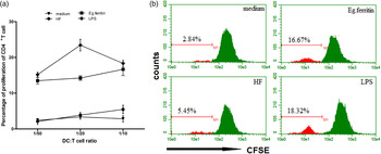
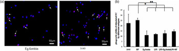
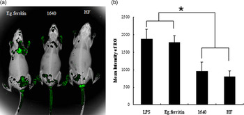
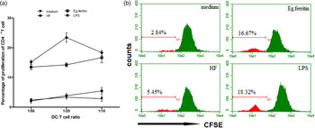
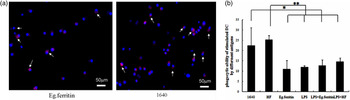
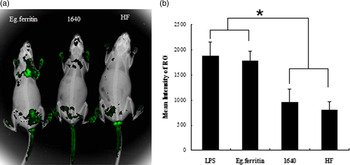
Antigens can stimulate iDCs transformation to mature DCs. The ability of DCs to induce a syngeneic CD4+ T-cell proliferative response was investigated using a MLR, as this test provides information concerning the overall antigen-presenting function of DCs. The effect of different antigens on DC function was assayed using DCs as a stimulus and CD4+T cells as effectors. Our initial results indicated that a DC: T ratio of 1:10 was optimal for activation of CD4+T-cell proliferation. In the presence of HF, DCs induced slightly increase T proliferative response compared with control cells, but no value. In contrast, ferritin-stimulated DCs induced a significantly greater proliferative response than cells stimulated with medium and HF (P < 0·001), HF-treated DC did not induce CD4+T-cell proliferation higher than medium (P > 0·05). These data suggest that ferritin can elicit T-cell proliferative response (Fig. 2A and B). To evaluate the influence of ferritin on DC receptor-mediated endocytosis, we performed a mini-rubby dextran uptake assay in iDCs in the presence or absence of ferritin, HF or LPS. Ferritin reduced the uptake capacity compared with control or HF (P < 0·0001). In the presence of HF, DCs were more efficient in uptake of dextran when compared with other antigens (P < 0·0001), although the effect of HF plus LPS for 20 h on DCs significantly reduced ingestion ability than cells stimulated with HF (Fig. 3A and B). DC Migration Assays, the picture showed that the more green fluorescence were seen on the groin and oxter of the mice injected with ferritin-stimulated DC than other group mice injected with HF or medium (Fig. 4A). DCs exposed to ferritin had higher intensity in mice compared with mice injected with HF-treated DCs and medium (P < 0·0001) (Fig. 4B). Thus, ferritin increases the antigen-presenting activity of the DCs, whereas HF reduces it.
Effect of differentiated DC on T-cell proliferation. (A) On day 8, iDCs stimulated with ferritin (1 μg ml−1), HF (30 μg ml−1) and LPS (100 ng ml−1) or 1640 RPMI medium were treated with mitomycin C and extensively washed and cultured with syngeneic CD4+T lymphocytes (1 × 10⁵ cells/well) labelled with CFSE for 5 days at different stimulator/responder ratios (DC/T cell ratio, 1:10, 1:20 and 1:50). (B) Proliferation of syngeneic T cells was measured by determining the CFSE fluorescence intensity through FACS. The data are means ± s.d. for triplicate cultures.
Uptake ability of DCs stimulated with ferritin and HF. The uptake ability of DC treated with LPS, ferritin, HF and medium was observed by fluorescence microscopy. (A) Nuclei of differentiated monocytes were labelled with Hoechst33258 and cultured with mini-rubby dextran 1 mg ml−1 in RPMI-1640 medium. White arrows point to DC ingestion of dextran labelled with Texas-red. (B) Phagocytosis activity of DCs cultivated under previously optimized conditions, at least 200 cells per sample and ten different fields were analysed. DCs exposed to ferritin alone or in the presence of LPS for 20 h, exhibited a significantly decreased uptake compared with DCs stimulated with medium and HF as determined by Student's paired t-test (P < 0·0001). HF alone had no effect on uptake when compared with medium as determined by Student's paired t-test (P > 0·05).
Migration capacity of DCs stimulated with ferritin and HF. The migration capacity of DCs was detected using an in vivo imaging system. (A) DCs (2 × 10⁵) were stimulated with ferritin, HF, LPS and medium alone, then incubated with CFSE and injected into the footpads of mice. After 48 h, the mice were anesthetized and cells labelled with CFSE that had migrated into the draining lymph nodes were quantified. Green fluorescence is proportional to the amount of DC migration. (B) The same areas were selected on different mice and the mean intensity of ROIs was analysed using the imaging system software. The migration capacity of DCs was stimulated with ferritin; this was significantly higher than in DCs stimulated with HF and medium (Student's paired t-test, P < 0·002).
Effect of E. granulosus antigens on DC morphology
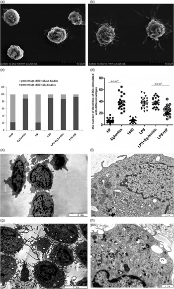
We next evaluated morphologic changes of DCs after stimulation with different antigens. SEM analysis indicated that mature DCs stimulated with antigen were larger and had rougher appearance and more dendrites which were thinner and longer on their surface than iDCs stimulated with medium (Fig. 5A and B). Comparison of the percentage of DCs with dendrites between treatments revealed that HF had essentially no effect. One hundred DCs were counted for each group under SEM (Fig. 5C). Although the group of DCs stimulated with HF plus LPS had similar ratio with the groups treated with ferritin, LPS and ferritin plus LPS, the number of dendrites of each DC treated with HF plus LPS were significantly less than the other three groups (P = 2 × 10⁻⁶). Twenty DCs with dendrites were randomly chosen and analysed for each group (Fig. 5D). The form of DCs present in medium and ferritin were observed through TEM, had consistent result with SEM (Fig. 5E and F). Numerous organelles were seen in cells of two groups. More vesicles were visible in cytoplasm and the vesicles were pinocytotic vesicles which were medium or self debris of cells (Fig. 5G). However, numerous lysosome and phagolysosome apparent were visible in cytoplasm and cluster of RER arranged on edge of cell (Fig. 5H).
Echinococcus granulosus antigens effect on DC morphologic change. DCs stimulated with medium (A) and ferritin antigen (B) were observed with a SEM. Comparison of dendrite number and form between DCs stimulated with medium and ferritin. (C) Quantification of the number of stimulated DCs that contain dendrites. DCs were stimulated with medium, LPS, ferritin and HF. Comparison of the ratio of DCs with or without dendrites among the treatment groups (100 DCs were counted in each group). (D) DCs were cultured as above, and the number of dendrites of each DC was counted under an SEM. The average dendrite number was compared between different groups. Bars represent means ± s.d. (n = 20). Ultrastructure of DC was observed with TEM. (E and G) DCs were stimulated with medium. (F and H) DCs were stimulated with ferritin antigen. The form of dendrites of DCs in medium and ferritin were showed with white arrow on (E) and (F). (G) Abundant organelle were seen, nuclei (N), mitochondria (M), golgi apparatus b (G) and rough endoplasmic reticulum (RER). A large number of vesicles were visible in cytoplasm and these vesicles were pinocytotic vesicle (PV). (H) Numerous lysosome (L) and phagolysosome (PL) apparent were visible in cytoplasm. Cluster of RER arranged on edge of cell.

Effect of E. granulosus antigens on the expression and secretion of cytokines by DCs
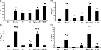
We found that DCs stimulated with HF generated IL-6 and a small amount of TNF-α, and no IL-10 and IL-12p70, whereas cells treated with HF plus LPS produced a highest level of IL-6 in all groups, TNF-α and IL-12p70 higher than DC exposed in HF and medium.(P < 0·05). Ferritin or ferritin plus LPS treatment led to abundant DC production of IL-6, TNF-α, IL-10 and IL-12 when compared to DCs exposed to HF, LPS and HF plus LPS. The level of IL-12 of DC treated with ferritin plus LPS were higher than DC exposed in ferritin (P < 0·002) (Fig. 6).
Effect of E. granulosus antigens on the expression and secretion of cytokines by DCs. Cytokine concentrations in pg ml−1 were measured by ELISA for IL-6 (A) TNF-α (B) and IL-10 (C) and IL-12p70 (D) in supernatants from bone-marrow-derived DC. Cells were stimulated with medium, LPS (mDCs) or with different antigens (HF or ferritin) for 20 h, or plus LPS for an additional 20 h. Significant differences compared to medium stimulated DCs are indicated by asterisks (P < 0·0001); significant differences in comparison with DCs plus HF are indicated by ‘$‘(P < 0·0001); significant differences in comparison with DCs in presence of HF plus LPS additional are indicated by ‘&’ (P < 0·0001); significant differences for comparisons with ferritin stimulated DCs are indicated by ‘+’ (P < 0·05).

DISCUSSION
Iron plays a role in DNA synthesis, electron transfer and oxygen activation, and is an essential requirement for animals, plants and microorganisms (Williams, Reference Williams1988). Owing to its high insolubility under physiological conditions, the ferritins are needed to maintain iron in a soluble form and also protect against the toxic effects of excess iron. Under conditions of iron deficiency, pathogen growth is reduced, DNA synthesis and cell division is suppressed and the microorganism eventually dies (Clarke et al. Reference Clarke, Tari and Vogel2001).
Since ferritin plays a key role in the metabolism of organisms, the possibility that it could be used as a vaccine for prevention of helminth infection has received considerable attention. Some parasite ferritins have previously been cloned and expressed (Clemens and Basch, Reference Clemens and Basch1989; Ersfeld and Craig, Reference Ersfeld and Craig1995; Schussler et al. Reference Schussler, Potters, Winnen, Bottke and Kunz1995; Wang et al. Reference Wang, Li, Bo and Zhao2009). Through our previous studies, we found that ferritin vaccination was successful in mice, as they were significantly protected from PSC infection. Furthermore, the antiserum of rabbits immunized with ferritin significantly inhibited the PSC of E. granulosus (data not shown). These data suggest that ferritin induces an effective antibody-mediated response to parasites. However, cellular immunity also plays an important role although, until our present study, the underlying mechanism(s) have been unclear. About concentration selection of Eg.ferritin effected on DC, on post-6 and 48 h, the surface marker of DC was expressed lower than post 20 h, because the most immature DC did not transmit to mature on 6 h. However, on 48 h the expression of surface molecular on DC should not less than 20 h but on the contrary. Why? We found that a number of cells become small and cell debris increased. So we inferred that DC did not tolerated antigen for long time, a mass of cells were death or apoptosis.
HF, which contains complex and multifunctional components, creates an environment for parasite survival and metabolism and also a place for parasite and host interaction. Recent research into human CE has shown that HF controls the immunomodulatory activities of E. granulosus, boosting its survival within a mammalian host (Chemale et al. Reference Chemale, Ferreira, Barrett, Brophy and Zaha2005; Lorenzo et al. Reference Lorenzo, Last and Gonzalez-Sapienza2005; Mamuti et al. Reference Mamuti, Sako, Nakao, Xiao, Nakaya, Ishikawa, Yamasaki, Lightowlers and Ito2006). HF elicits both T helper-1 (Th1) and Th2 cell activation (Thery and Amigorena, Reference Thery and Amigorena2001; Rigano et al. Reference Rigano, Buttari, De Falco, Profumo, Ortona, Margutti, Scotta, Teggi and Siracusano2004). In our studies, we found that DCs exposed to HF retained an immature phenotype, failed to upregulate MHCII, CD80, CD86 and CD40 surface markers, and had a weak stimulatory effect on T-cell proliferation. Co-stimulatory signals are crucial for T-cell activation, and it is well known that failure to express one of the components of the receptor/ligand pairs may severely impair T-cell activation and induce tolerance (Steinman and Nussenzweig, Reference Steinman and Nussenzweig2002; Ermann and Fathman, Reference Ermann and Fathman2003). The results revealed that HF had a weaker effect on inducing iDC into mature DC. By contrast, HF plus LPS significantly induced CD40, induced CD80, CD86 and MHC-II surface markers, although this did not reach significance, decreased the phagocytic ability of DCs, and promoted DC maturation. Furthermore, the number of dendrites of DC was significantly increased. CD40 is particularly important in this process; it is a member of the TNF receptor family that triggers DC maturation, increases CD80 and CD86 expression, and improves co-stimulatory signalling (O'Sullivan and Thomas, Reference O'Sullivan and Thomas2003; Steinman, Reference Steinman2003; Hanig and Lutz, Reference Hanig and Lutz2008). In this regard, ferritin is a more potent inducer of DC maturation, expression of co-stimulatory molecules and increasing migration capacity than is LPS. Our result is consistent with that of Jene (Jenne et al. Reference Jenne, Arrighi, Sauter and Kern2001) and Margos (Margos et al. Reference Margos, Grandgirard, Leib and Gottstein2011). They found that DCs in presence of the Echinococcus multilocularis 14-3-3 antigen transform into mature DC that were slightly more effective inducers of T-cell proliferation than DC exposed to crude non-fractionated E. multilocularis antigen or Em2 (G11) antigen. The term ‘mature’ in a functional sense has always implied immunogenicity, e.g., the differentiation and expansion of effector functions and memory. The concept of maturation was one of the first to distinguish antigen handling by iDCs, which is essential for antigen specific tolerance, from the many other accessory functions used by DCs and other cells to control immune responsiveness (Hawiger et al. Reference Hawiger, Inaba, Dorsett, Guo, Mahnke, Rivera, Ravetch, Steinman and Nussenzweig2001; Steinman et al. Reference Steinman, Hawiger and Nussenzweig2003; Thomson and Robbins, Reference Thomson and Robbins2008).
DCs produce abundant of IL-12 and TNF-α, and a small amount of IL-6, which induces differentiation of Th0 cells to Th1 cells; however, DCs that producing abundant IL-6 and a small amount of IL-12 induce differentiation of Th0 to Th2 cells (Liu, Reference Liu2001). In our study, DCs exposed to HF released IL-6 and a small amount of TNF-α, but no IL-10 and IL-12p70 (Demeure et al. Reference Demeure, Tanaka, Mateo, Rubio, Delespesse and Sarfati2000; He et al. Reference He, Gurunathan, Iwasaki, Ash-Shaheed and Kelsall2000; Latour et al. Reference Latour, Tanaka, Demeure, Mateo, Rubio, Brown, Maliszewski, Lindberg, Oldenborg, Ullrich, Delespesse and Sarfati2001; Fukao et al. Reference Fukao, Tanabe, Terauchi, Ota, Matsuda, Asano, Kadowaki, Takeuchi and Koyasu2002). Whereas in the additional presence of LPS, IL-6 concentration was increased remarkably, and reached levels higher than those observed with ferritin treatment, TNF-α and IL-12p70 were also increased. As we all know LPS induce Th1 immune responses. It indicated that HF-treated DC inhibited secretion of IL-12p70, and prevented Th0 cells differentiation to Th1 cells rather than induced Th2 cells which were related to susceptibility to disease and to escape mechanisms. The results were agreed with Rigano and Kanan (Kanan and Chain, Reference Kanan and Chain2006; Rigano et al. Reference Rigano, Buttari, Profumo, Ortona, Delunardo, Margutti, Mattei, Teggi, Sorice and Siracusano2007). Strikingly, either ferritin alone or ferritin plus LPS stimulated DCs to secrete high levels of all cytokines. We speculated that ferritin can elicited both Th1 and Th2 cell activation which induced different immune response in different time, the assumption was confirmed with MLR, ferritin induced higher CD4+T-cell proliferation which included Th1 and Th2, As were consisted with the result in mice immunized with Eg.ferritin were infected with PSC, we found that the CD4+T of spleen were increased remarkable to 31·27% by flow cytometry assay (the data now show). However, regard of the mechanism was not clearly. Through SEM, we found that the number of dendrites on DC varied widely according to the specific stimulus they received. Among all treatments, ferritin induced the highest number of dendrites on DCs. The dendrite may induce potential as a maturation-related sign and it able to reflect to the degree of DC maturation and increased capacity of presentation antigen. We infer DC dendrites transmit signal, the more dendrites can present the more antigen and trigger the more T-cell activation and proliferation, as well as the emergence of distinct effector T-cell subsets that modulate the immune response. Through TEM, we found numerous lysosome and phagolysosome appeared in DC which were stimulated with antigen. It revealed that DCs captured some exogenous antigen and these exogenous antigens were decomposed to some small fragment peptides by a large of phagolysosome. Abundant of organelles such as mitochondria, golgi apparatus and rough endoplasmic reticulum were seen in cell and cluster of rough endoplasmic reticulum arranged the edge of DCs. We inferred these numerous organelles maybe supplement for energy for that a great deal of peptides of antigen coupled with MHCII were transformed to cell surface.
Finally we speculate that exposure to HF would block DC maturation (Rescigno and Borrow, Reference Rescigno and Borrow2001), expressed higher level of IL-6 but not IL-12p70. These would limit DCs to an immature phenotype, and thus prevent their migration and antigen presentation activities. Since this also leads to a failure of inflammatory cytokine production. The additional of LPS induced DC maturation. However, ferritin was a robust stimulator of DC maturation, and exerted a similar effect as LPS. Ferritin stimulated DC maturation, which lead to the release of cytokines, and triggered CD4+T lymphocyte proliferation. Eg.ferritin induced immune responses of host were complex. It was necessary to further research the subsets of T cell which were dominant in the processing, which would contribute for illuminating the mechanism.
ACKNOWLEDGEMENTS
The authors thank the Center of Scientific Technology of Ningxia Medical University for sharing instruments, Yan Zhang for help in electron microscopy and Jie Wang and Jia Zhu for help in cytokine detection.
FUNDING
This work was supported by grants from the National Natural Science Foundation of China (Grant Number 81160203).