Case report
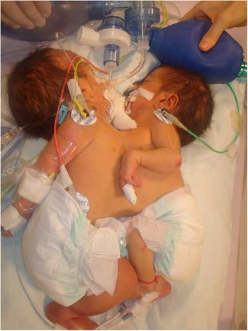
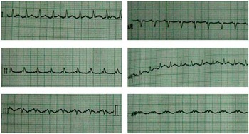
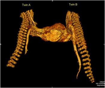
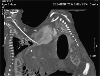
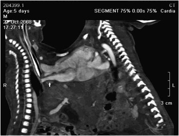
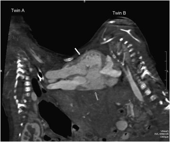
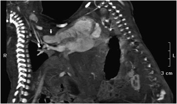
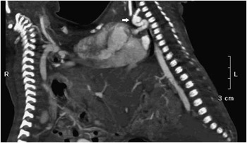
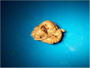
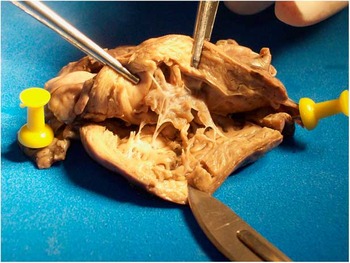
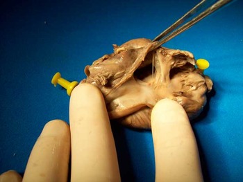
We report the case of thoracopagus conjoined twins (Fig 1) born with cyanosis and hypoxaemia, which improved upon prostaglandin infusion and mechanical ventilation. The electrocardiogram (Fig 2) showed an inverted P wave and unique QRS complex. Both colour Doppler echocardiogram and invasive angiography confirmed cardiac fusion; however, these studies were not conclusive, and thus a 64-slice computed tomography angiography (Aquilion™, Toshiba Medical Systems) was performed to establish a later conduct. The volumetric image (Fig 3) showed a unique heart with two left aortas arteries. The sagittal multiplanar images show systemic veins (Fig 4), pulmonary veins (Fig 5), a common atrium with a complete atrioventricular channel and common ventricle (Fig 6), and the ventricular outflows (Fig 7). In addition, pulmonary atresia and ductus-dependent pulmonary circulation were observed in both (Fig 8). The twins subsequently died 5 days after birth, and at autopsy (Figs 9–11) we confirmed the above details; therefore, the diagnosis for each segment was made correctly by computed tomography – that is, two systems of pulmonary venous return, a common atrium connected to a common ventricle across a complete atrioventricular channel, two ventricular outflows, two aortas, and pulmonary atresia with ductus permeable in both twins. The incidence of conjoined twins is estimated at 1 in 50,000–200,000 live births,Reference Hansen1 of which 40% are at the thoracic level (thoracopagus).Reference Spitz and Kiely2 Successful separation depends on the severity of cardiac involvement; we report the effectiveness of 64-slice computed tomography angiography in defining accurately the intra- and extracardiac anatomy in the postnatal evaluation of conjoined twins with a high degree of cardiac fusion to determine the feasibility of separation. Despite the fact that this methodology allows a rapid and complete evaluation of the cardiovascular system, the weakness of the computed tomography in these cases is the high dose of irradiation the patients are subjected to.
Thoracopagus Conjoined Twins.

The electrocardiography shows an inverted P wave and unique QRS complex.

The volumetric image by 64-slice computed tomography shows a high degree of cardiac fusion and two left aortas arteries.

The sagittal multiplanar image by 64-slice computed tomography shows the systemic veins in twin A (arrows).

The sagittal multiplanar image of the pulmonary veins return in twin A (arrow).

The sagittal multiplanar image shows the common atrium (pink arrow) with a complete atrioventricular channel and common ventricle (yellow arrow).

The sagittal multiplanar image shows the ventricular outflow in twin A (arrow).

Patent ductus arteriosus in twin B (arrow).

Autopsy.

The autopsy shows the common ventricle, the complete atrioventricular channel, and the exit of both aortas (pink yellow).

The autopsy shows the common atrium, the complete atrioventricular channel, and the exit of both aortas (pink yellow).