Introduction
Genome engineering is a major strategy for the development of new biological models of interest in academia and the applied sciences, such as biotechnological, biomedical, and agronomic research. Remarkably, this constitutes the basis of gene therapy aiming to correct an endogenous, mutated, defective gene and to restore normal physiological functions. DNA is the central biomolecule bearing the genetic information that is transmitted from one generation to another. Hence, methods aimed at extracting and purifying DNA and/or transferring DNA into living cells represent central issues in biotechnology and biomedical applications.
Notably, the development of vectors based on biohybrid materials for DNA transfer, which allows the avoidance of virus-based vectors, represents an appealing approach for the treatment of different genetic disorders (Choy et al., Reference Choy, Kwak, Jeong and Park2000; Lin et al., Reference Lin, Chen, Cheng and Kuo2006; Shi et al., Reference Shi, Tian, Zhang, Shen and Jia2011; Choi et al., Reference Choi, Kwon, Oh, Yun and Choy2014; Wu et al., Reference Wu, Shi, Huang, Zhang, Wu, Shen and Jia2014). Among the various micro/nanoparticles, sepiolite represents an alluring solution.
Sepiolite is a natural magnesium silicate clay mineral with a micro-fibrous morphology. The size of sepiolite fibers varies according to geographical origin. For example, the length of sepiolite fibers from Taxus Basin deposits in Spain ranges from 0.2 to 0.8 μm (Castro-Smirnov et al., Reference Castro-Smirnov, Piétrement, Aranda, Bertrand, Ayache, Le Cam, Ruiz-Hitzky and Lopez2016; Piétrement et al., Reference Piétrement, Castro-Smirnov, Le Cam, Aranda, Ruiz-Hitzky and Lopez2018). Interestingly, sepiolite is able to bind different kinds of biological molecules, including polysaccharides (Alcântara et al., Reference Alcântara, Darder, Aranda and Ruiz-Hitzky2014), lipids (Wicklein et al., Reference Wicklein, Darder, Aranda and Ruiz-Hitzky2010), proteins (Alcântara et al., Reference Alcântara, Darder, Aranda and Ruiz-Hitzky2012), and virus particles (Ruiz-Hitzky et al., Reference Ruiz-Hitzky, Darder, Wicklein, Fernandes, Castro-Smirnov, Martín del Burgo, del Real and Aranda2012). These abilities make sepiolite a promising micro/nanovector for the non-viral transfer of biomolecules (Piétrement et al., Reference Piétrement, Castro-Smirnov, Le Cam, Aranda, Ruiz-Hitzky and Lopez2018). In particular, when combining analyses from physics, chemistry, and biology, sepiolite has been shown to bind to DNA (Castro-Smirnov et al., Reference Castro-Smirnov, Piétrement, Aranda, Bertrand, Ayache, Le Cam, Ruiz-Hitzky and Lopez2016). These capabilities can be leveraged for biotechnology applications for future biomedical strategies.
This aim of the present review was to discuss the potential biotechnological and biomedical uses of sepiolite, based on the physicochemical characterization of sepiolite/DNA interactions. Such interactions allow consideration of sepiolite as a promising substrate for DNA extraction from bacteria, for improving sepiolite-mediated bacterial transformation, and for mammalian cell DNA transfection. A further objective was to envision the putative toxicity of sepiolite versus its advantages and future perspectives.
SEPIOLITE AND ITS INTERACTION WITH DNA
Sepiolite Structure
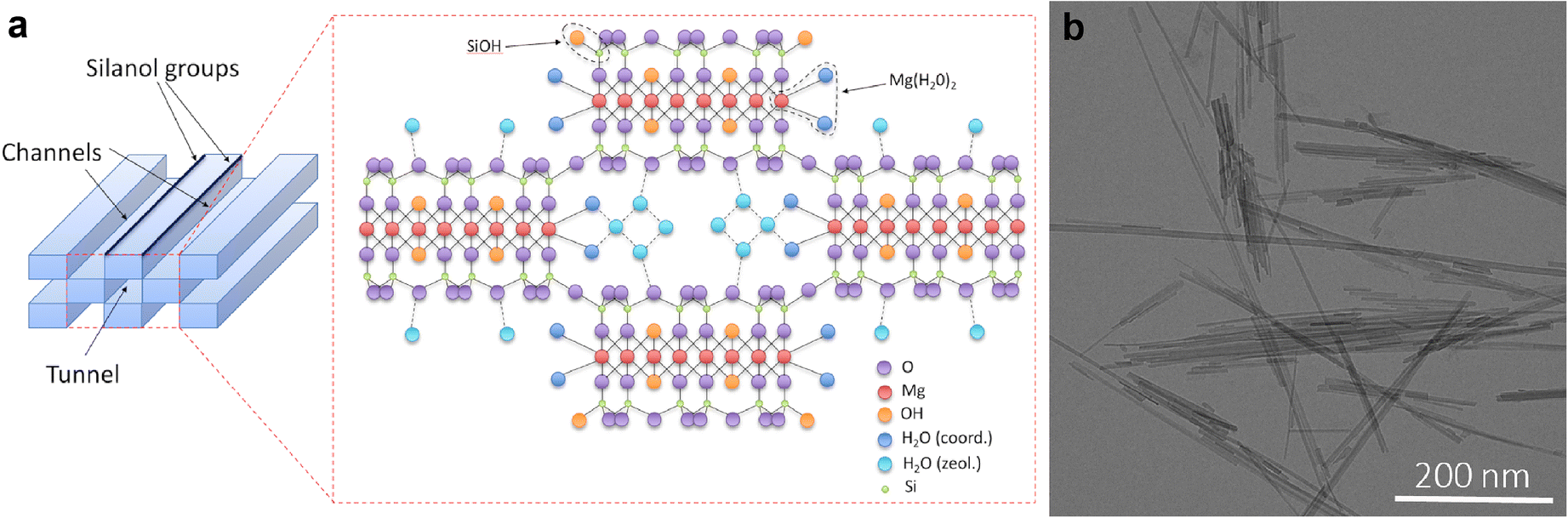
DNA/sepiolite interactions depend mainly on the sepiolite nanostructure and surface properties. Sepiolite belongs to a family of clays called pseudo-layered hydrated fibrous magnesium phyllosilicates. Its chemical formula is Si12Mg8O30(OH)4(H2O)4(H2O)8 and it has a trioctahedral structure: two layers of tetrahedral silica sandwiching a central octahedral magnesium oxide-hydroxide layer (TOT family). Unlike other phyllosilicates, sepiolite, like palygorskite, exhibits a three-dimensional crystalline organization with surface channels and internal tunnels. The crystal structure of sepiolite was studied by Brauner and Preisinger (Reference Brauner and Preisinger1956) and is shown in Fig. 1. The internal channels have dimensions of close to (0.37×1.06 nm).
Sepiolite structure (adapted from Brauner and Preisinger, Reference Brauner and Preisinger1956)

Many active sites on the sepiolite surface consist of both silanol groups (SiOH) located at the periphery of the fibers and magnesium hydroxyl type (Mg-(H2O)2) groups located on either side of the channels (Fig. 1). These sites play an essential role in DNA adsorption (Castro-Smirnov et al., Reference Castro-Smirnov, Piétrement, Aranda, Bertrand, Ayache, Le Cam, Ruiz-Hitzky and Lopez2016) and allow the grafting of numerous chemical groups (García et al., Reference García, Guzmán, Benito, Esteban-Cubillo, Aguilar, Santarén and Tiemblo2011; Moreira et al., Reference Moreira, Ciuffi, Rives, Vicente, Trujillano, Gil, Korili and de Faria2017; Undabeytia et al., Reference Undabeytia, Madrid, Vázquez and Pérez-Martínez2019) and are responsible for the remarkable physicochemical properties of sepiolite. Sepiolite also has a sorption capacity due mainly to the edge surfaces: the tunnels can accept molecules and retain them in a three-dimensional space, whereas the surface channels interact with the outside. Both participate in very different sorption mechanisms.
Like all clays, sepiolite has the property of retaining and exchanging cations with its environment, which is called the cation exchange capacity (CEC). However, the structure of sepiolite makes the internal faces of the channels difficult to access, and its three-dimensional structure prevents swelling, unlike many of the two-dimensional phyllosilicate clays. Sorption is, therefore, limited to the capacity of binding to the faces of the edges of the sheets that emerge at the periphery of the fibers. For sepiolite, the CEC is quite small (20 to 30 meq/100 g of mineral) compared to other clays, but nevertheless plays an essential role in the ability of sepiolite to adsorb various biomolecules, such as polysaccharides (Darder et al., Reference Darder, López-Blanco, Aranda, Aznar, Bravo and Ruiz-Hitzky2006), lipids (Wicklein et al., Reference Wicklein, Martín del Burgo, Yuste, Darder, Llavata, Aranda, Ortin, del Real and Ruiz-Hitzky2012), proteins (Alcântara et al., Reference Alcântara, Darder, Aranda and Ruiz-Hitzky2012), and viruses (Ruiz-Hitzky et al., Reference Ruiz-Hitzky, Darder, Aranda, del Burgo and del Real2009), giving rise to a great diversity of bio-nanocomposites. Finally, as a consequence of its structure, sepiolite has a large interaction surface that can be split into two contributions; the internal surface is close to 300 m2/g, and the external surface ranges from 200 to 300 m2/g (mainly depending on the fineness of the fibers). This large interaction surface (both internal and external) partly gives an understanding of how sepiolite can interact strongly with biomolecules.
This ability to adsorb cations has helped to develop the theory that clays could be the cradle of life on Earth, or, more specifically, of complex biochemical compounds that made life possible (Yang et al., Reference Yang, Peng, Hartman, Gupton-Campolongo, Rice, Chang, Gu, Lu and Luo2013). The hypothesis is that the clay hydrogel, thanks in part to its structure organized in microcavities, plays a containment role for basic chemical elements that could have carried out the complex reactions at the origin of the formation of proteins, DNA, and ultimately all of the machinery involved in the functions of a living cell. The clay hydrogels could also then have confined and protected these chemical processes until the formation of the membrane surrounding the living cells.
In addition, the adsorption of DNA on mineral surfaces might favor horizontal gene transfer into living organisms, which may play a significant role in the evolution of living species. All these studies suggest a specific interaction between clays and nucleic acids.
Sepiolite and DNA
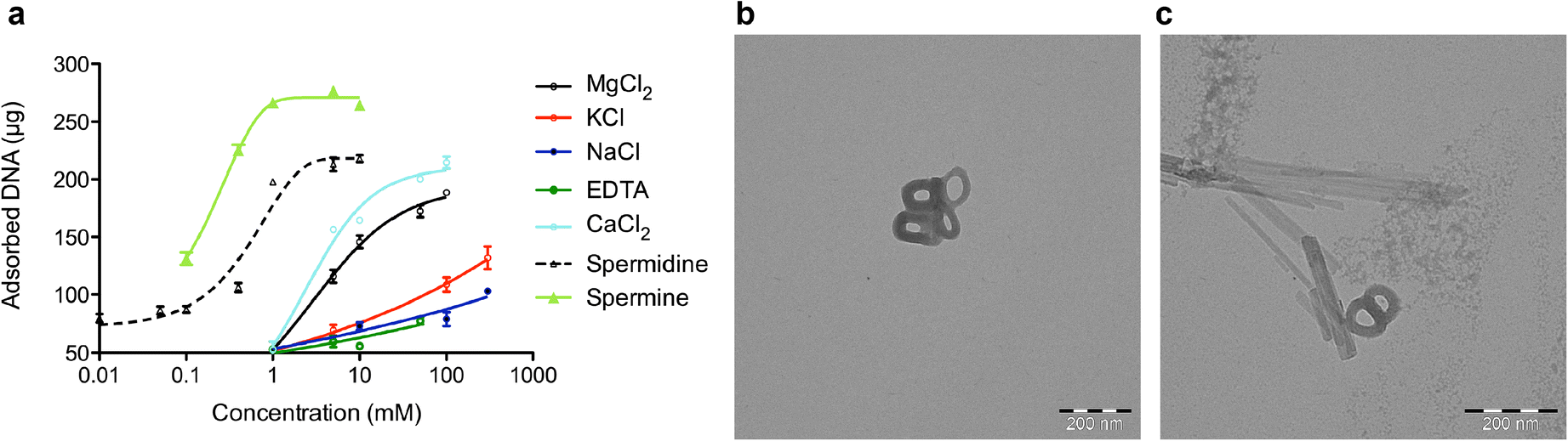
With the aim of using sepiolite as a new DNA vector, a study has shown that DNA could bind efficiently to sepiolite, notably highlighting the role of multivalent cations as enhancers of the DNA binding process (Castro-Smirnov et al., Reference Castro-Smirnov, Piétrement, Aranda, Bertrand, Ayache, Le Cam, Ruiz-Hitzky and Lopez2016). However, while monovalent cations reduce the adsorption of DNA molecules to mica (Pastré et al., Reference Pastré, Pietrement, Fusil, Landousy, Jeusset, David, Hamon, Le Cam and Zozime2003), surprisingly, these cations promote the adsorption to sepiolite fibers. While monovalent cations are less effective than multivalent cations, they contribute unequivocally to the adsorption process (Fig. 2). Interestingly, the presence of polyvalent cations (Mg2+, Ca2+, spermidine, or spermine) stimulates strongly the adsorption of DNA depending on the valence of the cation (Fig. 2). These results suggest an adsorption mechanism mediated by an electrostatic bridge through hydrogen bonds between the DNA phosphate groups and the silanol groups located on the outer surface of the sepiolite, which is unlike the mica surface, where counterion correlations occur (Pastré et al., Reference Pastré, Pietrement, Fusil, Landousy, Jeusset, David, Hamon, Le Cam and Zozime2003, Reference Pastré, Hamon, Landousy, Sorel, David, Zozime, Le Cam and Piétrement2006). Fourier-transform infrared (FTIR) spectroscopy analysis thus showed that, indeed, the interaction of DNA with sepiolite was mediated by only the external silanol groups, whereas the hydroxyl groups of the octahedral sheets of sepiolite play no role (Castro-Smirnov et al., Reference Castro-Smirnov, Piétrement, Aranda, Bertrand, Ayache, Le Cam, Ruiz-Hitzky and Lopez2016).
a The effects of the presence of cations with various valences on DNA adsorption. Reaction conditions: 10 mM Tris-HCl pH 7.5, salmon sperm DNA and sepiolite concentrations of 615 ng/μL and 1 mg/mL, respectively (from Castro-Smirnov et al., Reference Castro-Smirnov, Piétrement, Aranda, Bertrand, Ayache, Le Cam, Ruiz-Hitzky and Lopez2016). TEM images of DNA condensed with b spermidine and c the interaction with sepiolite fibers. Transmission electron microscopy (TEM) imaging: DNA molecules (5 nM) were condensed in 10 mM Tris-HCl pH 8, 50 mM NaCl, and 1 mM spermidine. 5 μL of reaction mixture was deposited onto a 600 mesh copper grid coated with a thin carbon film previously activated by glow discharge in the presence of 1-aminopentane (Merck, Saint Quentin Fallavier, France) (Dupaigne et al., Reference Dupaigne, Tavares, Piétrement and Le Cam2018). After 1 min, the grids were washed with aqueous 2 wt.% uranyl acetate (Merck, Saint Quentin Fallavier, France) and then dried with ashless filter paper (VWR, Rosny sous bois, France). TEM observations were carried out using Zeiss 912AB transmission electron microscope in bright field mode. Electron micrographs were obtained using a ProScan 1024 HSC digital camera and iTEM software (Olympus, Soft Imaging Solutions, Munster, Germany)

If the DNA is adsorbed onto the sepiolite fibers, the question of its desorption also arises in the context of a project to create a nano-cargo for cell transfection. In cellulo, the mechanisms that could induce this desorption remain unknown, but the possibility of DNA desorption in vitro has been tested through the desorption of DNA from the bionanocomposite by incubating it with EDTA, a well known metal ion-chelating agent. By using different DNA plasmids, the quality of the DNA recovered after its attachment to sepiolite was examined and the various DNA isoforms (super-coiled, open circle, or linear) were unmodified after their binding to sepiolite. This shows that sepiolite does not alter the quality of DNA and that this method can be considered a new method of DNA purification (Castro-Smirnov et al., Reference Castro-Smirnov, Piétrement, Aranda, Bertrand, Ayache, Le Cam, Ruiz-Hitzky and Lopez2016).
All these results confirmed the potential of sepiolite as an efficient nano/micro cargo for DNA transfer into cells. Moreover, the capacity of desorption suggests the possibility of using sepiolite for DNA extraction.
EXTRACTION OF PLASMID DNA FROM BACTERIA
DNA extraction and purification, especially of plasmids, is the basis of molecular biology. Plasmids are circular DNA molecules that replicate autonomously in bacteria and are essential tools in molecular biology and biotechnology, from bacteria to mammalian cells. Indeed, they are used as DNA vectors/backbones in gene transfer technology. Hence, fusion of an exogenous DNA (e.g. a piece of a human DNA) allows replication and amplification into bacteria. After this replication and amplification process, their extraction is necessary for subsequent experiments.
Several methods of plasmid extraction are available commercially. These methods are more or less expensive and time-consuming. The DNA binding capability of sepiolite, associated with the possibility of DNA release, makes sepiolite a simple and inexpensive alternative method (Castro-Smirnov et al., Reference Castro-Smirnov, Piétrement, Aranda, Le Cam, Ruiz-Hitzky and Lopez2020). Indeed, the binding of DNA onto sepiolite requires cations, notably divalent cations such as Mg2+ or Ca2+ (Castro-Smirnov et al., Reference Castro-Smirnov, Piétrement, Aranda, Bertrand, Ayache, Le Cam, Ruiz-Hitzky and Lopez2016). The chelation of cations with EDTA, therefore, enables the release of DNA from sepiolite, which is not covalently bound (Castro-Smirnov et al., Reference Castro-Smirnov, Piétrement, Aranda, Le Cam, Ruiz-Hitzky and Lopez2020). The general protocol (Castro-Smirnov et al., Reference Castro-Smirnov, Piétrement, Aranda, Le Cam, Ruiz-Hitzky and Lopez2020) can be summarized as follows:
(a) Bacteria-bearing plasmids are grown in an appropriate medium with a suitable selection of antibiotics.
(b) At the mid-saturation state (as monitored by spectrophotometry), bacteria are pelleted by centrifugation and then re-suspended in specific buffers. Bacteria are lysed, and the DNA is denatured (the two complementary strands are separated) in alkaline buffers.
(c) The addition of neutralizing buffer allows plasmid DNA (which are small DNA molecules) to renature (return to the bicatenary DNA structure), remaining soluble. In contrast, bacterial genomic DNA (which are long DNA molecules) cannot renature properly and so they precipitate.
(d) Centrifugation precipitates genomic DNA pellets and bacterial debris.
(e) The supernatant (containing the plasmid DNA) is recovered. Sepiolite plus Mg2+ or Ca2+ is added, allowing plasmid DNA to bind to sepiolite, allowing the subsequent washing steps to eliminate contaminants.
(f) Sepiolite-bearing DNA is centrifuged and washed with Tris buffer.
(g) EDTA is then added to the sepiolite/DNA pellets, releasing the plasmid DNA from the sepiolite.
(h) Plasmid DNA and sepiolite are then separated by centrifugation (sepiolite in the pellet and plasmid DNA in the supernatant).
Steps a to d are common to almost all methods of plasmid extraction.
Importantly, the structures of plasmid DNA molecules are not altered by this procedure, as shown by electrophoresis and transmission electron microscopy analyses. Moreover, plasmid DNA can be processed by enzymes such as restriction enzymes (Castro-Smirnov et al., Reference Castro-Smirnov, Piétrement, Aranda, Le Cam, Ruiz-Hitzky and Lopez2020). These facts attest to the good quality of the plasmid DNA extracted by the sepiolite-based method.
IMPROVEMENT OF THE YOSHIDA EFFECT ON BACTERIAL TRANSFORMATION
In bacteria and non-animal eukaryotic cells, the non-viral transfer of DNA is called transformation. In animal cells, it is called transfection.
Efficient transformation first requires rendering of the bacteria to be competent for DNA transfer. The protocols to prepare competent bacteria are generally laborious and time consuming, however. One alternative is to purchase commercially competent bacteria, but this method is expensive. One advantage of the Yoshida effect protocol for bacterial transformation is that it does not require the use of competent bacteria.
In the Yoshida effect protocol, the solution containing the bacteria and the acicular material forms a colloidal solution. Upon spreading on solid agar medium in Petri dishes using a solid spreader, the sliding friction stimulates bacterial transformation (Yoshida et al., Reference Yoshida, Ikeda, Yoshida, Sengoku and Ogawa2001; Yoshida, Reference Yoshida2007; Yoshida & Sato, Reference Yoshida and Sato2009). The Yoshida effect was first described with asbestos, however, thus raising health and toxicity concerns for its use. Sepiolite, which is considered non-toxic and non-carcinogenic (see below), can circumvent this health worry. Indeed, the fibrous nature of sepiolite favors bacterial transformation through a Yoshida-like effect (Yoshida, Reference Yoshida2007; Yoshida & Sato, Reference Yoshida and Sato2009; Tan et al., Reference Tan, Fu and Seno2010; Wilharm et al., Reference Wilharm, Lepka, Faber, Hofmann, Kerrinnes and Skiebe2010; Rodríguez-Beltrán et al., Reference Rodríguez-Beltrán, Elabed, Gaddour, Blázquez and Rodríguez-Rojas2012, Reference Rodríguez-Beltrán, Rodriguez-Rojas, Yubero and Blazquez2013).
The Yoshida effect does not absolutely require the pre-assembly of DNA with the fibers. Combining various capabilities of sepiolite (e.g. disaggregation by sonication, and the capacity to bind both DNA and bacteria), a 100-fold improvement in transformation efficiency has been observed (Castro-Smirnov et al., Reference Castro-Smirnov, Piétrement, Aranda, Le Cam, Ruiz-Hitzky and Lopez2020). The general protocol can be summarized as follows.
(a) The use of sonicated sepiolite (sSep). Indeed, sepiolite aggregates spontaneously and can be dissociated by sonication, increasing its spreading efficiency.
(b) Pre-assembly of DNA and sSep, using the characteristics (incubation in the presence of divalent cations, then centrifugation) described above, before adding the bacteria.
(c) Pre-incubation of the sSep/DNA biohybrid material with bacteria (at the mid-phase of growth) before spreading. Indeed, sepiolite can bind spontaneously to bacteria (Castro-Smirnov et al., Reference Castro-Smirnov, Piétrement, Aranda, Bertrand, Ayache, Le Cam, Ruiz-Hitzky and Lopez2016, Reference Castro-Smirnov, Piétrement, Aranda, Le Cam, Ruiz-Hitzky and Lopez2020) and it can thus be used for liquid decontamination. In addition, this procedure favors bacterial transformation (Castro-Smirnov et al., Reference Castro-Smirnov, Piétrement, Aranda, Le Cam, Ruiz-Hitzky and Lopez2020). The Yoshida effect is still necessary, however, because sepiolite-bacteria assembly alone is not sufficient for efficient internalization.
Adapting these three conditions to the Yoshida effect, transformation efficiencies reached close to 106 transformants per microgram of DNA (Castro-Smirnov et al., Reference Castro-Smirnov, Piétrement, Aranda, Le Cam, Ruiz-Hitzky and Lopez2020). Such transformation efficiency is lower than that of the commercially available competent bacteria but is largely sufficient for most molecular biology applications at a lower cost. This method is also much less time-consuming than the classical protocols for preparing competent bacteria.
TRANSFECTION OF MAMMALIAN CELLS
Transfer of DNA into mammalian cells is an essential issue in biotechnology and biomedical applications. Indeed, this constitutes the basis of strategies aimed at designing new model organisms for academic, biomedical, or agronomical research and gene therapy. Mammalian cells do not incorporate exogenous DNA efficiently, however.
The Yoshida effect cannot work with mammalian cells because frictional forces kill them. Sepiolite can be internalized spontaneously into mammalian cells without requiring friction, mainly through endocytosis and macropinocytosis pathways that involve the invagination of the cell membrane around the sepiolite fibers, resulting in its incorporation in the endosomes (intracellular organelles embedded into membranes) of the cells (Castro-Smirnov et al., Reference Castro-Smirnov, Ayache, Bertrand, Dardillac, Le Cam, Piétrement, Aranda, Ruiz-Hitzky and Lopez2017). Interestingly, this process has little effect on viability (Castro-Smirnov et al., Reference Castro-Smirnov, Ayache, Bertrand, Dardillac, Le Cam, Piétrement, Aranda, Ruiz-Hitzky and Lopez2017; Ragu et al., Reference Ragu, Dardillac, Adame Brooks, Castro-Smirnov, Aranda, Ruiz-Hitzky and Lopez2020a). Given that sepiolite can bind DNA, it can serve as a vector for stable DNA transfection into mammalian cells (Castro-Smirnov et al., Reference Castro-Smirnov, Piétrement, Aranda, Bertrand, Ayache, Le Cam, Ruiz-Hitzky and Lopez2016). This application relies on the binding of DNA to sSep prior to the addition of the sSep/DNA bio-hybrid material to the cell media. The uptake of the sSep/DNA bio-hybrid also acts through the endocytosis pathway, and the intracellular sSep/DNA bio-hybrid is then embedded in endosomes. The addition of drugs that destabilize endosome membranes, such as chloroquine, results in a 2-fold increase in the transfection efficiency (Castro-Smirnov et al., Reference Castro-Smirnov, Piétrement, Aranda, Bertrand, Ayache, Le Cam, Ruiz-Hitzky and Lopez2016).
These methods of cell transfection are somewhat less efficient than the use of commercial reagents but are much less expensive. Different strategies can be envisioned to increase the efficiency of intake of the nanoparticles into the cell. These strategies might be based on the structure of the cell membrane, which consists of a bi-layer of lipids. Techniques that could favour the delivery from the endosomes, such as those fostering endosome collapse into the cells, are addressed here. The pH of the endosome is very acid and thus might lead to hydrolysis of the DNA. Methods controlling this issue are under investigation. The injection of sepiolite in vivo should be analyzed precisely. Indeed, it could be injected into the blood micro-vessel and/or accumulated in the liver. The production of ROS (reactive oxygen species) and of inflammatory cytokines might generate unwanted inflammation and/or premature ageing. The behavior and the fate of sepiolite, as well as the consequences of sepiolite injection (through different procedures such as in blood versus intra-peritoneal), should be addressed and analyzed in depth, and in vivo in a mouse model, before attempting this transfer in humans.
TOXICITY OF SEPIOLITE IN HUMAN CELLS
Because of the multiple potential uses of sepiolite, including its biomedical applications, the question of its toxicity has become an important issue. Its fibrous nature has led to concerns of asbestos-like effects. This can be supported by the fact that, like asbestos, sepiolite can generate a Yoshida effect and, in addition, generate DNA damage in bacteria (González-Tortuero et al., Reference González-Tortuero, Rodríguez-Beltrán, Radek, Blázquez and Rodríguez-Rojas2018). However, translating these conclusions to mammalian cells would represent an over-interpretation. Indeed, bacteria are much smaller than mammalian cells (approximately 1000-fold). Therefore, while the length of a sepiolite fiber is similar to that of a bacterial cell, the sepiolite fiber is much smaller than that of a human cell (Castro-Smirnov et al., Reference Castro-Smirnov, Ayache, Bertrand, Dardillac, Le Cam, Piétrement, Aranda, Ruiz-Hitzky and Lopez2017; Ragu et al., Reference Ragu, Dardillac, Adame Brooks, Castro-Smirnov, Aranda, Ruiz-Hitzky and Lopez2020a). In addition, the genome of eukaryotes (including human cells) is embedded into a nuclear compartment, while in prokaryotes (bacteria) no nucleus exists, and DNA is contained directly in the cytoplasm. Therefore, a sepiolite fiber that penetrates bacteria can interact directly with its genomic DNA to alter it. In contrast, in mammalian cells, the sepiolite fiber that enters the cell will be in the cytoplasm and not in the nucleus. Hence, the sepiolite is not in contact with the genomic DNA (Castro-Smirnov et al., Reference Castro-Smirnov, Ayache, Bertrand, Dardillac, Le Cam, Piétrement, Aranda, Ruiz-Hitzky and Lopez2017; Ragu et al., Reference Ragu, Dardillac, Adame Brooks, Castro-Smirnov, Aranda, Ruiz-Hitzky and Lopez2020a). For DNA interaction, the sepiolite fiber should be transported into the nucleus, which appears to be an infrequent process (Castro-Smirnov et al., Reference Castro-Smirnov, Ayache, Bertrand, Dardillac, Le Cam, Piétrement, Aranda, Ruiz-Hitzky and Lopez2017; Ragu et al., Reference Ragu, Dardillac, Adame Brooks, Castro-Smirnov, Aranda, Ruiz-Hitzky and Lopez2020a). In agreement with this observation, the interaction of human cells with sepiolite does not trigger the DNA damage response (Ragu et al., Reference Ragu, Dardillac, Adame Brooks, Castro-Smirnov, Aranda, Ruiz-Hitzky and Lopez2020a) supporting the fact that sepiolite does not attack genomic DNA in mammalian cells. Sepiolite appears to be weakly toxic in cultured mammalian cells (Castro-Smirnov et al., Reference Castro-Smirnov, Ayache, Bertrand, Dardillac, Le Cam, Piétrement, Aranda, Ruiz-Hitzky and Lopez2017; Ragu et al., Reference Ragu, Dardillac, Adame Brooks, Castro-Smirnov, Aranda, Ruiz-Hitzky and Lopez2020a), especially at the doses used for DNA transfection, which are much lower than other classical transfection methods.
Sepiolite also induces the production of ROS in cells (Ragu et al., Reference Ragu, Dardillac, Adame Brooks, Castro-Smirnov, Aranda, Ruiz-Hitzky and Lopez2020a). Because ROS can potentially alter any biological component, including DNA, and lead to mutagenesis, this has raised some concerns about the potential toxicity of sepiolite. The respiratory chains that synthetize ATP and provide the energy required for various metabolic pathways generate ROS as by-products. However, cells can also control the production of ROS that act as secondary messengers in physiological processes (Ameziane-El-Hassani et al., Reference Ameziane-El-Hassani, Schlumberger and Dupuy2016). In this situation, ROS are beneficial to the organism. Notably, while antioxidants have been proposed to protect against tumorigenesis, in contrast, treatment with antioxidants favors the development of lung carcinomas and metastasis (Le Gal et al., Reference Le Gal, Ibrahim, Wiel, Sayin, Akula, Karlsson, Dalin, Akyürek, Lindahl, Nilsson and Bergo2015; Breau et al., Reference Breau, Houssaini, Lipskaia, Abid, Born, Marcos, Czibik, Attwe, Beaulieu, Palazzo, Flaman, Bourachot, Collin, Tran Van Nhieu, Bernard, Mechta-Grigoriou and Adnot2019; Wiel et al., Reference Wiel, Le Gal, Ibrahim, Jahangir, Kashif, Yao, Ziegler, Xu, Ghosh, Mondal, Kanduri, Lindahl, Sayin and Bergo2019), thus underlying the potential benefit of ROS production. In addition, ROS can also trigger the induction of apoptosis, eliminating stressed cells, to the benefit of the whole organism. Hence, an intracellular increase in ROS can result from the detection of sepiolite by cells and their response but does not automatically imply that the fate of the cell or organism will be jeopardized.
Finally, ROS can oxidize lipid cell membranes, leading to lipid peroxidation (LP). However, sepiolite represses LP from the supernatants of rat brain homogenates, suggesting a putative antioxidant role for sepiolite (Cervini-Silva et al., Reference Cervini-Silva, Nieto-Camacho and Gómez-Vidales2015). One hypothesis is that cells respond to sepiolite interaction through the production of controlled ROS but, in parallel, sepiolite could also scavenge ROS, thus protecting against potential noxiousness.
One important component of the potential toxicity of fibers (such as asbestos) relies on the fact that they remain stuck in tissues, generating chronic inflammation, which can become pathogenic with time. These concerns connect two issues: the size of the fibers and the ability of the cells to exclude the fibers. Small fibers should be excluded more easily, thereby avoiding a potential asbestos-like effect. The large majority of the sepiolite fibers from Vallecas-Vicalvaro deposits in Spain range in length from 200 to 800 nm (Castro-Smirnov et al., Reference Castro-Smirnov, Piétrement, Aranda, Bertrand, Ayache, Le Cam, Ruiz-Hitzky and Lopez2016), values which are smaller than those reported in the literature (Bellmann et al., Reference Bellmann, Muhle and Ernst1997). Such sizes are much smaller than mammalian cells; the ability of the cells themselves to exclude the sepiolite fibers is thus key in rendering the sepiolite harmless. Interestingly, the stable, natural, intrinsic fluorescence of the sepiolite used in the present study (from Vallecas-Vicalvaro deposit) enabled examination of its fate in cells (Castro-Smirnov et al., Reference Castro-Smirnov, Ayache, Bertrand, Dardillac, Le Cam, Piétrement, Aranda, Ruiz-Hitzky and Lopez2017; Ragu et al., Reference Ragu, Dardillac, Adame Brooks, Castro-Smirnov, Aranda, Ruiz-Hitzky and Lopez2020a). In particular, time-lapse fluorescent video microscopy analysis revealed the spontaneous exclusion of these sepiolite fibers from mammalian cells, probably through an exocytosis process that involved the membranes of endosomes (Castro-Smirnov et al., Reference Castro-Smirnov, Ayache, Bertrand, Dardillac, Le Cam, Piétrement, Aranda, Ruiz-Hitzky and Lopez2017). These data can account for the low cellular toxicity of sepiolite (Denizeau et al., Reference Denizeau, Marion, Chevalier and Cote1985; Castro-Smirnov et al., Reference Castro-Smirnov, Piétrement, Aranda, Bertrand, Ayache, Le Cam, Ruiz-Hitzky and Lopez2016; Ragu et al., Reference Ragu, Dardillac, Adame Brooks, Castro-Smirnov, Aranda, Ruiz-Hitzky and Lopez2020a). More generally, and in agreement with the size-effect hypothesis, both epidemiological studies and in vitro and in vivo analyses have led to the conclusion that sepiolite, especially with fiber lengths of <5 μm, does not present a health risk (Maisanaba et al., Reference Maisanaba, Pichardo, Puerto, Gutiérrez-Praena, Cameán and Jos2015). Previous biological and epidemiological analyses have led the International Agency for Research on Cancer (IARC) to classify sepiolite as non-hazardous and non-carcinogenic (McConnochie et al., Reference McConnochie, Bevan, Newcombe, Lyons, Skidmore and Wagner1993; Santarén & Alvarez, Reference Santarén and Alvarez1994; Wagner et al., Reference Wagner, Griffiths and Munday1987).
PERSPECTIVES FOR IMPROVEMENT OF IMMUNOTHERAPY
Additional biomedical applications can be derived from the interaction of sepiolite with cells, e.g. immunotherapy. This strategy takes advantage of the capacity of the immune system to eliminate stressed cells. It has been applied to cancer therapy (Esfahani et al., Reference Esfahani, Roudaia, Buhlaiga, Del Rincon, Papneja and Miller2020) and was recognized by the Nobel Academy (Nobel prize in Physiology or Medicine to James Allison and Tasuku Honjo, in 2018). This process is triggered by the production and excretion of inflammatory cytokines that modify the microenvironment of the cell. This leads to activation of the innate immune response, which recruits immune system cells (e.g. natural killer cells, macrophages, and lymphocytes). Tumors are classified as hot or cold, depending on whether they respond to immunotherapy. Unfortunately, the majority of tumors are cold. Strategies that are able to change the status of tumors from cold to hot should represent important progress in immunotherapy, therefore (Duan et al., Reference Duan, Zhang, Zheng and Zhang2020). Sepiolite is internalized into cells leading to the production of inflammatory cytokines (Ragu et al., Reference Ragu, Dardillac, Adame Brooks, Castro-Smirnov, Aranda, Ruiz-Hitzky and Lopez2020a). This offers hope for using hot/cold tumors for immunotherapy purposes.
In addition, grafting DNA onto sepiolite can be expected to optimize the inflammatory response. Indeed, cytoplasmic DNA is recognized by cells that induce inflammatory cytokines (Ragu et al., Reference Ragu, Matos-Rodrigues and Lopez2020b). Thus, one hope would be that the bio-hybrid sepiolite/DNA material that delivers DNA into the cytoplasm of the cell might synergize the efficiency of cytokine production and thus the immune response.
Conclusions
DNA is negatively charged and the first obstacle is the cell membrane, which is made of lipid bilayers that have charges incompatible with the spontaneous uptake of DNA by cells. Non-viral methods use physical and chemical strategies to bypass the membrane barrier but are generally toxic to the cell. One limitation of both viral and physicochemical methods is the fact that they can transfer DNA only. One ideal strategy would be based on transport systems that can co-convey different kinds of molecules. The development of novel nanocarriers using biohybrid materials for non-viral gene transfer thus constitutes a promising approach. In this context, sepiolite is an enticing candidate for use as a nanocarrier for the non-viral vectorization of DNA, because it is biocompatible and is internalized spontaneously and excreted by mammalian cells. Thanks to its spontaneous fluorescence, sepiolite can be detected in cells (and maybe hopefully in animals) without the requirement for fluorescent chemical grafting and progress can be followed in cells. Importantly, sepiolite is only weakly toxic to mammalian cells and is classified as non-hazardous and non-carcinogenic by the IARC (World Health Organization). Strategies based on natural internalization/externalization capacities of the mammalian cells are preferable to limit cell toxicity. These strategies implied that DNA should first bind to sepiolite to form a sepiolite/DNA (Sep/DNA) biohybrid. Indeed, sepiolite can adsorb different molecules very efficiently, thus allowing the development of many elaborate strategies, and, as a natural clay, sepiolite represents an answer to societal concerns regarding challenging nanotechnologies.
ACKNOWLEDGMENTS
The authors thank Prof. Eduardo Ruiz-Hiztky and Dr Pilar Aranda for sharing their expertise, particularly on sepiolite, and for stimulating discussions. OP acknowledges Dr Eric Le Cam for access to the electron microscope and helpful discussions. SR and BSL were supported by grants from the Ligue Nationale contre le cancer “Equipe labellisée”, ANR (Agence Nationale de la Recherche, ANR-16-CE12-0011-02, and ANR-16-CE18-0012-02), and INCa (Institut National du Cancer, PLBIO18-232). OP was supported by the CNRS Mission pour l’Interdisciplinarité (MI-DynAFM-DNARep 2018_273085) and the Bourgogne Franche-Comté Graduate School EUR-EIPHI (17-EURE-0002).
Funding
Funding sources are as stated in the Acknowledgments.
Compliance with Ethical Standards
Conflict of Interest
The authors declare that they have no conflict of interest.