




No CrossRef data available.
Article contents
Persistent infection of Mongolian jirds with a non-pathogenic trypanosome, Trypanosoma (Herpetosoma) grosi
Published online by Cambridge University Press: 17 October 2003
Abstract
Non-pathogenic trypanosomes of the subgenus Herpetosoma are normally host specific, and laboratory models include Trypanosoma lewisi in rats and Trypanosoma musculi in mice. Two isolates of Trypanosoma grosi, originating from Apodemus agrarius and Apodemus peninsulae, grew well in Mongolian jirds, Meriones unguiculatus, after intraperitoneal inoculation of 2×10⁵ or a minimum 500 bloodstream forms. The course of T. grosi infection in jirds resembled T. musculi infection in mice, rather than T. lewisi infection in rats. At week 2 to 3 p.i. trypanosomes disappeared from the bloodstream, and neither prednisolone treatment nor splenectomy prevented parasite elimination from the bloodstream. However, these treatments induced a marked increase in peak parasite counts. Regardless of prednisolone treatment or splenectomy, all jirds after day 21 p.i. became resistant to the reinfection. Although no trypanosomes were detected in the bloodstream of recovered jirds, dividing parasites persisted in the medullary capillaries of the kidney, like T. musculi infection in mice. We propose the T. grosi infection in jirds as an additional laboratory model for the study of non-pathogenic trypanosomes.
Information
- Type
- Research Article
- Information
- Copyright
- 2003 Cambridge University Press
INTRODUCTION
Stercorarian trypanosomes of the subgenus Herpetosoma are host specific for their vertebrate hosts where they live extracellularly, primarily in the bloodstream (Hoare, 1972; Santos-Gomes et al. 1993; Noyes et al. 2002). Among these stenoxenous trypanosomes, Trypansoma lewisi and Trypanosoma musculi have been well studied in the laboratory, because they naturally parasitize rats of the genus Rattus and mice of the genus Mus, respectively. Both T. lewisi and T. musculi produce self-limiting asymptomatic infections and elicit life-long host immunity (Hoare, 1972; Mansfield, 1977; Viens, 1985; Teixeira, 1987). Infections caused by these 2 species evoke 2 serial immunological responses; the first response results in the arrest of parasite reproduction or eliminates the dividing forms from the peripheral blood (so-called ‘first crisis’), while the second response eliminates the parasite from the circulation (so-called ‘second crisis’). The mechanisms responsible for both responses have been studied intensively, and shown to differ between T. lewisi and T. musculi infections (Viens, 1985; Teixeira, 1987; Albright & Albright, 1991).
Trypanosoma (Herpetosoma) grosi is a non-pathogenic trypanosome species parasitizing field and wood mice, Apodemus agrarius and Apodemus sylvaticus (Hoare, 1972). Information on the nature of T. grosi infection is limited due to limited availability of the natural hosts bred in the laboratory (Krampitz, 1959; Maraghi & Molyneux, 1989; Maraghi, Wallbanks & Molyneux, 1995). Recently, we succeeded in maintaining this protozoan species in the laboratory using Mongolian jirds, Meriones unguiculatus, without special manipulation of the parasite or the host. The course of infection with T. grosi in this laboratory host resembled T. musculi infection in mice, rather than T. lewisi infection in rats.
MATERIALS AND METHODS
Animals and parasites
Mongolian jirds were bred in the Institute for Animal Experiments, Hirosaki University School of Medicine. Six-week-old female SCID mice of CB-17 or BALB/c backgrounds were bred in the Central Institute for Experimental Animals, Japan. Female Hartley-strain guinea-pigs, 13 months old, were purchased from a commercial breeder (Funabashi Farm, Shizuoka, Japan). Fischer rats, 4 months old, were bred and provided by Professor T. Sato, Medical Technology College, Hirosaki University. All animal experiments were performed according to the Guidelines on Animal Experimentation as set out by Hirosaki University.
T. grosi were obtained as contaminants in primary cell cultures of tails from A. agrarius and A. peninsulae caught in Vladivostok, Russia, and maintained in vitro using RPMI 1640 medium (Nissui Pharmaceutical Co., Sugamo, Tokyo, Japan) supplemented with 0·3% L-glutamine, 100 U/ml penicillin, 100 μg/ml streptomycin, 0·25 μg/ml amphotericin B, and 10% heat-inactivated foetal bovine serum (Filtron, Export Dve, Brooklyn, Australia) under sterile conditions of 5% CO2 at 37 °C. The original cultures containing trypanosomes were kindly provided by Professor Y. Obara, Faculty of Agriculture and Life Science, Hirosaki University, and referred as SESUJI and HANTO isolates, that originated in tissue cultures from A. agrarius (Sesuji-nezumi in Japanese) and A. peninsulae (similarly, Hanto-nezumi), respectively. After confirmation that T. grosi can proliferate in jirds, the parasite was maintained by weekly intraperitoneal injection of 2·0×105 bloodstream-form trypanosomes into jirds that were also injected s.c. with 10 mg of prednisolone tertiary-butylacetate (Suspension of Codelcortone® – T.B.A., Merck & Co., Inc., Rahway, New Jersey, USA) at infection.
Immunosuppressive treatment
To evaluate the effects of immunosuppressive treatments on the growth of T. grosi, jirds were splenectomized and/or treated with prednisolone. Splenectomy was carried out 1 week before infection. Jirds were treated with 10 mg of prednisolone on the day of infection as described above. In addition to a single prednisolone treatment, a group of jirds was repeatedly treated with 10 mg of prednisolone on days 10, 7, 4 and 0 prior to infection.
Infection and monitoring of parasitaemia
Parasite preparations for infection of jirds were isolated from prednisolone-treated jirds on day 7 p.i. and resuspended in heparin-containing supplemented RPMI 1640 medium without foetal bovine serum at a concentration of 1·0×106 organisms/ml. Animals were injected i.p. with 0·2 ml of this suspension (2·0×105 organisms/animal), unless otherwise stated. Those for infection of mice, rats and guinea-pigs were isolated from in vitro cultures to avoid the graft vs host reaction due to contaminated leukocytes in parasite preparations from jird blood. We have never experienced noticeable differences in infectivity and the course of infection between parasite preparations from jird bloods and in vitro cultures when infection was made to jirds. Two groups of 5 SCID mice each of different genetic background were inoculated i.p. with 5·0×106 trypanosomes. A group of 3 rats was inoculated i.p. with 1·0×106 trypanosomes, and a group of 2 guinea-pigs was injected i.p. with 1·5×105. These experiments were carried out for both isolates. Details of the experiments using jirds were mentioned below in the Results section.
The course of parasitaemia was monitored by counting the number of trypanosomes in blood on a haemocytometer. The blood sample was collected from the orbital venous plexus and diluted in 0·83% NH4Cl–Tris buffer with heparin. Morphological examination and measurements of the dimensions of the 2 isolates were conducted using a thin blood smear obtained from prednisolone-treated jirds after staining with Giemsa's solution, with the aid of camera lucida. To determine the presence of dividing parasites in the kidney, impression smears were obtained and stained with Giemsa's solution.
In vitro culture
In vitro cultures of bloodstream forms of Herpetosoma trypanosomes are successfully maintained for long periods by using mammalian feeder cells or a conditioned medium from murine macrophages grown in the presence of 2-mercaptoethanol (Albright & Albright, 1980; Vincendeau et al. 1986; Mohamed et al. 1988; Behr, Mathews & D'Alesandro, 1990). In RPMI 1640 medium supplemented with heat-inactivated 10% foetal bovine serum, T. grosi grew well attaching to the primary culture of tail cells from Apodemus spp. by means of their flagella. Based on this property, we prepared the primary cell culture of kidneys taken from 2-week-old jirds in culture flasks (SUMILON 250 ml flask; Sumitomo Bakelite Co., Ltd, Shinagawa, Tokyo, Japan) for maintenance of the parasites. With use of the feeder cells, trypanosomes proliferated well, and a huge number of trypanosomes appeared freely in the medium. The culture without feeder cells resulted in no increase of parasites. Growth of trypanosomes was checked under an inverted microscope, and the medium was changed every 3–4 days. Parasites used for infection were prepared from trypanosomes free in the medium.
Antiserum to T. grosi
To obtain anti-trypanosome sera, guinea-pigs examined for their susceptibility to T. grosi were repeatedly immunized by reinfection with 1·0×105 to 2·0×107 of each isolate on days 15, 22 and 36 after the primary injection. Sera were collected 8 days after the last injection. Elevation of specific IgG titres was confirmed by an enzyme-linked immunosorbent assay (ELISA) using crude trypanosome antigens decribed below and peroxidase-conjugated goat serum to guinea-pig IgG (E-Y Laboratories, Inc., San Mateo, California, USA). The assay was carried out according to a standard procedure, as described previously (Sato et al. 1996).
Parasite antigens
Cultured trypanosomes of SESUJI and HANTO isolates were washed several times in supplemented RPMI medium without foetal bovine serum, and immersed in 50 mM Tris buffer, pH 8·2, containing 1% Triton X-100, 150 mM NaCl, 10 mM EDTA and 0·5 mM phenylmethylsulfonyl fluoride. After 3 cycles of freezing and thawing, the samples were subjected to intermittent ultrasonication for 5 min. Supernatant was collected by centrifugation at 6000 g for 30 min, and the protein concentration was determined using Bio-Rad protein assay solution (Bio-Rad Lab., Richmond, CA, USA).
Immunohistochemistry
Cryostat sections, 6 μm thick, of the kidney, liver and spleen were air-dried and fixed for 10 min in cold acetone. To reduce the activity of endogenous peroxidase, sections were immersed in acetone containing 0·02% H2O2. To block non-specific binding sites, sections were incubated for 1 h with phosphate-buffered saline (PBS) containing 10% normal goat serum. Anti-serum to trypanosomes and the conjugate were diluted in PBS containing 0·05% (v/v) Tween 20® and 10% normal goat serum. Each reaction was continued for 1 h at room temperature. Bound antibody was detected using colour development by 3,3′-diaminobenzidine, followed by light counterstaining with haematoxylin. In order to assess non-specific staining, control sections were prepared as above but with serum from naive guinea-pigs.
Statistical analysis
Differences between 2 groups were examined for significance using the Student's t-test. A P value less than 0·05 denoted statistical significance.
RESULTS
Intraperitoneal inoculation of T. grosi caused parasitaemia exclusively in jirds, whereas SCID mice, rats and guinea-pigs were completely resistant to the infection with either isolate of trypanosomes. Therefore, further observations were conducted using only jirds.
Course of parasitaemia in jirds after intraperitoneal inoculation
Although jirds were susceptible to both SESUJI and HANTO isolates of T. grosi, the course of parasitaemia with these 2 isolates differed (Fig. 1). After a latent period of approximately 3 days, an exponential growth phase of the parasites was noted. Parasitaemia peaked on days 5–9 or days 5–7 after intraperitoneal inoculation of SESUJI and HANTO isolates, respectively. This was followed by a rapid clearance of the parasites from the blood to levels below those detectable by microscopy, which was completed by day 14 p.i. in jirds infected with HANTO isolate, in contrast to day 21 p.i. in jirds infected with SESUJI isolate (Fig. 1). Challenge inoculation of the homologous isolate to these jirds on day 21 p.i. produced no parasitaemia when blood samples were checked on days 3, 5 and 7 after the challenge infection.

Fig. 1. Time-course of parasitaemia following intraperitoneal inoculation of 2×10⁵Trypanosoma grosi of SESEJI (A) and HANTO (B) isolates in 12-week-old female jirds with/without immunosuppressive treatments; naive ([bull ]), prednisolone-treated (○), splenectomized ([utri ]), and prednisolone-treated, splenectomized (□). Each value is the average of log trypanosomes/ml of blood from 2 animals/group.
Fig. 2 compares the effects of single treatment or serial pre-treatment of jirds with prednisolone on the level and duration of parasitaemia. Serial treatment with prednisolone from day 10 prior to infection did not affect significantly the latent period and the duration of parasitaemia, but it did increase the level of peak parasitaemia. Although these 2 isolates showed different courses of infection even under conditions of immunosuppression, morphological examination and measurement of trypanosomes obtained from prednisolone-treated jirds on day 6 p.i. showed no difference between the 2 isolates (Table 1 and Fig. 3).

Fig. 2. Time-course of parasitaemia following intraperitoneal inoculation of 2×10⁵ of Trypanosoma grosi of SESEJI (A) and HANTO (B) isolates in 12-week-old male jirds with single or serial prednisolone treatment. Jirds were treated singly with 10 mg of prednisolone at infection (○), or repeatedly with 10 mg of prednisolone from day 10 prior to the infection ([bull ]). Each value is the average of log trypanosomes/ml of blood of 5 animals/group.

Fig. 3. Trypanosoma grosi in the peripheral blood of prednisolone-treated jirds on day 6 p.i. (A) SESUJI isolate; and (B) HANTO isolate. Giemsa's stain.

To examine for host-sex differences in the susceptibility to T. grosi infection, 14-week-old female and male jirds were inoculated i.p. with 2·0×105 and 2·0×104 parasites of SESUJI isolate (4 animals/group), and parasitaemia was checked on days 6, 10 and 14 p.i. Higher parasite counts were detected in female jirds than in males, but the difference was not statistically significant at both inoculation doses. To determine the minimum dose necessary to induce parasitaemia of T. grosi, 20-week-old male jirds were inoculated i.p. with 1·0×106, 2·0×105, 2·0×104, 2·0×103 and 500 parasites of HANTO isolate (3–4 animals/group), and parasitaemia was checked on days 6, 10, 14, 18 and 22 p.i. Parasitaemia was observed in all animals on day 6 p.i., but no parasites were detected on day 10 p.i. in blood samples of jirds inoculated with fewer than 2×10⁵ parasites. No jirds showed parasitaemia on day 14 p.i. or later.
Persistence of proliferating parasites in the kidney
To check for the persistence of dividing parasites in the kidney, which is known to occur in T. musculi infection in mice (Viens et al. 1972; Wilson et al. 1973; Targett & Viens, 1975), two 12-week-old female or male jirds used in the laboratory maintenance of each isolate of T. grosi were sacrificed at weekly intervals from weeks 1 to 5 p.i. Cryostat sections of the kidney, liver and spleen, as well as impression smears of the kidney, were prepared for immunohistochemistry and Giemsa's staining. Aggregates of parasites were noted in the medullary capillaries of kidneys from all animals (Fig. 4), and rarely in the spleen. Occurrence of such packed parasites in the medullary capillaries of prednisolone-treated jirds became less frequent with time, and was infrequent in untreated jirds when compared with immunosuppressed jirds (data not shown). No parasites were detected in the liver. The impression smears of the kidney contained dividing parasites showing variable morphology (Fig. 5).

Fig. 4. Persistent Trypanosoma grosi (SESUJI isolate) in the medullary capillaries of the kidney of a prednisolone-treated jird at week 5 p.i., as demonstrated by immunohistochemistry. Asterisks indicate the margin of pyramis renales, and multiple dark patches scattering in the medulla are aggregations of dividing forms (an arrow indicates one example).

Fig. 5. Impression smear of dividing forms of Trypanosoma grosi (SESUJI isolate) in the medullary capillaries of the kidney from a prednisolone-treated jird at week 5 p.i. Giemsa's stain.
DISCUSSION
T. lewisi, which naturally infects rats, can also infect other rodents such as mice, Mongolian jirds and guinea-pigs, when large inocula of dividing bloodstream forms are used, coupled with daily administration of rat serum (Mühlpfordt, 1968, 1969). However, the duration of parasitaemia in such infections is shorter than in the natural host. Furthermore, even after serial passages in mice for 16 months, T. lewisi did not adapt to or survive in mice without daily administration of rat serum (Mühlpfordt, 1968). In contrast to the above findings with T. lewisi, our results demonstrate that T. grosi can infect Mongolian jirds without special manipulation of the parasite or the host.
Mühlpfordt (1969) succeeded in establishing parasitaemia in jirds using 18 serial passages of T. lewisi for 67 days, with inoculum doses of mostly 1·6–9·3×106. The mean duration of the patent period was 5·3 days (range 3–7 days). Parasitaemia in these animals was characterized by polymorphism of the parasite, lack of a chronic stage in which a uniform size of trypanosomes was seen, and abrupt disappearance of the parasite from the bloodstream. In contrast, we have demonstrated that T. grosi can produce parasitaemia in jirds using an inoculum dose of 2·0×105, or a minimum 500, and a chronic stage is established, which closely resembles the infection course of T. musculi in the natural host. Our results also indicate that the duration of parasitaemia of T. grosi in jirds is not shorter than that reported in the natural host, A. sylvaticus (Maraghi & Molyneux, 1989). Although different courses of parasitaemia were noted in jirds inoculated with SESUJI and HANTO isolates of T. grosi, these two had identical morphology and dimensions, coincident with the previous description of T. grosi in the natural host (Krampitz, 1961). The same kind of a difference in the course of parasitaemia has been reported in different isolates of T. musculi (Chiejina et al. 1993). Alternatively, since Noyes et al. (2002) recently detected Herpetosoma trypanosomes of a novel genotype distinct from T. grosi from A. sylvaticus, we should consider such a possibility for our 2 isolates until genetic confirmations have done.
To examine the effects of immunosuppression on the course of T. grosi infection in jirds, we investigated the effects of treatment with prednisolone and/or splenectomy. Prednisolone treatment and/or splenectomy resulted in higher levels of peak parasitaemia, but did not prolong remarkably the duration of patent parasitaemia. Treated animals still showed resistance to the homologous reinfection in a manner similar to that observed in untreated animals. It has been reported that both cortisone treatment and splenectomy of rats prolong the reproductive phase and increase the level of parasitaemia of T. lewisi (Sherman & Ruble, 1967; Greenblatt, Spira & Tyroler, 1972; Dusanic, 1975).
Dividing forms of T. musculi have been detected in the kidney of immune mice almost 1 year after recovery from patent parasitaemia (Viens et al. 1972; Wilson et al. 1973; Targett & Viens, 1975). Similar forms have been found in the kidney of rats infected with T. lewisi, but only during the patent reproductive phase in the blood and not during the chronic phase or after recovery from parasitaemia (Wilson et al. 1973; Targett & Viens, 1975). Drug-induced elimination of T. musculi remaining in the vasa recta of the kidney enabled reinfection, indicating that the long-term survival of T. musculi in the kidney of the immune host plays an important role in maintaining immunity against reinfection (Oliver & Viens, 1985). When the recovered mice become pregnant, the parasites leave their refuge in the kidney, causing patent parasitaemia (Viens, Roger & Dubois, 1983). Persistent dividing forms of T. grosi in jirds that recovered from parasitaemia suggests that the immune mechanism(s) acting against T. grosi infection probably resemble those against T. musculi infection in mice rather than those against T. lewisi in rats.
In both T. musculi and T. lewisi infections in the natural hosts, humoral immune responses play a major role in inhibiting parasite reproduction and their elimination from the bloodstream (Giannini & D'Alesandro, 1982; D'Alesandro, 1975, 1986; Desbiens & Viens, 1981; Vargas et al. 1984; Wechsler & Kongshavn, 1985, 1986; Viens, 1985; Teixeira, 1987; Albright & Albright, 1991). Thymus-dependence of the response is noted only in infections caused by T. musculi (Viens et al. 1974; Hanson & Chapman, 1974; Targett & Viens, 1975; House & Dean, 1988 a,b). The role played by T-cells in T. grosi infection in jirds seems, however, to be different from T. musculi infection in mice; parasitaemia of T. musculi continued in T-cell-depleted mice, whereas T-cell depletion could not prevent the recovery of infected jirds from parasitaemia with T. grosi (Sato, Ito & Kamiya, unpublished observations).
Intriguing points to be pursued still remain in rodent trypanosomes of the subgenus Herpetosoma as has been mentioned previously by Albright & Albright (1991), but all published information is based on the only 2 laboratory models so far described, T. lewisi in rats and T. musculi in mice. We propose the T. grosi infection in jirds as an additional laboratory model for the study of Herpetosoma trypanosomes.
This work was supported in part by Grants-in-Aid (Nos. 13575039 and 13670240) from the Japan Society for the Promotion of Science. We would like to acknowledge the Japan Health Science Foundation for a scholarship to H. Sato, that enabled valuable discussion with Professor John R. Kusel, IBLS, University of Glasgow on this work.
References
REFERENCES
ALBRIGHT, J. W. &
ALBRIGHT, J. F.
(1980).
In vitro growth of Trypanosoma musculi in cell-free medium conditioned by rodent macrophages and mercaptoethanol.
International Journal for Parasitology 10, 137–142.CrossRefGoogle Scholar
ALBRIGHT, J. W. &
ALBRIGHT, J. F.
(1991).
Rodent trypanosomes: their conflict with the immune system of the host. Parasitology Today 7, 137–140.CrossRefGoogle Scholar
BEHR, M. A.,
MATHEWS, S. A. &
D'ALESANDRO, P. A.
(1990).
A medium for the continuous cultivation of bloodstream forms of Trypanosoma lewisi at 37 °C.
Journal of Parasitology 76, 711–716.CrossRefGoogle Scholar
CHIEJINA, S. N.,
STREET, J.,
WAKELIN, D. &
BEHNKE, J. M.
(1993).
Response of inbred mice to infection with a new isolate of Trypanosoma musculi.
Parasitology 107, 233–236.CrossRefGoogle Scholar
D'ALESANDRO, P. A.
(1975).
Ablastin: the phenomenon.
Experimental Parasitology 38, 303–308.CrossRefGoogle Scholar
D'ALESANDRO, P. A.
(1986).
Elicitation of the reproduction-inhibiting antibody ablastin by serum exoantigens of Trypanosoma lewisi.
Journal of Parasitology 72, 939–943.CrossRefGoogle Scholar
DESBIENS, C. &
VIENS, P.
(1981).
Trypanosoma musculi in CBA mice: trypanocidal mechanism eliminating dividing forms.
Parasitology 83, 109–113.CrossRefGoogle Scholar
DUSANIC, D. G.
(1975).
Immunosuppression and ablastin.
Experimental Parasitology 38, 322–337.CrossRefGoogle Scholar
GIANNINI, M. S. &
D'ALESANDRO, P. A.
(1982).
Trypanostatic activity of rat IgG purified from the surface coat of Trypanosoma lewisi.
Journal of Parasitology 68, 765–773.CrossRefGoogle Scholar
GREENBLATT, C. L.,
SPIRA, D. T. &
TYROLER, E.
(1972).
Trypanosoma lewisi: immune spleen cell transfer in rats.
Experimental Parasitology 32, 131–139.CrossRefGoogle Scholar
HANSON, W. L. &
CHAPMAN, W. L., Jr.
(1974).
Comparison of the effects of neonatal thymectomy on Plasmodium berghei, Trypanosoma lewisi and Trypanosoma cruzi infection in the albino rat.
Zeitschrift für Parasitenkunde 44, 227–238.CrossRefGoogle Scholar
HOARE, C. A.
(1972).
The Trypanosomes of Mammals. Blackwell Scientific Publications,
Oxford.
HOUSE, R. V. &
DEAN, J. H.
(1988 a).
Trypanosoma musculi: characterization of the T-lymphocyte dependency of immunity by selective immunomodulation of the mouse, Mus musculus.
Experimental Parasitology 67, 104–115.Google Scholar
HOUSE, R. V. &
DEAN, J. H.
(1988 b).
Adoptive cell transfer studies to examine the role of T lymphocytes in immunity to Trypanosoma musculi.
Journal of Parasitology 74, 819–827.Google Scholar
KRAMPITZ, H. E.
(1959).
Über das europäische Waldmaustrypanosom, Trypanosoma grosi Laveran et Pettit 1909 (Protomonadina, Trypanosomidae).
Zeitschrift für Parasitenkunde 19, 232–258.Google Scholar
KRAMPITZ, H. E.
(1961).
Kritisches zur Taxonomie und Systematik parasitischer Säugetier-Trypanosomen mit besonderer Beachtung einiger der in Wühlmäusen verbreiteten spezifischen Formen.
Zeitschrift für Tropenmedizin und Parasitologie 12, 117–137.Google Scholar
MANSFIELD, J. M. (1977). Nonpathogenic trypanosomes of mammals. In Parasitic Protozoa, vol. I, Taxonomy, Kinetoplastids, and Flagellates of Fish (ed. Kreier, J. P.), pp. 297–327. Academic Press, New York.CrossRef
MARAGHI, S. &
MOLYNEUX, D. H.
(1989).
Studies on cross-immunity in Herpetosoma trypanosomes of Microtus, Clethrionomys and Apodemus.
Parasitology Research 75, 175–177.CrossRefGoogle Scholar
MARAGHI, S.,
WALLBANKS, K. R. &
MOLYNEUX, D. H.
(1995).
Oral transmission of trypanosomes of the subgenus Herpetosoma from small mammals.
Parasitology Research 81, 693–695.CrossRefGoogle Scholar
MOHAMED, H. A.,
MARAGHI, S.,
WALLBANKS, K. R. &
MOLYNEUX, D. H.
(1988).
In vitro cultivation of Herpetosoma trypanosomes on embryonic fibroblasts and in semidefined cell-free medium.
Journal of Parasitology 74, 421–426.CrossRefGoogle Scholar
MÜHLPFORDT, H.
(1968).
Untersuchungen über die experimentelle Trypanosoma lewisi-Infektion der Maus.
Zeitschrift für Tropenmedizin und Parasitologie 19, 73–82.Google Scholar
MÜHLPFORDT, H.
(1969).
Die Entwicklung von Trypanosoma lewisi in Meriones unguiculatus bei zusätzlicher Bartonella-Infection.
Zeitschrift für Tropenmedizin und Parasitologie 20, 136–144.Google Scholar
NOYES, H. A.,
AMBROSE, P.,
BARKER, F.,
BEGON, M.,
BENNET, M.,
BOWN, K. J. &
KEMP, S. J.
(2002).
Host specificity of Trypanosoma (Herpetosoma) species: evidence that bank voles (Clethrionomys glareolus) carry only one T. (H.) evotomys 18S rRNA genotype but wood mice (Apodemus sylvaticus) carry at least two polyphyletic parasites.
Parasitology 124, 185–190.Google Scholar
OLIVER, M. &
VIENS, P.
(1985).
Trypanosoma musculi persisting in the kidneys of mice are responsible for maintaining the host immunity.
Transactions of the Royal Society of Tropical Medicine and Hygiene 79, 516.CrossRefGoogle Scholar
SANTOS-GOMES, G. M.,
ABRANCHES, P.,
MARAGHI, S.,
DIRIE, M. F.,
SILVA-PEREIRA, M. C.,
VALVERDE, D. &
MOLYNEUX, D. H.
(1993).
Laboratory and field studies on Herpetosoma trypanosomes from Portugal.
Annales de Parasitologie Humaine et Comparée 68, 163–168.CrossRefGoogle Scholar
SATO, H.,
KAMIYA, H.,
GRAUERT, M.-R.,
STERN, D.,
ALTAMIRANO, Z.,
PERDOMO, R.,
CARMONA, C.,
CARBÓ, A.,
ALVAREZ, C.,
MONTI, J.,
SAKAI, H.,
OKU, Y. &
KAMIYA, M.
(1996).
Comparison of serodiagnostic tests and ultrasonography for cystic hydatidosis in an epidemiological study of rural Uruguay.
Journal of Parasitology 82, 852–854.CrossRefGoogle Scholar
SHERMAN, I. W. &
RUBLE, J. A.
(1967).
Virulent Trypanosoma lewisi infections in cortisone-treated rats.
Journal of Parasitology 53, 258–262.CrossRefGoogle Scholar
TARGETT, G. A. T. &
VIENS, P.
(1975).
The immunological response of CBA mice to Trypanosoma musculi: elimination of the parasite from the blood.
International Journal for Parasitology 5, 231–234.CrossRefGoogle Scholar
TEIXEIRA, A. R. L.
(1987).
The stercorarian trypanosomes. In
Immune Responses in Parasitic Infections: Immunology, Immunopathology, and Immunoprophylaxis, Vol. III: Protozoa (ed. Soulsby, E. J. L.), pp. 25–118.
CRC Press,
Boca Raton, Florida.
VARGAS, F.,
DEL, C.,
VIENS, P. &
KONGSHAVN, P. A. L.
(1984).
Trypanosoma musculi infection in B-cell-deficient mice.
Infection and Immunity 44, 162–167.Google Scholar
VIENS, P.
(1985).
Immunology of nonpathogenic trypanosomes of rodents. In
Immunology and Pathogenesis of Trypanosomiasis
(ed. Tizard, I.), pp. 201–223.
CRC Press,
Boca Raton, Florida.
VIENS, P.,
ROGER, M. &
DUBOIS, R.
(1983).
Influence of pregnancy on mouse immunity to Trypanosoma musculi.
Transactions of Royal Society of Tropical Medicine and Hygiene 77, 274–275.CrossRefGoogle Scholar
VIENS, P.,
TARGETT, G. A. T.,
LEUCHARS, E. &
DAVIES, A. J. S.
(1974).
The immunological response of CBA mice to Trypanosoma musculi. I. Initial control of the infection and the effect of T-cell deprivation.
Clinical and Experimental Immunology 16, 279–294.Google Scholar
VIENS, P.,
TARGETT, G. A. T.,
WILSON, V. C. L. C. &
EDWARDS, C. I.
(1972).
The persistence of Trypanosoma (Herpetosoma) musculi in the kidneys of immune CBA mice.
Transactions of the Royal Society of Tropical Medicine and Hygiene 66, 669–670.CrossRefGoogle Scholar
VINCENDEAU, P.,
GUILLEMAIN, B.,
DAULOUEDE, S. &
RIPERT, C.
(1986).
In vitro growth of Trypanosoma musculi: requirements of cells and serum free culture medium.
International Journal for Parasitology 16, 387–390.CrossRefGoogle Scholar
WECHSLER, D. S. &
KONGSHAVN, P. A. L.
(1985).
Characterization of antibodies mediating protection and cure of Trypanosoma musculi infection in mice.
Infection and Immunity 48, 787–794.Google Scholar
WECHSLER, D. S. &
KONGSHAVN, P. A. L.
(1986).
Heat-labile IgG2a antibodies affect cure of Trypanosoma musculi infection in C57BL/6 mice.
Journal of Immunology 137, 2968–2972.Google Scholar
WILSON, V. C. L. C.,
VIENS, P.,
TARGETT, G. A. T. &
EDWARDS, C. I.
(1973).
Comparative studies on the persistence of Trypanosoma (Herpetosoma) musculi and T. (H.) lewisi in immune hosts.
Transactions of the Royal Society of Tropical Medicine and Hygiene 67, 271–272.Google Scholar
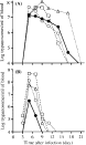
Fig. 1. Time-course of parasitaemia following intraperitoneal inoculation of 2×10⁵Trypanosoma grosi of SESEJI (A) and HANTO (B) isolates in 12-week-old female jirds with/without immunosuppressive treatments; naive ([bull ]), prednisolone-treated (○), splenectomized ([utri ]), and prednisolone-treated, splenectomized (□). Each value is the average of log trypanosomes/ml of blood from 2 animals/group.
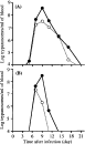
Fig. 2. Time-course of parasitaemia following intraperitoneal inoculation of 2×10⁵ of Trypanosoma grosi of SESEJI (A) and HANTO (B) isolates in 12-week-old male jirds with single or serial prednisolone treatment. Jirds were treated singly with 10 mg of prednisolone at infection (○), or repeatedly with 10 mg of prednisolone from day 10 prior to the infection ([bull ]). Each value is the average of log trypanosomes/ml of blood of 5 animals/group.

Fig. 3. Trypanosoma grosi in the peripheral blood of prednisolone-treated jirds on day 6 p.i. (A) SESUJI isolate; and (B) HANTO isolate. Giemsa's stain.

Table 1. Morphological features of different isolates of Trypanosoma grosi (measurements in μm)

Fig. 4. Persistent Trypanosoma grosi (SESUJI isolate) in the medullary capillaries of the kidney of a prednisolone-treated jird at week 5 p.i., as demonstrated by immunohistochemistry. Asterisks indicate the margin of pyramis renales, and multiple dark patches scattering in the medulla are aggregations of dividing forms (an arrow indicates one example).

Fig. 5. Impression smear of dividing forms of Trypanosoma grosi (SESUJI isolate) in the medullary capillaries of the kidney from a prednisolone-treated jird at week 5 p.i. Giemsa's stain.
 You have
Access
- 10
- Cited by