




No CrossRef data available.
Article contents
A comparison of episodic memory deficits in neuropathologically-confirmed Dementia with Lewy bodies and Alzheimer's disease
Published online by Cambridge University Press: 01 September 2004
Abstract
Little is known about possible differences in the memory deficits that occur in Dementia with Lewy bodies (DLB) and Alzheimer's disease (AD). We compared 24 autopsy-confirmed DLB and 24 age-, education-, and MMSE-matched autopsy-confirmed AD patients on the California Verbal Learning Test (CVLT) and the Wechsler Memory Scale-Revised Logical Memory subtest. The DLB and AD groups were similarly impaired on CVLT Total Learning (Trials 1–5) and Long Delayed Free Recall, but the DLB group demonstrated relative improvement in Savings scores and on recognition testing compared to the AD group. Likewise, the patient groups were equally impaired on Logical Memory immediate and delayed recall, but the DLB group's Saving scores were significantly better than those of the AD patients. These results indicate that while both DLB and AD patients exhibit significant memory impairment, the ability to consolidate information may be less severely impaired in DLB patients than in AD patients. (JINS, 2004, 10, 689–697.)
Information
- Type
- Research Article
- Information
- Journal of the International Neuropsychological Society , Volume 10 , Issue 5 , September 2004 , pp. 689 - 697
- Copyright
- 2004 The International Neuropsychological Society
INTRODUCTION
Dementia with Lewy bodies (DLB) is a clinical–pathological condition characterized by a progressive dementia syndrome that is associated with neurodegenerative changes including neuron loss and Lewy body deposition in subcortical areas usually affected in Parkinson's disease (Hansen et al., 1990), and widespread cortical Lewy bodies most prominent in limbic and neocortical association areas (Gomez-Tortosa et al., 1998; Lennox et al., 1989; Rezaie et al., 1996). In a large majority of cases, Alzheimer's disease (AD) pathology (e.g., neurofibrillary tangles and, especially, neuritic plaques) is also present in sufficient amounts to warrant a concurrent diagnosis of that disease (Hansen & Samuel, 1997). Key clinical features of DLB include progressive cognitive decline, spontaneous motor features of parkinsonism, recurrent well-formed visual hallucinations, and fluctuations in cognition, attention and alertness (McKeith et al., 1996). These clinical features form the primary consensus criteria for the clinical diagnosis of DLB and were largely adopted to help distinguish the disorder from the pathologically “pure” form of AD. Unfortunately, the diagnostic sensitivity and specificity of the criteria remain problematic (Hohl et al., 2002; Lopez et al., 2000; Merdes et al., 2003), but might be improved by the identification of additional features that distinguish between DLB and AD. The accurate differential diagnosis of DLB is important because the disease appears to progress more rapidly than AD (Olichney et al., 1998) and may respond differently to cholinergic therapy and neuroleptic medications (Levy et al., 1994; McKeith et al., 1996).
Retrospective neuropsychological studies of patients with autopsy-confirmed DLB have attempted to delineate distinct patterns of cognitive deficits that may differentiate the disorder from “pure” AD. Although these studies have included DLB cases with concomitant AD pathology (i.e., Lewy body variant of AD; Connor et al., 1998; Förstl et al., 1993; Galasko et al., 1996; Hansen et al., 1990; Heyman et al., 1999) or without concomitant AD pathology (i.e., pure diffuse Lewy body disease; Galasko et al., 1996; Salmon et al., 1996), they have led to similar conclusions. Patients with DLB have greater impairments in attention (Hansen et al., 1990), executive functions (Connor et al., 1998; Galasko et al., 1996; Hansen et al., 1990; Salmon et al., 1996), and visuospatial abilities (Galasko et al., 1996; Salmon et al., 1996) than AD patients who are at a similar level of dementia as measured by standard mental status examinations (e.g., Mini-Mental State Exam). These abnormally severe cognitive deficits have been attributed to additional frontostriatal pathology in DLB compared to AD, and to the disorder's particularly severe impact on the cholinergic system (Langlais et al., 1993; Tiraboschi et al., 2000) and on metabolic activity in the occipital cortex (Albin et al., 1996; Minoshima et al., 2001). In contrast, studies comparing the episodic memory deficits associated with DLB and AD suggest that DLB patients have impairment that is similar to (Förstl et al., 1993; Galasko et al., 1996; Hansen et al., 1990) or less severe than (Connor et al., 1998; Heyman et al., 1999) that of patients with AD. Furthermore, the two disorders may have a differential impact on episodic memory processes such as learning, retention, and recognition (Salmon et al., 1996). These findings are consistent with neuropathologic studies demonstrating less severe hippocampal atrophy (Lippa et al., 1994; 1998) and more severe frontostriatal dysfunction in DLB than in AD (Hansen et al., 1990), but remain speculative given that the studies employed small samples (Förstl et al., 1993; Galasko et al., 1996; Hansen et al., 1990; Salmon et al., 1996), severely demented patients (Heyman et al., 1999), or very simple tests of memory (Connor et al., 1998).
The present study compared the effects of DLB and AD on episodic memory in order to determine if there are differences in the patterns of specific memory processes that these diseases most affect. The analyses were limited to DLB patients with concomitant AD pathology (a condition also referred to as the Lewy Body Variant of AD; Hansen et al., 1990) as this is the more common form of DLB and the form that is likely to be the most difficult to clinically differentiate from “pure” AD. Episodic memory was assessed with two rigorous, standardized verbal memory measures, the California Verbal Learning Test (CVLT) and the Logical Memory subtest of the Wechsler Memory Scale–Revised (WMS–R), that have been used extensively with patients with AD to show that the disorder is characterized by poor learning and retention, abnormally rapid forgetting, and poor recognition memory (Delis et al., 1991; Tröster et al., 1993). This pattern of deficits suggests a primary impairment of encoding and storage of new information in AD. These memory tests have also been used to examine the episodic memory deficits associated with dementing disorders that have their primary site of pathology in subcortical structures (e.g., the striatum and fronto-striatal circuits), such as Huntington's disease, and show that these disorders present with poor learning and free recall of information, but only mildly impaired retention and recognition memory (Delis et al., 1991; Tröster et al., 1993). Given the disparate patterns of episodic memory deficits associated with AD and fronto-striatal disorders, the significant subcortical dysfunction and milder hippocampal damage in DLB compared to AD suggest that DLB may be characterized by better recognition memory and less rapid forgetting than AD, even though the disorders may engender similar overall deficits in learning and free recall.
METHODS
Research Participants
Data from 24 patients with autopsy-confirmed DLB, 24 patients with autopsy-confirmed AD, and 24 healthy normal control (NC) participants were retrospectively examined. All study participants were drawn from the University of California, San Diego (UCSD) Alzheimer's Disease Research Center (ADRC). Participants and/or their caregivers had provided written informed consent for participation after the study was explained to them. Informed consent for autopsy was obtained at the time of death from the next of kin. Cases were specifically excluded if autopsy revealed any significant pathological process (e.g., stroke, hippocampal sclerosis, metabolic encephalopathy) other than DLB or AD.
The patients with DLB represented all patients from the UCSD ADRC autopsy series who had completed both the CVLT and the Logical Memory subtest of the WMS–R and satisfied clinico-pathological criteria for the disease. Four patients with autopsy-confirmed diffuse Lewy body disease (i.e., no concomitant AD pathology) who otherwise satisfied these criteria were not included. Each patient with DLB was matched 1:1 to a patient with AD on the basis of age, years of education, and Mini-Mental State Examination (MMSE) score at the time of testing, and to a NC subject on the basis of age and years of education. When more than one patient with AD from the UCSD ADRC autopsy series, or more than one NC subject from the ongoing longitudinal study, matched a patient with DLB and had completed both memory measures, the one included in the present analysis was selected randomly. As expected from the matching procedure, the groups did not differ in age or years of education nor did they differ in sex distribution (all ps > .10). The NC participants obtained higher MMSE scores than the DLB and AD patients (p < .001), but the patient groups did not differ significantly.
Although not specifically matched for the course of disease, the DLB and AD groups did not differ significantly in the intervals between estimated onset of dementia and time of testing or between time of testing and death. To further demonstrate that these patient groups were well matched with respect to disease severity at the time of testing, no group differences were found in caregiver ratings of functional capacity as measured by the Pfeffer Outpatient Disability Scale. Finally, it is not simply the case that the DLB group was less cognitively disabled than the AD group as there were no significant or meaningful differences between the groups' total scores on the Mattis Dementia Rating Scale or a 30-item Boston Naming Test, and the DLB group performed significantly worse than the AD group on the Wechsler Intelligence Scale for Children-Revised Block Design subtest (p < .05). Table 1 presents these demographic and clinical data.
Demographic characteristics and clinical data for patients with dementia with Lewy bodies (DLB), patients with Alzheimer's disease (AD), and normal control (NC) participants

All patients were diagnosed with dementia at their clinical evaluation by a senior staff neurologist. Of the 24 DLB patients, four were clinically diagnosed with DLB, 19 with possible (n = 4) or probable (n = 15) AD, and one with mixed dementia. All 24 AD patients were clinically diagnosed with possible (n = 6) or probable (n = 18) AD. The diagnosis of possible AD was made when dementia was present in the absence of other neurologic, psychiatric, or systemic disorders sufficient or likely to cause dementia, but disease onset, presentation, or course was atypical for AD. Such variations are expected in the individuals who were later confirmed by autopsy to have Lewy body pathology, and not unusual in the individuals with definite AD, many of whom were tested early in the disease course when MMSE scores were still high. Upon autopsy, only two DLB cases and two AD cases had generalized vascular changes (e.g., atherosclerosis, arteriosclerosis), but they lacked infarcts or hemorrhages. None of the cases in this study had hippocampal sclerosis or other confounding pathology. Therefore, clinical diagnoses other than DLB in the DLB group or AD in the AD group were inaccurate.
Neuropathologic Diagnosis
The procedures for histological analysis of the patients' brains have been previously described in detail (Hansen & Samuel, 1997). Briefly, plaques and neurofibrillary tangles were stained with thioflavin-S and counted by the same examiner (L.A.H.) using consistent criteria. Lewy bodies were detected using hematoxylin–eosin (H & E) stained preparations. Cases were only construed as DLB if Lewy bodies were found in the locus ceruleus, substantia, nigra, and/or nucleus basalis of Meynert, as well as in the superior temporal cortex. Because all cases categorized as DLB had neocortical as well as brainstem Lewy bodies, they would all fall into either the limbic (transitional) or neocortical categories proposed in the 1996 consensus guidelines for the pathologic diagnosis of DLB (McKeith et al., 1996). Further, all DLB cases were neocortical stage 5 or 6 according to the recently proposed Lewy-body based staging of brain pathology related to sporadic Parkinson's disease (Braak et al., 2003). In accordance with recommendations of the 3rd International Conference on Parkinson's disease with Dementia/Dementia with Lewy bodies, cases were not classified as DLB if Lewy bodies were only found in the amygdala with alpha-synuclein stain. None of the AD cases had Lewy bodies recognized in pigmented brainstem nuclei where they are readily apparent with H & E stain and where they would appear prior to neocortical involvement (Braak et al., 2003), except in “amygdala-only” cases. Therefore, it is unlikely that cases were misclassified; however, such misclassifications would serve only to attenuate group differences.
All DLB patients had concomitant AD pathology sufficient to meet National Institute on Aging (NIA) and Consortium to Establish a Registry for Alzheimer's Disease (CERAD) criteria for definite AD. Thus, all DLB patients conformed to what Hansen et al. (1990) have called Lewy Body Variant of Alzheimer's disease. Patients with AD satisfied NIA and CERAD criteria for definite AD and had no cortical or subcortical Lewy bodies. A modified Braak stage (see Hansen & Samuel, 1997) was obtained for each case by counting the number of neurofibrillary tangles in at least five neuron clusters in layer two of the entorhinal cortex and then averaging the results. Cases with modified Braak stage I to IV have fewer than 18 tangles on average in layer two of the entorhinal cortex and sparse neocortical tangles. Cases assigned to modified Braak stage V have moderate numbers of tangles in at least two neocortical sections, and in modified Braak stage VI, all neocortical areas assessed have at least moderate numbers of tangles. The modified Braak stage of the DLB patients was significantly lower than that of the patients with AD (Mann-Whitney U = 104.5, p < .001). Table 2 details the modified Braak stage and the neuritic plaque and neurofibrillary tangle counts in the hippocampus and midfrontal, superior temporal, and inferior parietal cortex.
Modified Braak staging and neuritic plaque and neurofibrillary tangle counts in patients with Dementia with Lewy Bodies (DLB) or Alzheimer's disease (AD)

Measures
The California Verbal Learning Test (CVLT) and the Wechsler Memory Scale–Revised (WMS–R) Logical Memory subtest were administered to each participant as part of a larger battery of tests used to annually evaluate participants at the UCSD ADRC. Each participant was tested individually by a trained psychometrist in a quiet, well-lit room. The two memory tests are described below.
California Verbal Learning Test
The CVLT is a standardized memory test that was developed to assess a variety of memory processes identified through cognitive psychological studies of normal memory (Delis et al., 1987). The CVLT assesses rate of learning, retention after short- and long-delay intervals, semantic encoding ability, recognition memory (i.e., discriminability), intrusion and perseverative errors, and response biases. Five presentation/free recall trials for a list (List A) of 16 items (four items in each of four semantic categories) are administered followed by a single interference trial using a second, different list (List B) of 16 items. Immediately after the List B trial, free-recall and then cued-recall (utilizing the names of the four categories) of the items on the initial list (List A) are elicited. Twenty minutes later, free recall and cued recall of the List A items are again elicited, followed by a yes–no recognition test consisting of the 16 List A items and 28 randomly-interspersed distracter items. A savings score was computed by dividing the number of items recalled on the Long Delay Free Recall trial by the number recalled on Trial 5 of the initial presentation/free recall trials. This proportion was then multiplied by 100 to provide a percentage score.
Logical Memory subtest
The Logical Memory subtest of the WMS–R (Wechsler, 1987) is a standardized test that assesses free recall of two short stories that each consist of 25 bits of information. The two stories are presented sequentially in the immediate recall condition, and free recall of the story is elicited after each presentation. Thirty minutes later, free recall of the two stories is again elicited. The total bits of information recalled immediately and after the delay interval are recorded and a savings score is computed by dividing the total score achieved during delayed recall by the total score achieved during immediate recall. This proportion was then multiplied by 100 to provide a percentage score.
Data Analysis
The performances of the DLB patients, AD patients, and NC participants on measures of learning (i.e., CVLT Trials 1–5 raw score and the Logical Memory Immediate Recall score), retention (i.e., the CVLT Long-Delay Free Recall and Logical Memory Delayed Recall scores, and the CVLT and Logical Memory Savings scores), and recognition memory (i.e., CVLT Discriminability score) were compared using one-way Analysis of Variance (ANOVA) with Tukey's Least Significant Difference (LSD) test for post-hoc pair-wise comparisons set at a significance level of p < .05. Because of concern that the CVLT discriminability measure was insensitive to response bias, recognition accuracy and decision bias were also assessed with metrics derived from the two-high-threshold model of recognition memory (Snodgrass & Corwin, 1988). This model postulates that a subject uses one memory threshold for endorsing old items and another memory threshold for identifying new items. These two thresholds delineate three possible memory states for a given test item: certain recognition of an old item as old, certain recognition of a new item as new, and uncertainty. Items in the uncertain state will be classified as either old or new depending on the subject's response bias. The measure of recognition accuracy derived from the two-high-threshold model is a discrimination index, or Pr, which is calculated as the hit rate minus the false alarm rate. The measure of decision bias is Br, which is calculated as the false alarm rate divided by the quantity 1 minus Pr. Because the calculation of Br can lead to division by zero in the case where Pr equals 1, Snodgrass and Corwin (1988) suggest that the hit rate (i.e., true positive rate) and false alarm rate be adjusted as follows prior to the calculation of Pr and Br: hit rate = (hits + 0.5)/(the number of target stimuli + 1); false alarm rate = (false alarms + 0.5)/(the number of distractor stimuli + 1).
Pr provides a measure of recognition accuracy that is relatively independent of decision bias. Pr increases up to 1.0 as the overall accuracy of recognition memory test performance increases. Random guessing that results in 50% accuracy for targets and 50% accuracy for distracters would produce a Pr of .0. Br reflects the proportion of trials on which the subject guesses “yes” when uncertain that the test item is a target or distractor. Br greater than 0.5 indicates a liberal response bias (i.e., a tendency to say “yes”), less than 0.5 indicates a conservative response bias (i.e., a tendency to say “no”), and equal to 0.5 indicates a neutral response bias.
RESULTS
Learning
The mean scores achieved by the DLB patients, AD patients, and NC participants on the CVLT Trials 1–5 and Logical Memory Immediate Recall are shown in the upper panel of Figure 1. One-way ANOVAs revealed significant group differences on both measures of learning [CVLT Trials 1–5: F(2,69) = 73.62, p < .001; Logical Memory Immediate Recall: F(2,69) = 84.10, p < .001]. Post-hoc pair-wise comparisons demonstrated that the DLB and AD groups were significantly impaired compared to the NC group on both learning measures, but did not differ significantly from each other.

Upper panel: Immediate Recall. The mean scores achieved by patients with Dementia with Lewy bodies (DLB), patients with Alzheimer's disease (AD), and normal control (NC) participants on the California Verbal Learning Test (CVLT) Trials 1–5 and Logical Memory Immediate Recall measures. Lower panel: Delayed Recall. The mean scores achieved by each group on CVLT Long Delay Free Recall and Logical Memory Delayed Recall. Error bars represent standard error of the mean.
Retention
The mean scores achieved by the DLB patients, AD patients, and NC participants on the CVLT Long Delay Free Recall and Logical Memory Delayed Recall are shown in the lower panel of Figure 1. One-way ANOVAs revealed significant group differences on both of these delayed recall measures [CVLT Long Delay Free Recall: F(2,69) = 77.79, p < .001; Logical Memory Delayed Recall: F(2,69) = 84.48, p < .001]. Post-hoc pair-wise comparisons revealed that the DLB and AD patient groups performed significantly worse than the NC group on both delayed recall measures, but did not differ significantly from each other.
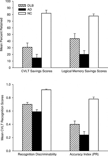
The mean Savings scores achieved on the CVLT and Logical Memory subtest by each group are shown in the upper panel of Figure 2. Savings scores could not be calculated for three patients (1 DLB and 2 AD patients) because they were unable to generate any items during immediate recall. The three groups differed significantly on both of these measures of retention [CVLT savings score: F(2,68) = 41.46, p < .001; Logical Memory savings score: F(2,67) = 26.77, p < .001]. Post-hoc pair-wise comparisons showed that the DLB and AD groups had significantly lower savings scores than the NC participants on both measures. In addition, the CVLT and Logical Memory savings scores of the AD patients were significantly lower than those of the DLB patients. It should be noted that the magnitude of the difference in savings scores for the DLB and AD patients could be attenuated by floor effects on delayed recalled measures. The delayed recall scores of AD patients were generally lower than those of the DLB patients, but the floor effect may have restricted the lower boundary of their savings scores leading to an underestimation of the discrepancy in the retention deficits of the two groups.

Upper panel: savings scores. The mean California Verbal Learning Test (CVLT) and Logical Memory Savings scores achieved by patients with Dementia with Lewy bodies (DLB), patients with Alzheimer's disease (AD), and normal control (NC) participants. Lower panel: CVLT Recognition Memory. The mean scores achieved by each group on the recognition discriminability measure and the Accuracy Index (Pr) derived from the CVLT recognition trial. Error bars represent standard error of the mean.
Recognition Memory
The mean scores achieved by the DLB patients, AD patients, and NC participants on the standard Discriminability measure and the Accuracy Index (Pr) derived from the CVLT recognition test are shown in the lower panel of Figure 2. The three groups differed significantly on both of these measures of recognition memory [CVLT Discriminability score: F(2,69) = 49.29, p < .001; Accuracy Index (Pr): F(2,69) = 40.66, p < .001]. Post-hoc pair-wise comparisons showed that the DLB and AD patients scored lower than the NC participants on both of these recognition memory measures. In addition, the AD patients exhibited significantly poorer recognition memory than the DLB patients on both measures.
Paired t tests demonstrated that while the patient groups did not differ significantly in the number of hits they achieved [DLB: M = 11.8, SD = 3.4; AD: M = 12.3, SD = 3.1; t(23) = −0.64, p = .53], AD patients committed significantly more false positive errors than the patients with DLB [DLB: M = 8.8, SD = 6.0; AD: M = 14.3, SD = 6.1; t(23) = −3.42, p < .01] and had a significantly greater positive response bias [DLB: M = 0.28, SD = 0.49; AD: M = 0.51, SD = 0.33; t(23) = −2.36, p < .05]. Patients with AD also had a significantly higher Decision Bias Index (Br) than the patients with DLB [DLB: M = 0.55, SD = 0.26; AD: M = 0.68, SD = 0.20; t(23) = −2.37, p < .05], and this index was indicative of a liberal response bias in the AD patients (i.e., a tendency to respond “yes”) and a neutral response bias in the DLB patients.
CVLT Intrusion Errors
Because the CVLT intrusion data significantly departed from normality, nonparametric tests were used to identify group differences. A significant group difference in the number of Free Recall intrusions was identified [DLB: M = 8.0, SD = 14.1; AD: M = 9.8, SD = 13.0; NC: M = 2.41, SD = 2.3; Kruskal-Wallis χ2 = 7.5, p < .05]. Pairwise comparisons revealed that the NC group made significantly fewer intrusion errors than the DLB and AD groups, but the patient groups did not significantly differ. A significant group difference for Cued Recall intrusions was also identified [DLB: M = 6.0, SD = 6.9; AD: M = 9.3, SD = 5.7; NC: M = 1.8, SD = 1.7; Kruskal-Wallis χ2 = 6.9, p < .001]. Cued recall intrusion rates were significantly lower in the NC group than in the DLB group (p < .01) and significantly lower in the DLB group than in the AD group (p < .05).
DISCUSSION
The results of the present study demonstrate that DLB and AD produce consistent differences in the patterns of episodic memory deficits in the early stages of the diseases. Although both patient groups were severely impaired relative to NC participants on all memory measures and were equally impaired in their ability to learn new verbal information, patients with autopsy-confirmed DLB exhibited better retention and recognition memory than patients with autopsy-confirmed pure AD. This different pattern of memory performance occurred despite equivalent severity of dementia in the two groups as determined by cognitive (MMSE, DRS, Boston Naming Test), functional (Pfeffer Outpatient Disability Scale), and clinical (disease course) measures. Furthermore, the patients with DLB had more severe visuospatial deficits than the patients with AD arguing against the possibility that the DLB patients were simply less demented than the AD patients. These results are consistent with the notion that the episodic memory deficit associated with DLB is generally less severe than that of AD, substantiating previous studies that used less rigorous memory tests with smaller samples of autopsy-confirmed patients (Connor et al., 1998; Heyman et al., 1999; Salmon et al., 1996) or that used potentially cross-contaminated clinically-diagnosed patient groups (Ballard et al., 1996; Calderon et al., 2001; Graham et al., 1997; Shimomura et al., 1998; Walker et al., 1997). Unlike a previous study that reported an increased frequency of environmentally cued intrusion errors in patients clinically diagnosed with DLB compared to AD (Doubleday et al., 2002), patients with DLB in the present study generated fewer intrusion errors on the CVLT compared to patients with AD. This discrepancy could result from methodological differences or from the possibility that some clinically diagnosed DLB and AD patients were misclassified by Doubleday et al. (2002).
The differences exhibited by the DLB and AD patients on the retention and recognition measures of the verbal memory tests suggest that specific memory processes may be differentially impaired in the two disorders. Consistent with previous studies (Delis et al., 1991; Tröster et al., 1993), the patients with AD exhibited a severe learning deficit, rapid forgetting of information over a delay interval (indicated by low savings scores), and severely impaired recognition memory. This pattern of deficits suggests a primary impairment of encoding and storage in which information is unavailable even when retrieval support is provided by the recognition memory format (Delis et al., 1991). Despite similarly severe impairment in the learning and delayed recall conditions when free recall was required, the patients with DLB had better savings over the delay interval than patients with AD, and performed better on the recognition memory component of the CVLT. These differences in performance suggest that a deficit in retrieval plays a greater role in the memory impairment of patients with DLB than in that of patients with AD. Of course, the pattern of results does not rule out the possibility that an encoding deficit contributes to memory impairment in the two disorders. It may be the case that encoding is too shallow in DLB and AD to allow adequate free recall, but deep enough in DLB to allow for assisted retrieval. Supporting this possibility, patients with DLB utilized the efficient semantic clustering strategy significantly more than the AD patients, but less than the NC participants, and utilized the inefficient serial clustering strategy significantly more than AD patients (data not shown). In any event, when retrieval demands are reduced through the use of the recognition format, it becomes clear that DLB patients have better retention than patients with AD.
It is important to note that the difference in the performances of DLB and AD patients on the recognition memory test of the CVLT was not solely the result of a greater positive response bias in the patients with AD. Although AD patients made significantly more false positive errors and obtained higher scores on indices of response bias than patients with DLB, their recognition accuracy was significantly lower than that of the DLB patients even after these factors were controlled by using an unbiased accuracy index derived from the two-high-threshold model (Snodgrass & Corwin, 1988). Thus, the particularly poor accuracy of the patients with AD on the recognition memory component of the CVLT appears to reflect a true memory deficit and not simply an inability to perform the test because of impaired set-shifting or other executive dysfunction.
The observed differences in the severity of impairment of specific memory processes in DLB and AD are consistent with the particular pathological changes that occur in the two disorders. A number of studies using neuropathologic (Lippa et al., 1994, 1998) or magnetic resonance imaging (Barber et al., 2000, 2001; Hashimoto et al., 1998) procedures have shown that the medial temporal lobe structures important for memory (e.g., hippocampus, entorhinal cortex, parahippocampal gyrus; Squire & Zola-Morgan, 1991) are more severely affected in AD than in DLB. The extensive damage to these structures that occurs in AD is thought to mediate the severe amnesia that characterizes the disorder (Hyman et al., 1990). In contrast, damage to subcortical structures that are affected in Parkinson's disease and other disorders of the basal ganglia (e.g., Huntington's disease) are more affected in DLB than in AD. Damage to these structures has been implicated in a memory disorder characterized by a general deficit in the ability to initiate and carry out the systematic retrieval of successfully stored information (Butters et al., 1985; 1986; Delis et al., 1991; Martone et al., 1984; Moss et al., 1986). This retrieval deficit is demonstrated by studies showing that patients with basal ganglia disorders have a relatively normal rate of forgetting of information over a delay interval (Granholm et al., 1985; Martone et al., 1986; Moss et al., 1986) and that their memory impairment appears greatly reduced (Butters et al., 1985; 1986; Martone et al., 1984; Massman et al., 1993), and in some cases eliminated (Martone et al., 1984; Moss et al., 1986; but see Lang et al., 2000), when memory is tested with recognition procedures that decrease retrieval demands. In light of these findings, the less severe retention deficit and the greater impact of deficient retrieval processes in DLB than in AD is consistent with the combination of moderate medial temporal lobe damage and frontostriatal dysfunction that occurs in the disease.
There are several limitations of the present study that should be noted. First, the relationship between the severity of specific episodic memory deficits and Lewy body counts could not be examined because Lewy body density was not quantified (qualitative presence or absence of pathology was used to classify a patient as DLB or AD) and because of the long interval between testing and death. Second, the DLB patients examined in this study were a sample referred to an Alzheimer's Disease Research Center and may not be representative of the disorder in the general population. A referral bias towards dementia may account for the high proportion of DLB patients (19/24) who were initially diagnosed with probable or possible AD. Importantly, such a referral bias would most likely lead to an underestimation of differences in the severity and pattern of episodic memory deficits in DLB and AD since DLB patients with particularly mild memory impairment may not have been referred to the Center and included in the present sample. Despite these limitations, these results clearly demonstrate differences in the episodic memory deficits associated with DLB and AD. Additional studies designed to identify reliable cognitive and clinical markers that differentiate between the disorders early in their course are needed.
ACKNOWLEDGMENTS
The results of this study were presented by J.M.H. at the February, 2002, meeting of the International Neuropsychological Society in Toronto, Canada. The study was supported by NIH Grants AG12963 and AG05131 to the University of California, San Diego. We thank the participants and staff of the UCSD Alzheimer's Disease Research Center.
References
REFERENCES
Albin, R.L.,
Minoshima, S.,
D'Amato, C.J.,
Frey, K.A.,
Kuhl, D.A., &
Sima, A.A.F.
(1996).
Fluoro-deoxyglucose positron emission tomography in diffuse Lewy
body disease.
Neurology,
47,
462–466.CrossRefGoogle Scholar
Ballard, C.,
Patel, A.,
Oyebode, F., &
Wilcock, G.K.
(1996).
Cognitive decline in patients with Alzheimer's disease,
vascular dementia, and senile dementia of the Lewy body type.
Age and Aging,
25,
209–213.CrossRefGoogle Scholar
Barber, R.,
Ballard, C.,
McKeith, I.G.,
Gholkar, A., &
O'Brien, J.T.
(2000).
MRI volumetric study of dementia with Lewy bodies: A comparison
with AD and vascular dementia.
Neurology,
54,
1304–1309.CrossRefGoogle Scholar
Barber, R.,
McKeith, I.G.,
Ballard, C.,
Gholkar, A., &
O'Brien, J.T.
(2001).
A comparison of medial and lateral temporal lobe atrophy in
dementia with Lewy bodies and Alzheimer's disease: Magnetic
resonance imaging volumetric study.
Dementia & Geriatric Cognitive Disorders,
12,
198–205.CrossRefGoogle Scholar
Braak, H.,
Del Tredici, K.D.,
Rüb, U.,
de Vos, R.A.I.,
Jansen Steur, E.N.H., &
Braak, E.
(2003).
Staging of brain pathology related to sporadic Parkinson's
disease.
Neurobiology of Aging,
24,
197–211.CrossRefGoogle Scholar
Butters, N.,
Wolfe, J.,
Martone, M.,
Granholm, E., &
Cermak, L.
(1985).
Memory disorders associated with Huntington's disease: Verbal
recall, verbal recognition and procedural memory.
Neuropsychologia,
23,
729–743.CrossRefGoogle Scholar
Butters, N.,
Wolfe, J.,
Granholm, E., &
Martone, M.
(1986).
An assessment of verbal recall, recognition and fluency abilities
in patients with Huntington's Disease.
Cortex,
22,
11–32.CrossRefGoogle Scholar
Calderon, J.,
Perry, R.J.,
Erzinclioglu, S.W.,
Berrios, G.E.,
Dening, T.R., &
Hodges, J.R.
(2001).
Perception, attention, and working memory are disproportionately
impaired in dementia with Lewy bodies compared with Alzheimer's
disease.
Journal of Neurology, Neurosurgery and Psychiatry,
70,
157–164.CrossRefGoogle Scholar
Connor, D.J.,
Salmon, D.P.,
Sandy, T.J.,
Galasko, D.,
Hansen, L.A., &
Thal, L.J.
(1998).
Cognitive profiles of autopsy-confirmed Lewy body variant vs. pure
Alzheimer's disease.
Archives of Neurology,
55,
994–1000.CrossRefGoogle Scholar
Delis, D.C.,
Kramer, J.H.,
Kaplan, E., &
Ober, B.A.
(1987).
California Verbal Learning Test.
New York:
Psychological Corporation.
Delis, D.C.,
Massman, P.J.,
Butters, N.,
Salmon, D.P.,
Cermak, L.S., &
Kramer, J.H.
(1991).
Profiles of demented and amnesic patients on the California Verbal
Learning Test: Implications for the assessment of memory
disorders.
Psychological Assessment,
3,
19–26.CrossRefGoogle Scholar
Doubleday, E.K.,
Snowden, J.S.,
Varma, A.R., &
Neary, D.
(2002).
Qualitative performance characteristics differentiate dementia with
Lewy bodies and Alzheimer's disease.
Journal of Neurology, Neurosurgery, and Psychiatry,
72,
602–607.CrossRefGoogle Scholar
Förstl, H.,
Burns, A.,
Luthert, P.,
Cairns, N., &
Levy, R.
(1993).
The Lewy-body variant of Alzheimer's disease: Clinical and
pathological findings.
British Journal of Psychiatry,
162,
385–392.CrossRefGoogle Scholar
Galasko, D.,
Katzman, R.,
Salmon, D.P., &
Hansen, L.
(1996).
Clinical and neuropathological findings in Lewy body dementias.
Brain and Cognition,
31,
166–175.CrossRefGoogle Scholar
Gomez-Tortosa, E.,
Newell, K.,
Irizarry, M., &
Hyman, B.T.
(1998).
Clinical and neuropathological features of dementia with Lewy
bodies.
American Journal of Alzheimer's Disease,
13,
284–290.CrossRefGoogle Scholar
Graham, C.,
Ballard, C., &
Saad, K.
(1997).
Variables which distinguish patients fulfilling clinical criteria
for dementia with Lewy bodies from those with Alzheimer's
disease.
International Journal of Geriatric Psychiatry,
12,
314–318.3.0.CO;2-7>CrossRefGoogle Scholar
Granholm, E.,
Wolfe, J., &
Butters, N.
(1985).
Affective-arousal factors in the recall of thematic stories by
amnesic and demented patients.
Developmental Neuropsychology,
1,
317–333.CrossRefGoogle Scholar
Hansen, L.,
Salmon, D.,
Galasko, D.R.,
Masliah, E.,
Katzman, R.,
DeTeresa, R.,
Thal, L.J.,
Pay, M.M.,
Hofstetter, R.,
Klauber, M.,
Rice, V.,
Butters, N., &
Alford, M.
(1990).
The Lewy body variant of Alzheimer's disease: A clinical and
pathologic entity.
Neurology,
40,
1–8.CrossRefGoogle Scholar
Hansen, L.A. &
Samuel, W.
(1997).
Criteria for Alzheimer's disease and the nosology of dementia
with Lewy bodies.
Neurology,
48,
126–132.CrossRefGoogle Scholar
Hashimoto, M.,
Kitagaki, H.,
Imamura, T.,
Hirono, N.,
Shimomura, T.,
Kazui, H.,
Tanimukai, S.,
Hanihara, T., &
Mori, E.
(1998).
Medial temporal and whole-brain atrophy in dementia with Lewy
bodies: A volumetric MRI study.
Neurology,
51,
357–362.CrossRefGoogle Scholar
Heyman, A.,
Fillenbaum, G.G.,
Gearing, M.,
Mirra, S.S.,
Welsh-Bohmer, K.A.,
Peterson, B., &
Pieper, C.
(1999).
Comparison of Lewy body variant of Alzheimer's disease with
pure Alzheimer's disease: Consortium to establish a registry for
Alzheimer's disease, part XIX.
Neurology,
52,
1839–1844.CrossRefGoogle Scholar
Hohl, U.,
Tiraboschi, P.,
Hansen, L.,
Thal, L.J., &
Corey-Bloom, J.
(2002).
Diagnostic accuracy of dementia with Lewy bodies.
Archives of Neurology,
57,
347–351.Google Scholar
Hyman, B.T.,
Van Hoesen, G.W., &
Damasio, A.R.
(1990).
Memory-related neural systems in Alzheimer's disease: An
anatomic study.
Neurology,
40,
1721–1730.CrossRefGoogle Scholar
Lang, C.J.G.,
Majer, M.,
Balan, P., &
Reischies, F.M.
(2000).
Recall and recognition in Huntington's disease.
Archives of Clinical Neuropsychology,
15,
361–371.CrossRefGoogle Scholar
Langlais, P.J.,
Thal, L.J.,
Hansen, L.A.,
Galasko, D.,
Alford, M., &
Masliah, E.
(1993).
Neurotransmitters in basal ganglia and cortex of Alzheimer's
disease with and without Lewy bodies.
Neurology,
43,
1927–1934.CrossRefGoogle Scholar
Lennox, G.,
Lowe, J.,
Landon, M.,
Byrne, E.J.,
Mayer, R.J., &
Godwin-Austen, R.B.
(1989).
Diffuse Lewy body disease: Correlative neuropathology using
anti-ubiquitin immunocytochemistry.
Journal of Neurology, Neurosurgery & Psychiatry,
52,
1236–1247.CrossRefGoogle Scholar
Levy, R.,
Eagger, S., &
Griffiths, M.
(1994).
Lewy bodies and response to Tacrine in Alzheimer's disease.
Lancet,
343,
176–176.CrossRefGoogle Scholar
Lippa, C.F.,
Johnson, R., &
Smith, T.W.
(1998).
The medial temporal lobe in dementia with Lewy bodies: A
comparative study with Alzheimer's disease.
Annals of Neurology,
43,
102–106.CrossRefGoogle Scholar
Lippa, C.F.,
Smith, T.W., &
Swearer, J.M.
(1994).
Alzheimer's disease and Lewy body disease: A comparative
clinicopathological study.
Annals of Neurology,
35,
81–88.CrossRefGoogle Scholar
Lopez, O.L.,
Hamilton, R.L.,
Becker, J.T.,
Wisniewski, S.,
Kaufer, D.I., &
DeKosky, S.T.
(2000).
Severity of cognitive impairment and the clinical diagnosis of AD
with Lewy bodies.
Neurology,
54,
1780–1787.CrossRefGoogle Scholar
Martone, M.,
Butters, N.,
Payne, M.,
Becker, J.T., &
Sax, D.S.
(1984).
Dissociations between skill learning and verbal recognition in
amnesia and dementia.
Archives of Neurology,
41,
965–970.CrossRefGoogle Scholar
Martone, M.,
Butters, N., &
Trauner, D.
(1986).
Some analyses of forgetting of pictorial material in amnesic and
demented patients.
Journal of Clinical and Experimental Neuropsychology,
8,
161–178.CrossRefGoogle Scholar
Massman, P.J.,
Delis, D.C., &
Butters, N.
(1993).
Does impaired primacy recall equal impaired long-term storage?
Serial position effects in Huntington's disease and
Alzheimer's disease.
Developmental Neuropsychology,
9,
1–15.Google Scholar
McKeith, I.G.,
Galasko, D.,
Kosaka, K.,
Perry, E.K.,
Dickson, D.W.,
Hansen, L.A.,
Salmon, D.P.,
Lowe, J.,
Mirra, S.S.,
Byrne, E.J.,
Lennox, G.,
Quinn, N.P.,
Edwardson, J.A.,
Ince, P.G.,
Bergeron, C.,
Burns, A.,
Miller, B.L.,
Lovestone, S.,
Collerton, D.,
Jansen, E.N.,
Ballard, C.,
de Vos, R.A.,
Wilcock, G.K.,
Jellinger, K.A., &
Perry, R.H.
(1996).
Consensus guidelines for the clinical and pathologic diagnosis of
dementia with Lewy bodies (DLB): report of the consortium on DLB
international workshop.
Neurology,
47,
1113–1124.CrossRefGoogle Scholar
Merdes, A.R.,
Hansen, L.A.,
Jeste, D.V.,
Galasko, D.,
Hofstetter, C.R.,
Ho, G.J.,
Thal, L.J., &
Corey-Bloom, J.
(2003).
Influence of Alzheimer's pathology on clinical diagnostic
accuracy in dementia with Lewy bodies.
Neurology,
60,
1586–1590.CrossRefGoogle Scholar
Minoshima, S.,
Foster, N.L.,
Sima, A.,
Frey, K.A.,
Albin, R.L., &
Kuhl, D.E.
(2001).
Alzheimer's disease versus dementia with Lewy bodies: Cerebral
metabolic distinction with autopsy confirmation.
Annals of Neurology,
50,
358–365.CrossRefGoogle Scholar
Moss, M.B.,
Albert, M.S.,
Butters, N., &
Payne, M.
(1986).
Differential patterns of memory loss among patients with
Alzheimer's disease, Huntington's disease, and alcoholic
Korsakoff's syndrome.
Archives of Neurology,
43,
239–246.CrossRefGoogle Scholar
Olichney, J.M.,
Galasko, D.,
Salmon, D.P.,
Hofstetter, C.R.,
Hansen, L.A.,
Katzman, R., &
Thal, L.J.
(1998).
Cognitive decline is faster in Lewy body variant than in
Alzheimer's disease.
Neurology,
51,
351–357.CrossRefGoogle Scholar
Rezaie, P.,
Cairns, N.J.,
Chadwick, A., &
Lantos, P.L.
(1996).
Lewy bodies are located preferentially in limbic areas in diffuse
Lewy body disease.
Neuroscience Letters,
12,
114.CrossRefGoogle Scholar
Salmon, D.P.,
Galasko, D.,
Hansen, L.A.,
Masliah, E.,
Butters, N.,
Thal, L.J., &
Katzman, R.
(1996).
Neuropsychological deficits associated with diffuse Lewy body
disease.
Brain and Cognition,
31,
148–165.CrossRefGoogle Scholar
Shimomura, T.,
Mori, E.,
Yamashita, H.,
Imamura, T.,
Hirono, N.,
Hashimoto, M.,
Tanimukai, S.,
Kazui, H., &
Hanihara, T.
(1998).
Cognitive loss in dementia with Lewy bodies and Alzheimer disease.
Archives of Neurology,
55,
1547–1552.CrossRefGoogle Scholar
Snodgrass, J.G. &
Corwin, J.
(1988).
Pragmatics of measuring recognition memory: Applications to
dementia and amnesia.
Journal of Experimental Psychology: General,
117,
34–50.CrossRefGoogle Scholar
Squire, L.R. &
Zola-Morgan, S.
(1991).
The medial temporal lobe memory system.
Science,
253,
1380–1386.CrossRefGoogle Scholar
Tiraboschi, P.,
Hansen, L.A.,
Alford, M.,
Sabbagh, M.N.,
Schoos, B.,
Masliah, E.,
Thal, L.J., &
Corey-Bloom, J.
(2000).
Cholinergic dysfunction in diseases with Lewy bodies.
Neurology,
54,
407–411.CrossRefGoogle Scholar
Tröster, A.I.,
Butters, N.,
Salmon, D.P.,
Cullum, C.M.,
Jacobs, D.,
Brandt, J., &
White, R.F.
(1993).
The diagnostic utility of savings scores: Differentiating
Alzheimer's and Huntington's diseases with Logical Memory and
Visual Reproduction tests.
Journal of Clinical and Experimental Neuropsychology,
15,
773–788.CrossRefGoogle Scholar
Walker, Z.,
Allen, R.L.,
Shergill, S., &
Katona, C.L.
(1997).
Neuropsychological performance in Lewy body dementia and
Alzheimer's disease.
British Journal of Psychiatry,
170,
156–158.CrossRefGoogle Scholar
Wechsler, D.
(1987).
Wechsler Memory Scale–Revised.
New York:
The Psychological Corporation.

Demographic characteristics and clinical data for patients with dementia with Lewy bodies (DLB), patients with Alzheimer's disease (AD), and normal control (NC) participants

Modified Braak staging and neuritic plaque and neurofibrillary tangle counts in patients with Dementia with Lewy Bodies (DLB) or Alzheimer's disease (AD)

Upper panel: Immediate Recall. The mean scores achieved by patients with Dementia with Lewy bodies (DLB), patients with Alzheimer's disease (AD), and normal control (NC) participants on the California Verbal Learning Test (CVLT) Trials 1–5 and Logical Memory Immediate Recall measures. Lower panel: Delayed Recall. The mean scores achieved by each group on CVLT Long Delay Free Recall and Logical Memory Delayed Recall. Error bars represent standard error of the mean.
Upper panel: savings scores. The mean California Verbal Learning Test (CVLT) and Logical Memory Savings scores achieved by patients with Dementia with Lewy bodies (DLB), patients with Alzheimer's disease (AD), and normal control (NC) participants. Lower panel: CVLT Recognition Memory. The mean scores achieved by each group on the recognition discriminability measure and the Accuracy Index (Pr) derived from the CVLT recognition trial. Error bars represent standard error of the mean.
 You have
Access
- 76
- Cited by