


Introduction
Plasmodium malariae is one of the 4 recognized species of Plasmodium causing malaria in humans. This species is distributed in tropical and subtropical regions and is most frequently found in sympatry with other Plasmodium species. P. malariae is often found as a mixed infection with P. falciparum or P. vivax. Historically, this parasite has been considered of minor importance because of its moderate pathogenicity and low prevalence. In endemic regions, the prevalence of P. malariae, based on standard microscopic examination of Giemsa-stained blood smears, can vary from less than 4% to more than 20% in different epidemiological settings (Molineaux and Grammiccia, Reference Molineaux and Grammiccia1980; Greenwood et al. Reference Greenwood, Bradley, Greenwood, Byass, Jammeh, Marsh, Tulloch, Oldfield and Hayes1987; Smith et al. Reference Smith, Charlwood, Kihonda, Mwankusye, Billingsey, Meuwissen, Lyimo, Takken, Teuscer and Tanner1993, Reference Smith, Genton, Baea, Gibson, Narara and Alpers2001; Trape et al. Reference Trape, Rogier, Konate, Diagne, Bouganali, Canque, Legros, Badji, Ndiaye, Ndiaye, Brahimi, Faye, Druilhe and Pereira da Silva1994). These values are 3–15 times less than the prevalence of P. falciparum in the same geographical regions. However, longitudinal microscopy studies reveal cumulative prevalences in children of 50 to 80% for periods of 1–24 months (Molineaux and Grammiccia, Reference Molineaux and Grammiccia1980; Trape et al. Reference Trape, Rogier, Konate, Diagne, Bouganali, Canque, Legros, Badji, Ndiaye, Ndiaye, Brahimi, Faye, Druilhe and Pereira da Silva1994; Bruce et al. Reference Bruce, Donnelly, Packer, Lagog, Gibson, Narara, Walliker, Alpers and Day2000a). Studies using PCR-based diagnostic methods have shown that prevalences based on microscopy are grossly underestimated due to its inability to detect low-level P. malariae parasitaemia, particularly when this species is present with one or more other species of Plasmodium (see Kawamoto et al. Reference Kawamoto, Liu, Ferreira and Tantular1999; Singh et al. Reference Singh, Bobogare, Cox-Singh, Snounou, Shukri Abdullah and Abdul Rahman1999). In studies in Africa, high prevalences of P. malariae were revealed by PCR analyses in surveys in which infection was not detectable by microscopy; for example, 36% in Gabon (Blampain-Azzibrouck et al. Reference Blampain-Azzibrouck, Lekoulou, Snounou, Ravollet and Ntoumi1999) and 25% in Kenya (Tobian et al. Reference Tobian, Mehlotra, Malhotra, Wamachi, Mungai, Koech, Ouma, Zimmerman and King2000). In studies in which P. malariae was detected by microscopy and PCR, higher prevalences (8–30 times) were recorded using the latter technique (Zhou et al. Reference Zhou, Liu, Wongsrichanalai, Suwonkerd, Panart, Prajakwong, Pensiri, Kimura, Matsuoka, Ferreira, Isomura and Kawamoto1998; Mehlotra et al. Reference Mehlotra, Lorry, Kastens, Miller, Alpers, Bockarie, Kazura and Zimmerman2000; Scopel et al. Reference Scopel, Fontes, Nunes, Horta and Braga2004). No figures are published for the prevalence of P. malariae infection worldwide, but a minimum estimate of 67 million infections per year can be calculated by comparison with the prevalence of P. falciparum (see Greenwood et al. Reference Greenwood, Marsh and Snow1991; Breman et al. Reference Breman, Egan and Keusch2001).
P. malariae differs from other Plamodium species of humans in numerous biological features that underlie parasite transmission and virulence, including reduced growth rate (Gilles and Warrell, Reference Gilles and Warrell1993), mature red blood cell preference (Garnham, Reference Garnham1966), late gametocyte formation (Garnham, Reference Garnham1966), extended duration of infection (up to 40 years in the absence of re-infection) (Vinetz et al. Reference Vinetz, Li, McCutchan and Kaslow1998; Chadee et al. Reference Chadee, Tilluckdharry, Maharaj and Sinanan2000; Siala et al. Reference Siala, Khalfaoui, Bouratbine, Hamdi, Hili and Aoun2005) and the absence of hypnozoite stages in the liver. The clinical features associated with P. malariae fever paroxysms are more moderate relative to P. falciparum and P. vivax (see Wernsdorfer and McGregor, Reference Wernsdorfer and McGregor1988). Fevers show quartan periodicity (due to a 72 h erythrocytic cycle) and parasite density is low, usually considerably below 1000 parasites per μl of blood, being microscopically undetectable much of the time. Infection is rarely life threatening, but can result in childhood mortality due to a ‘nephrotic syndrome’, an immunological complication which persists even after successful antimalarial therapy (Kibukamusoke, Reference Kibukamusoke1986; Otieno and Mc'Ligeyo, Reference Otieno and Mc'Ligeyo1988; Abdurrahman et al. Reference Abdurrahman, Aikhionbare, Babaoye, Sathiakumar and Narayana1990). Despite its relatively low morbidity in humans, P. malariae, in most endemic regions, is likely to contribute to the overall morbidity caused by Plasmodium species, manifested as anaemia (May et al. Reference May, Falusi, Mockenhaupt, Ademowo, Olumese, Bienzle and Meyer2000), low birth weight and reduced resistance to other infections, but its individual contribution has rarely been considered. Importantly, a number of studies have described the effect of prior or concurrent P. malariae infection on those with other Plasmodium species. Interactions can modify the within-host dynamics of Plasmodium species (see Bruce et al. Reference Bruce, Donnelly, Alpers, Galinski, Barwell, Walliker and Day2000b; Bruce and Day, Reference Bruce and Day2003), reduce the density of asexual stages of P. falciparum (see Alifrangis et al. Reference Alifrangis, Lemnge, Moon, Theisen, Bygbjerg, Ridley and Jakobsen1999) and result in an increased production of P. falciparum gametocytes (McKenzie et al. Reference McKenzie, Jeffery and Collins2002). Prior infection with P. malariae has been shown to reduce fever episodes caused by experimental, malaria therapy P. falciparum infections in humans with limited malaria exposure (Collins and Jeffery, Reference Collins and Jeffery1999) and also in humans living in areas with intense malaria transmission (Black et al. Reference Black, Hommel, Snounou and Pinder1994; Smith et al. Reference Smith, Genton, Baea, Gibson, Narara and Alpers2001).
Due to the difficulty of undertaking laboratory studies of P. malariae, few research projects have focused on this parasite, despite its global importance. Long-term in vitro culture is not available for P. malariae (see Millet et al. Reference Millet, Collins, Fisk and Nguyen-Dinh1988; Lingnau et al. Reference Lingnau, Doehring-Schwerdtfeger and Maier1994) and large numbers of parasites can be obtained only from costly non-human primate infections (Collins et al. Reference Collins, McClure, Strobert, Filipski, Skinner, Stanfill, Richardson and Morris1990, Reference Collins, Richardson, Sullivan, Morris and Galland1997). Limited availability of P. malariae DNA has resulted in a paucity of information about the genetics or genome of this parasite (Carlton et al. Reference Carlton, Galinski, Barnwell and Dame1999). Phylogenetic analyses suggest that P. malariae is distantly related to both P. falciparum and P. vivax (see Qari et al. Reference Qari, Shi, Pieniazek, Collins and Lal1996), with estimated divergence times being up to 174 million years ago (Escalante et al. Reference Escalante, Barrio and Ayala1995). P. brasilianum, a malaria parasite of New World monkeys, is both morphologically and genetically indistinguishable from P. malariae (see Cochrane et al. Reference Cochrane, Barnwell, Collins and Nussenzweig1985; Ayala et al. Reference Ayala, Lal, Escalante, Rich and Sherman1998; Fandeur et al. Reference Fandeur, Volney, Peneau and de Thoisy2000). P. brasilianum and P. malariae are likely to represent the same species but appear to have undergone a host switch within the last 500 to 15 000 years (Coatney et al. Reference Coatney, Collins, Warren and Contacos1971; Ayala et al. Reference Ayala, Lal, Escalante, Rich and Sherman1998; Escalante et al. Reference Escalante, Freeland, Collins and Lal1998; Fandeur et al. Reference Fandeur, Volney, Peneau and de Thoisy2000), most likely as an anthroponosis in the earlier time-frame. Morphological and genetic variants of human P. malariae described in the Far East may represent subspecies (Liu et al. Reference Liu, Zhu, Mizuno, Kimura, Liu, Isomura, Wang and Kawamoto1998; Kawamoto et al. Reference Kawamoto, Win, Mizuno, Lin, Kyaw, Tantulart, Mason, Kimura and Wongsrichanalai2002).
In order to address knowledge gaps relating to P. malariae, we have isolated polymorphic genetic markers that can be used to investigate multiple aspects of the biology, epidemiology and population structure of this parasite. Microsatellite markers for other species of Plasmodium, including P. falciparum (see Anderson et al. Reference Anderson, Su, Bockarie, Lagog and Day1999) and P. vivax (see Gomez et al. Reference Gomez, McNamara, Bockarie, Baird, Carlton and Zimmerman2003; Leclerc et al. Reference Leclerc, Durand, Gauthier, Patot, Billotte, Menegon, Severini, Ayala and Renaud2004), have been used for understanding their population genetic structures (Anderson et al. Reference Anderson, Haubold, Williams, Estrada-Franco, Richardson, Mollinedo, Bockarie, Mokili, Mharakurwa, French, Whitworth, Velez, Brockman, Nosten, Ferreira and Day2000), in the study of the emergence and spread of drug resistance (Nair et al. Reference Nair, Williams, Brockman, Paiphun, Mayxay, Newton, Guthmann, Smithuis, Hien, White, Nosten and Anderson2003; Roper et al. Reference Roper, Pearce, Nair, Sharp, Nosten and Anderson2004; Nash et al. Reference Nash, Nair, Mayxay, Newton, Guthmann, Nosten and Anderson2005), and in distinguishing new and recrudescent infections (Nyachieo et al. Reference Nyachieo, Van Overmeir, Laurent, Dujardin and D'Alessandro2005; Mwangi et al. Reference Mwangi, Omar and Ranford-Cartwright2006). In this study, we isolated non-coding, microsatellite and minisatellite markers, as they are less likely to be under selection pressure from the immune system of humans, have a high degree of polymorphism and also show a high evolutionary rate relative to expressed genes (Goldstein and Schlotterer, Reference Goldstein and Schlotterer1999), thus providing useful information regarding genetic changes considered to be relevant to recent Plasmodium population dynamics. These markers provide a means of genetically characterizing P. malariae samples for the first time. Such characterization is considered essential for in vivo drug and vaccine efficacy studies, for addressing questions about the duration and rate of relapse of P. malariae in endemic regions, in the study of within-host dynamics of the parasite and for exploring the population biology of P. malariae. We have validated the markers using a panel of 76 P. malariae isolates from individuals with varying clinical presentation and from a diverse geographical range. We present herein the first molecular epidemiological data for P. malariae and use them to explore the genetic diversity within P. malariae in relation to geographical origin, give insights into its population structure and describe the relationship between multiplicity of infection and clinical presentation.
Materials and Methods
Parasite and host samples and DNA extraction
Two strains of Plasmodium malariae were isolated from patients who had acquired P. malariae infections in Uganda (Collins et al. Reference Collins, Schwartz, Skinner and Broderson1984) or Brazil (John Barnwell, unpublished data). The UgandaI/CDC strain, after isolation from an infant infected by blood transfusion, was maintained by passage in splenectomized chimpanzees (Pan troglodytes) and New World monkeys. The infected red blood cells from the Brazil isolate (Pm-Brazil I) were isolated directly from a patient in North-East Brazil. P. brasilianum isolates from Peru (Pb-Peru I/NYU) and Colombia (Pb-Colombia I/NYU) were isolated from infections arising in newly splenectomized New World monkeys (Saimiri peruviensis and S. sciureus, respectively) and maintained by passage in splenectomized Saimiri boliviensis boliviensis (a related species of primate). Blood leukocytes and platelets were removed from the samples using acid-washed glass beads and CF11 cellulose columns or Plasmodipur filters (Euro-Diagnostica, Arnhem, The Netherlands), and genomic DNA was extracted using standard phenol/chloroform extraction protocols (Barnwell et al. Reference Barnwell, Nichols and Rubinstein1983, Reference Barnwell, Howard, Coon and Miller1989). Laboratory strains of P. falciparum 3D7 and HB3 were used as controls. These isolates were grown in vitro (Trager and Jenson, Reference Trager and Jenson1976) and parasites were prepared using saponin lysis and a standard DNA phenol/chloroform extraction (Bruce et al. Reference Bruce, Galinski, Barnwell, Snouou and Day1999). DNA from the monkey-adapted P. vivax isolate Salvador I and P. vivax from symptomatic travellers returning to the UK was isolated as reported previously (Bruce et al. Reference Bruce, Galinski, Barnwell, Snouou and Day1999).
Blood samples from human patients with naturally acquired, asymptomatic infections of P. falciparum, P. malariae or P. ovale were collected by ‘fingerprick’ in Malawi from residents of villages within a 25 km radius of the Dedza and Mangochi District Hospitals. Samples from symptomatic human infections known to be positive by microscopy for only P. malariae (320–385 parasites per μl of blood) or P. malariae and P. falciparum (P. malariae range 20–5000 per μl, P. falciparum range 6000–8500 per μl) from Gambian children (1–9 years of age) were collected as part of a clinical trial (Sutherland et al. Reference Sutherland, Drakeley, Obisike, Coleman, Jawara, Targett, Milligan, Pinder and Walraven2003). Samples from Thai patients in the Maela Refugee Camp, Tak Province, presenting with symptomatic infections positive by microscopy for only for P. malariae (1440–8096 parasites per μl of blood) were collected as part of ongoing drug resistance studies (Alan Brockman, personal communication); DNA from humans with naturally acquired infection was extracted, using a Chelex method (Kyes et al. Reference Kyes, Craig, Marsh and Newbold1993), from blood stored either on filter paper (20–50 μl of blood resulting in an extract volume of 200 μl) or as whole blood or red blood cell pellets stored frozen. Host DNA samples from humans, chimpanzees (Pan troglodytes) and squirrel monkeys (Saimiri boliviensis) were isolated from leukocytes using standard phenol/chloroform extraction protocols (Sambrook and Russell, Reference Sambrook and Russell2001) and used as negative controls in the molecular analyses.
Ethics approval for experimental infections in animals was granted by the Institutional Animal Care and Use Committees of Emory University and the Centres for Disease Control and Prevention, Atlanta, USA, and by the Animal Ethics Committee of Glasgow University. Collection of parasite samples from humans in Malawi was approved by the National Health Sciences Research Committee, Ministry of Population and Health, Malawi, and the Ethics Committee for Non-clinical Research Involving Human Subjects of Glasgow University. Ethics approval for the use of the samples from Gambia was obtained from the Joint Gambia Government/MRC Laboratories Ethics Committee. Ethics approval for the collection of the samples from Thailand was granted by the Ethics Committees of the Faculty of Tropical Medicine, Mahidol University, Bangkok and the Karen Refugee committee, Mae Sot, Thailand.
Diagnosis of Plasmodium species in naturally acquired human infections
Each of the 4 human species of Plasmodium in naturally acquired infections was detected by microscopic examination of Giemsa-stained blood smears. The species present were determined by examination of thin smears, and their density was measured by counting the number of parasites corresponding to 200 or 500 leukocytes in thick blood smears. The number of parasites per μl of blood was calculated, assuming a leukocyte density of 8000 per μl of blood. To confirm the microscopy results and detect parasites present below the detection limit of microscopy (∼40 parasites per μl of blood), a nested PCR based on the amplification of small subunit ribosomal RNA (ssrRNA) was also carried out essentially as described previously by other authors (Snounou et al. Reference Snounou, Viriyakosol, Zhu, Jarra, Pinheiro, Rosário, Thaithong and Brown1993; Singh et al. Reference Singh, Bobogare, Cox-Singh, Snounou, Shukri Abdullah and Abdul Rahman1999). In brief, the genus-specific primers Plu1 and Plu5 were used in a primary reaction, followed by a nested reaction using specific primers for each of the 4 species of Plasmodium from humans. Primary reactions were carried out in 50 μl containing 1·25 U of Thermoprime Plus Taq polymerase (ABgene), and the nested reactions were carried out in 25 μl containing 0·6 U of Thermoprime Plus Taq polymerase (transferring 2 μl of primary product). Both reactions contained the same reagents in the same concentrations: 75 mm Tris-HCl (pH 8·8), 20 mm (NH4)2SO4, 4·0 mm MgCl2, 0·01% Tween 20 (Sigma), 0·2 mm each of dATP, dCTP, dGTP and dTTP (ABgene) and 250 nm of each primer pair. The approximate amounts of template added to the primary PCR were 0·1 ng of purified DNA or 5 μl of DNA extract from filter-paper blood samples. Amplicons were separated on 1·6% agarose gels in TBE buffer (0·045 m Tris-borate, 0·001 m EDTA, pH 8·0), stained with ethidium bromide and detected by ultraviolet transillumination. A 100 bp ladder (Promega) was used as a size marker on these gels.
Isolation of microsatellite loci
Sequences containing microsatellite loci were isolated from a genomic library of Plasmodium brasilianum of Colombian origin (Pb-Colombia I/NYU). Genomic DNA was digested with RsaI (New England Biolabs, Beverly, MA, USA). Fragments of 300–1500 bp were selected, gel-purified using β-agarase I, according to the manufacturer's instructions (New England Biolabs) and cloned into the vector pZERO (Invitrogen). Approximately 100 000 colonies were transferred to N+ nitrocellulose filters (Amersham Biosciences) and screened using radio-isotope labelled probes containing polyT, TA, CA and TAA repeats of 100–300 bp in length (Armour et al. Reference Armour, Neumann, Gobert and Jeffreys1994). Equimolar concentrations of each probe were mixed, and 200 ng of the mixture was labelled with α32P dCTP (Perkin Elmer) using random priming (Prime-It II Kit, Stratagene). Unincorporated nucleotides were removed using a Microspin G-25 column (Amersham Biosciences). Filters were washed to a stringency of 50°C, 2×SSC, 0·1% SDS. Inserts of positive colonies were sequenced in both directions using Big Dye chemistry on a 377 sequencer (Applied Biosystems).
Sequencing of P. malariae microsatellite alleles
Polymorphism of P. malariae microsatellite alleles was initially assessed by aligning sequence data from the P. brasilianum library strain and from PCR products from orthologous loci in P. malariae Uganda I/CDC and P. malariae Brazil I, for which large amounts of genomic DNA were available. PCR primers were designed from non-repetitive flanking regions of microsatellite loci using Staden software (Staden et al. Reference Staden, Beal and Bonfield2000). PCR products for sequencing were amplified using the primary reaction primers (see Table 2), with the following alterations: Extensor Hi-Fidelity PCR Enzyme mix (containing Thermoprime Taq and a proof-reading enzyme, ABgene) with buffer 1 and 40 ng of genomic DNA were used. Amplicons were cloned using TOPO TA Cloning® Kit (Invitrogen) according to the manufacturer's instructions. DNA from transformed colonies was prepared using the Minprep Kit (QIAgen), and inserts were sequenced in both directions using M13 primers. Sequences were assembled using the Staden software, and alignments were made using ClustalW (Chenna et al. Reference Chenna, Sugawara, Koike, Lopez, Gibson, Higgins and Thompson2003). Loci showing polymorphism in repetitive domains were selected for further analysis using a panel of P. malariae DNA samples and controls from various geographical origins. Sequence homology to other Plasmodium species in the non-repetitive regions flanking each polymorphic locus was assessed by comparison with sequences available in GenBank™ using BLASTn.
Assessing the extent of polymorphism and validation of the microsatellite markers
To assess the extent of polymorphism for the selected loci microsatellite alleles were amplified, using a semi-nested PCR reaction, from a panel of 93 test samples. The panel contained the following samples (see Table 1). (1) Positive controls: laboratory strains of P. malariae and P. brasilianum, (2) P. malariae from humans with asymptomatic, naturally acquired infections, (3) P. malariae from humans with symptomatic, naturally acquired infections, (4) heterologous controls, including P. falciparum, P. vivax and P. ovale from laboratory strains or humans with naturally acquired infections, in order to test the species-specificity of the primers and PCR conditions, (5) negative controls, including host DNA samples from human, chimpanzee and Saimiri monkey.
Number and geographical origin of Plasmodium species and strains used in the present study

The detection of Plasmodium species in humans with naturally acquired infections was based initially on microscopy and then confirmed by PCR. Due to the high parasitaemia in samples from Gambia and Thailand, DNA extracts were diluted 1:50 or 1:20 before use. The sequences of primary and semi-nested PCR primers are listed in Table 2, and the amplification conditions are as follows. Both primary and semi-nested reactions were carried out in 25 μl volumes containing 0·6 units of Thermoprime Plus Taq polymerase, 75 mm Tris-HCl, pH 8·8, 20 mm (NH4)2SO4, 3·0 mm MgCl2, 0·01% Tween®20, 0·2 mm each of dATP, dCTP, dGTP, dTTP (ABgene, UK). A reduction in the efficiency of amplification of these and other microsatellite loci occurs if other polymerase-buffer combinations are used (Marian Bruce and Annette MacCleod, Glasgow University, unpublished observations). Primers were employed at 40 nm each for primary reaction and 80 nm for the semi-nested PCR. Target DNA used in the primary reaction was 0·1 ng of purified DNA or 5 μl of DNA extract from filter paper blood samples. A volume of 2 μl of the primary reaction was used as template in the semi-nested reaction. Temperature cycling conditions were as follows: primary reaction (94°C for 4 min, 1 cycle; 94°C for 30 sec, 48°C for 30 sec, 68°C for 60 sec, 35 cycles; 68°C for 2 min, 1 cycle); semi-nested reaction (94°C for 4 min, 1 cycle; 94°C for 30 sec, 52°C for 30 sec, 68°C for 60 sec, 19 cycles; 68°C for 2 min, 1 cycle). PCR products were separated on a 3730 capillary sequencer (Applied Biosystems) following dilution (1/50–1/100). The analysis of electropherograms was carried out using GeneMapper v3.7 software (Applied Biosystems). Alleles were scored by eye and sizes measured by comparison with a size standard HD400 (Applied Biosystems). To prevent miss-scoring of ‘stutter peaks’ (Goldstein and Schlotterer, Reference Goldstein and Schlotterer1999), multiple alleles were scored only if the peak height was greater than one third of the dominant peak. Alleles were ‘binned’ according to size and bin sizes were assigned to within 2 bp. The dominant allele in samples containing multiple alleles was defined as that with the highest peak height. To test the reproducibility of amplification and sizing of alleles, semi-nested PCR amplification and analysis from all loci and all samples was carried out in duplicate. For laboratory isolates, this was done up to 8 times. Multilocus genotypes from the 6 polymorphic loci were constructed using the dominant allele from each locus. The number of distinct P. malariae genotypes detectable per sample, defined as the ‘multiplicity of infection’ (MOI; see Smith et al. Reference Smith, Beck, Kitua, Mwankusye, Felger, Fraser-Hurt, Irion, Alonso, Teuscher and Tanner1999), was considered to be the greatest number of alleles detected at any single locus.
Sequences of primers used in a semi-nested PCR for the amplification of microsatellite loci from Plasmodium malariae
(The first 2 sequences for each locus were used for the primary reaction. The second and third were used in the semi-nested PCR. The third primer was 5′-labelled with a fluorescent tag for the detection of PCR products.)

Results
Isolation, sequencing and PCR amplification of microsatellite loci
In order to isolate genetic markers for Plasmodium malariae, a library containing P. brasilianum genomic DNA was constructed. This genetically closely-related parasite species was used in place of P. malariae as the only available P. malariae genomic DNA was insufficiently pure for library construction (contaminated with ∼1 host leukocyte per 100 parasites). Phylogenetic analysis has shown that P. brasilianum (found in New World monkeys) and P. malariae are genetically indistinguishable (Escalante and Ayala, Reference Escalante and Ayala1994) and are likely to be a single species exhibiting host polymorphism (Ayala et al. Reference Ayala, Lal, Escalante, Rich and Sherman1998; Fandeur et al. Reference Fandeur, Volney, Peneau and de Thoisy2000).
The genomic library was screened with probes containing repetitive sequences, in order to isolate non-coding, microsatellite markers. Thirty-six positive colonies were isolated from the screen and their inserts sequenced. Six were found to have homology to host DNA, 2 contained no repeats and 1 had a repeat at the end of the sequence. PCR primers were designed to the sequences of the other 27 clones and orthologous sequences were amplified and sequenced from the DNA from each of 2 laboratory isolates of P. malariae (Pm-Uganda I/CDC; Collins et al. Reference Collins, Schwartz, Skinner and Broderson1984 and Pm-Brazil I). On the basis of observed and potential for polymorphism, 11 loci were selected for further use and analyses using a semi-nested PCR (see Table 2) employing a panel of 76 test samples of P. malariae from a wide geographical range, 2 P. brasilianum isolates and 15 controls including other Plasmodium species from humans and host DNA samples (Table 1).
Identification and validation of polymorphic markers
The amplification of alleles from the panel of test samples revealed that 6 of 11 loci tested displayed size polymorphism. The characteristics of polymorphic loci are shown in Table 3. Sequence data for these loci from P. brasilianum Colombia I/NYU are available at GenBank™ (Accession numbers DQ787845–DQ787850). The chromosomal position or the linkage of these markers is not known due to the absence of P. malariae genome data. None of the sequences adjacent to the markers show significant homology with any other genomic sequences from Plasmodium species for which extensive sequence data are available; therefore, they cannot be positioned by inferred synteny (Carlton et al. Reference Carlton, Galinski, Barnwell and Dame1999).
Allelic characteristics for 6 polymorphic microsatellite loci detected in 76 Plasmodium malariae and 2 P. brasilianum samples (cf. Table 1)
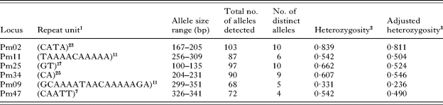
1 The number of repeat units is given for the lab isolate P. malariae Brazil I.
2 Heterozygosity cannot be measured directly from Plasmodium sp. Blood-stage parasites as they are haploid but is estimated as 1−(sum of the squares of the allele frequencies).
3 Adjusted heterozygosity was calculated from a restricted data set containing only the dominant allele in each sample.
The species-specificity of the PCR primers and conditions of amplification were tested using DNA samples from other species of Plasmodium and host DNA (Table 1). There was no amplification from DNA samples from humans, chimpanzees or Saimiri monkeys at any of the 6 loci. Similarly, no PCR products were detected from samples containing the other 3 human Plasmodium species, originating either from laboratory isolates (P. falciparum and P. vivax) or naturally acquired infections with varying levels of parasitaemia (P. falciparum 1840–760 000 parasites per μl of blood; P. vivax, 1400 and 2400 parasites per μl of blood; P. ovale, <40 parasites per μl of blood; mixed species samples <40–130 000 parasites per μl of blood).
The efficiency and reproducibility of the PCRs were investigated using results from replicate runs for each sample: All 4 laboratory isolates gave exactly the same single allele (sized to the nearest 1 bp) at each locus on up to 8 replicate runs. Of the 74 P. malariae samples from humans with naturally acquired infection (Table 1), 1 Gambian and 3 Malawian samples gave no results for any loci despite being positive for the P. malariae diagnostic PCR (Table 4). The efficiency of amplification for replicate runs was the lowest for loci Pm09 and Pm47 (79·7 and 78·4%, respectively) and the highest for Pm11 and Pm25 (both 91·9%). In samples for which results were obtained for both replicates, the concordance was based on a comparison of allele sizes. Concordance ranged from 71·1 to 94·3% for the 6 loci (Table 4), but this increased to >93% for all loci when samples with ‘consistent’ results were included. Consistent samples were considered to be those where alleles of the same size were detected for both replicates, but with the addition of 1 or more other alleles in one of the replicates. Blood stages of Plasmodium are haploid; therefore, the detection of multiple alleles in a sample from 1 individual human indicates the presence of multiple genotypes.
Efficiency of amplification and reproducibility of allele scoring in duplicate analysis at 6 polymorphic Plasmodium malariae loci in 74 naturally acquired human infections
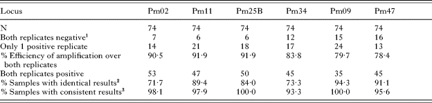
1 The number of samples negative for both replicates includes 4 samples that gave negative results for all 6 loci.
2 Calculated as a percentage of samples in which both replicates gave positive results.
3 Samples where allele(s) detected in one replicate was concordant with the other but with additional allele(s). Calculated as a percentage of samples in which both replicates gave positive results.
Multiple genotypes detected by analysis of up to 6 microsatellite loci in 70 samples of naturally acquired human Plasmodium malariae infections

Allelic diversity, geographical distribution of alleles
The number of distinct alleles detected at each of the 6 polymorphic loci was between 4 and 10 (Table 3). The repeat unit size for the 6 loci falls within the range of 2–17 bp. Sequencing of alleles revealed the presence of imperfect repeats at some loci; therefore, bin sizes do not follow exactly the repeat unit size. For some loci, all of the possible alleles within the allele size range were not detected (Fig. 1). Allele frequencies (Fig. 1) were calculated as a proportion of the total number of alleles detected for each locus using P. malariae and P. brasilianum samples (Table 3). The number of alleles detected was greater than the number of samples, due to the presence of multiple alleles per sample in some cases, indicative of the presence of multiple genotypes within a host individual. Heterozygosity was calculated using either all of the allelic data or a restricted data set in which the only dominant allele representing each sample was scored. Values ranged from 0·331 to 0·839 using the whole data set or 0·236 to 0·811 using frequencies only for the dominant allele (Table 3).
Allele frequencies for the 6 polymorphic Plasmodium malariae microsatellite loci.

Allelic diversity was restricted in the 3 samples from South America (Table 1). Two samples, P. brasilianum Colombia I/NYU and P. malariae Brazil I, were identical for all loci (i.e. Pm02-185, Pm11-288, Pm25-107, Pm34-220, Pm09-299, Pm47-331). The third sample, P. brasilianum Peru I, differed from the others at 1 locus (Pm02-181). All 3 samples had the allele Pm09-299 that was detected exclusively in samples from South America. The allele Pm47-331, recorded in all 3 South American samples, was rare amongst the other samples, being present in 2 other samples from Malawi. Allelic diversity was also low at some loci for samples from Thailand. All 18 samples from Thailand, for which results were available, were ‘mono-allelic’ for Pm09-325. Allele Pm25-107 was found in 23 of 27 Thai samples that amplified at this locus. In accordance with this ‘allelic restriction’, heterozygosity was lower in Thailand than in Malawi at all loci.
Multilocus genotyping
Genotypes based on all 6 loci were available for 56 of 78 samples. From the 56 samples, 48 different multilocus genotypes were recorded. Most genotypes (n=41) were detected only once, 6 different genotypes were detected in 2 samples and 1 genotype was recorded in 3 samples. In all cases, genotypes detected more than once were only found within the same geographical region. Unique genotypes were recorded in 29 of 33 samples from Malawi, 7 of 14 from Thailand, 3 of 5 from Gambia, and 1 of 3 from South America. The proportion of unique genotypes was significantly greater in Malawi compared with Thailand (χ2=7·88, P<0·005; only these two countries had sufficient sample sizes for statistical analysis). A restricted data set containing samples with a single genotype was also used in this analysis, and 22 different multilocus genotypes were detected in 27 samples: 18 of 22 were detected only once, 3 different genotypes were recorded in 2 samples and 1 genotype was detected in 3 samples. The proportion of samples with unique genotypes in this restricted data set was significantly greater in Malawi (7 of 7) than in Thailand (7 of 12) (χ2=3·96, P<0·01).
Multiple genotypes
Multiple genotypes of P. malariae were detected in 33 of 70 samples from humans with naturally acquired infection (Table 5).
The greatest number of genotypes in a single sample was 4. The frequency distribution of the MOI is shown in Fig. 2. The MOI was compared between samples from asymptomatic and symptomatic individuals, for which data was available at all 6 loci (Fig. 3). The mean MOI was significantly greater for asymptomatic infections (Malawi, 2·24; s.e.±0·16) compared with symptomatic ones (Gambia and Thailand, 1·21; s.e.±0·12) (t=−5·17, P<0·00001).
Number of Plasmodium malariae genotypes per sample recorded for 70 samples from humans with naturally acquired infections for which data for any of the 6 loci were available.

Comparison of the number of Plasmodium malariae genotypes per sample in symptomatic (n=19) and asymptomatic (n=33) samples of naturally acquired human infections for which data were available at all 6 microsatellite loci.

Discussion
In the present study, we have isolated 6 polymorphic genetic markers from Plasmodium malariae. These markers were validated using a series of P. malariae samples from laboratory isolates or from humans with naturally acquired infections. Previous genetic differences between P. malariae strains have been limited to 3 genes, namely the ssrRNA gene (Liu et al. Reference Liu, Zhu, Mizuno, Kimura, Liu, Isomura, Wang and Kawamoto1998; Kawamoto et al. Reference Kawamoto, Win, Mizuno, Lin, Kyaw, Tantulart, Mason, Kimura and Wongsrichanalai2002), the merozoite surface protein 1 gene (msp1) (Fandeur et al. Reference Fandeur, Volney, Peneau and de Thoisy2000) and the circumsporozoite gene (csp) (Escalante et al. Reference Escalante, Barrio and Ayala1995; Tahar et al. Reference Tahar, Ringwald and Basco1998; Ayala et al. Reference Ayala, Escalante and Rich1999). These genes have limited usefulness as genetic markers for studying populations. The ssrRNA gene is subjected to gene conversion and has few variants, whereas the gene products of msp1 and csp are major targets of immunity and are thus considered to be under ‘selection pressure’ from the immune system. Multiple, polymorphic, selectively neutral loci have been characterized to enable an assessment of the population genetic structure of P. malariae to be made. Microsatellite markers are useful for multilocus genotyping, and, in the present study, we have used them to distinguish multiple P. malariae genotypes in humans with naturally acquired infections, and have compared the number of genotypes (or multiplicity of infection, MOI) from asymptomatic and symptomatic individuals. This is the first molecular epidemiological study of P. malariae.
The markers are likely to be non-coding as they all lie within repetitive, highly AT-rich regions of the genome, in which there are numerous stop codons (Goldstein and Schlotterer, Reference Goldstein and Schlotterer1999). In such regions, primer sites are restricted and, therefore, amplicons were often significantly larger than the repetitive regions. Repeats units of 1–6 bp are considered microsatellites and those of a greater size are referred to as minisatellites. The size of the repeat unit is thought to relate to the mechanism/s by which such loci evolve (Armour et al. Reference Armour, Alegre, Miles, Williams, Badge, Goldstein and Schlotterer1999). For simplicity, we have used ‘microsatellites’ to describe these markers, although two markers Pm11 and Pm09 have repetitive units greater than 6 bp and, thus, formally represent minisatellites.
Differences between loci in the efficiency of amplification (Table 4) are considered to reflect variation in the efficiency of PCR reactions for each locus, as a result of the use of different primers, the quantity and quality of target DNA and potential errors at numerous micro-pipetting steps in the analysis protocol. Four P. malariae samples that gave negative results for all 6 loci were likely to have contained small amounts of target DNA that could be detected by nested PCR of the ssrRNA gene (which is repetitive), but was not sufficient for the detection at the (single copy) microsatellite loci. Differences in replicate results are thought to arise from variation in the dynamics of the PCR-amplification of multiple, low copy DNA targets and has been observed by other authors investigating P. falciparum (see Färnert et al. Reference Färnert, Arez, Babiker, Beck, Benito, Bjorkman, Bruce, Conway, Day, Henning, Mercereau-Puijalon, Ranford-Cartwright, Rubio, Snounou, Walliker, Zwetyenga and do Rosario2001). Almost half (47·1%) of natural human P. malariae samples were shown to contain multiple genotypes. Allele frequencies were calculated, allowing for multiple genotypes per sample. However, data from multiple genotypes can overestimate heterozygosity, because the frequency of minority alleles is overestimated. Therefore, heterozygosity was calculated from both the whole data set and from a restricted data set in which only the dominant allele recorded in each sample was scored. For some loci, the 2 values did not differ significantly (Pm02 and Pm11) but in others a substantial difference was observed. We did not assess the presence of null alleles in our study. Null alleles are difficult to distinguish from ‘pipetting errors’ without additional multiple amplification replicates or internal controls and cannot be adequately assessed in samples containing multiple genotypes.
Allelic diversity was restricted in the 3 samples from South America, and all contained the unique allele Pm09-299. Two of these samples showed identical multilocus genotypes, and the other varied from these only at 1 locus. This restricted allelic diversity may be a result of a ‘bottleneck’ from recent colonization events. Malaria is thought to have been brought to South America with European settlers and later with African slaves, and this has been suggested as an explanation of restricted diversity for P. falciparum in South America (Anderson et al. Reference Anderson, Haubold, Williams, Estrada-Franco, Richardson, Mollinedo, Bockarie, Mokili, Mharakurwa, French, Whitworth, Velez, Brockman, Nosten, Ferreira and Day2000). However, 2 of 3 samples from South America were P. brasilianum from New World monkeys, a species morphologically and genetically indistinguishable from P. malariae (see Cochrane et al. Reference Cochrane, Barnwell, Collins and Nussenzweig1985; Ayala et al. Reference Ayala, Lal, Escalante, Rich and Sherman1998; Fandeur et al. Reference Fandeur, Volney, Peneau and de Thoisy2000). The restricted diversity could also be the result of a single host-switch event of P. malariae from humans to New World Monkeys (Fandeur et al. Reference Fandeur, Volney, Peneau and de Thoisy2000) or a selective ‘sweep’ associated with a host-switch. In some settings in South America, it is thought that some P. malariae infections in humans may result from monkey to human transmission of P. brasilianum. Analysis of more South American samples from both monkey and human hosts in defined geographical settings is required to test these hypotheses.
The restriction of allelic diversity in Thai samples compared with Malawian samples was also seen at 2 loci. One explanation for the reduced diversity at some loci may be a ‘selective sweep’ due to linkage of the microsatellite loci to drug resistance loci. This phenomenon has been observed for P. falciparum microsatellite loci closely linked to the pyrimethamine resistance locus (Nair et al. Reference Nair, Williams, Brockman, Paiphun, Mayxay, Newton, Guthmann, Smithuis, Hien, White, Nosten and Anderson2003; Pearce et al. Reference Pearce, Malisa, Kachur, Barnes, Sharp and Roper2005). P. malariae is often clinically asymptomatic but will have increased exposure to drug pressure, as a result of treatment of humans with infections with multiple species of Plasmodium. The prevalence of mixed-species infections is high in South East Asia (Zhou et al. Reference Zhou, Liu, Wongsrichanalai, Suwonkerd, Panart, Prajakwong, Pensiri, Kimura, Matsuoka, Ferreira, Isomura and Kawamoto1998), and this is the only region in which drug resistance has been reported in P. malariae (see Maguire et al. Reference Maguire, Sumawinata, Masbar, Laksana, Prodjodipuro, Susanti, Sismadi, Mahmud, Bangs and Baird2002). Low genetic diversity could also be a result of subspeciation within P. malariae populations. Morphologically and genetically different forms of P. malariae have been detected in South East Asia (Kawamoto et al. Reference Kawamoto, Win, Mizuno, Lin, Kyaw, Tantulart, Mason, Kimura and Wongsrichanalai2002), but it is not known whether any of the Thai samples in this study represent one of these forms.
The extent of multispecies Plasmodium infections in asymptomatic patients could be indicative of an accumulation of chronic, long-lived, low-density infections over time rather than a high rate of acquisition of multiple genotypes in a short time-period. An analysis of how MOI is related to the age of individuals is required to further investigate this question. The lower number of genotypes in symptomatic compared with asymptomatic samples suggests that the clinical symptoms arising from increased P. malariae parasitaemia results from the growth of a single P. malariae genotype rather than the accumulation of multiple lower density infections. The samples from asymptomatic patients used herein contained ‘pure’ P. malariae infection or P. malariae in combination with other Plasmodium species. Most ‘asymptomatic samples’ from Malawi contain a high proportion of multiple species (>90%) and, when tested, gave similar results to those described here (multiple genotypes 74·6%, mean MOI 2·16; Marian Bruce, unpublished observations). Most ‘symptomatic samples’ originated from a low endemicity region in Thailand (Luxemburger et al. Reference Luxemburger, Thwai, White, Webster, Kyle, Maelankirri, Chongsuphajaisiddhi and Nosten1996) where most malaria infections are symptomatic. In contrast, the ‘asymptomatic samples’ originated from Malawi, which has much higher rates of malaria transmission, where individuals are commonly infected with multiple species and genotypes from infancy and where there is a high level of naturally acquired immunity in humans. However, the 4 samples from symptomatic patients from Gambia were also due to a single genotype. This strengthens the argument that symptoms arise from single genotypes, even between different endemic regions. A more rigorous comparison would include samples from symptomatic and asymptomatic individuals living in the same endemic region, although obtaining sufficient numbers for such a comparison would be challenging.
Direct comparison of the MOI data for P. malariae with those for other Plasmodium species is not informative, due to multiple confounding factors, including the age structure of the population sampled, level of malaria endemicity and the type, number and heterozygosity of the genetic markers used. However, MOI values for P. malariae described here fall within documented ranges for P. falciparum (see Babiker and Walliker, Reference Babiker and Walliker1997) and P. vivax (see Cole-Tobian et al. Reference Cole-Tobian, Biasor and King2005), despite the significant differences in the epidemiology of P. malariae. The MOI is a useful indicator of the degree of malaria transmission and is important for understanding the potential for recombination in Plasmodium species. Only when different genotypes are ingested by the mosquito vector in a single bloodmeal can recombination occur. The rate of recombination between different genotypes influences the generation of diversity and the rate of spread of drug resistance through a population (Hastings and Watkins, Reference Hastings and Watkins2005). A major aim of molecular and population genetic studies of Plasmodium is to determine how the rate of recombination, genetic diversity and the rate of transmission are linked to the acquisition of clinical immunity in human populations living in endemic regions. This description of genetic markers for P. malariae enables, for the first time, the investigation of the molecular epidemiology and population genetics of this little studied, yet widely distributed pathogen of humans. The first molecular epidemiology data for P. malariae presented herein, shows a high level of multi-genotype carriage in humans, suggesting that regular genetic exchange within P. malariae populations is probable, despite the lower prevalence and density of this parasite relative to other species of Plasmodium. Further analysis of a larger number of samples from a greater geographical range, different epidemiological settings, a variety of age groups and from potential primate reservoirs is necessary to further characterize the epidemiology and population structure of P. malariae.
We are grateful to Colin Sutherland, Ali Alloueche, Rosalynn Ord, Alan Brockman and Geoff Pasvol for the provision of P. malariae samples. Thanks to Malcolm Molyneux for assistance in the collection of Malawian samples. We are indebted to Keith Scott, Liz Peat and Julie Galbraith for excellent technical support. Our thanks go to Andy Tait and Annette MacLeod, Glasgow University, for useful discussions. Thanks also to Nick Helps and Joanne Quinney at the University of Dundee Sequencing Service for processing of microsatellite samples. The technical post of Keith Scott was funded by the University of Glasgow. This research was funded by a Wellcome Trust Junior Biodiversity Fellowship (Reference 060446) to Marian Bruce.


