I. INTRODUCTION
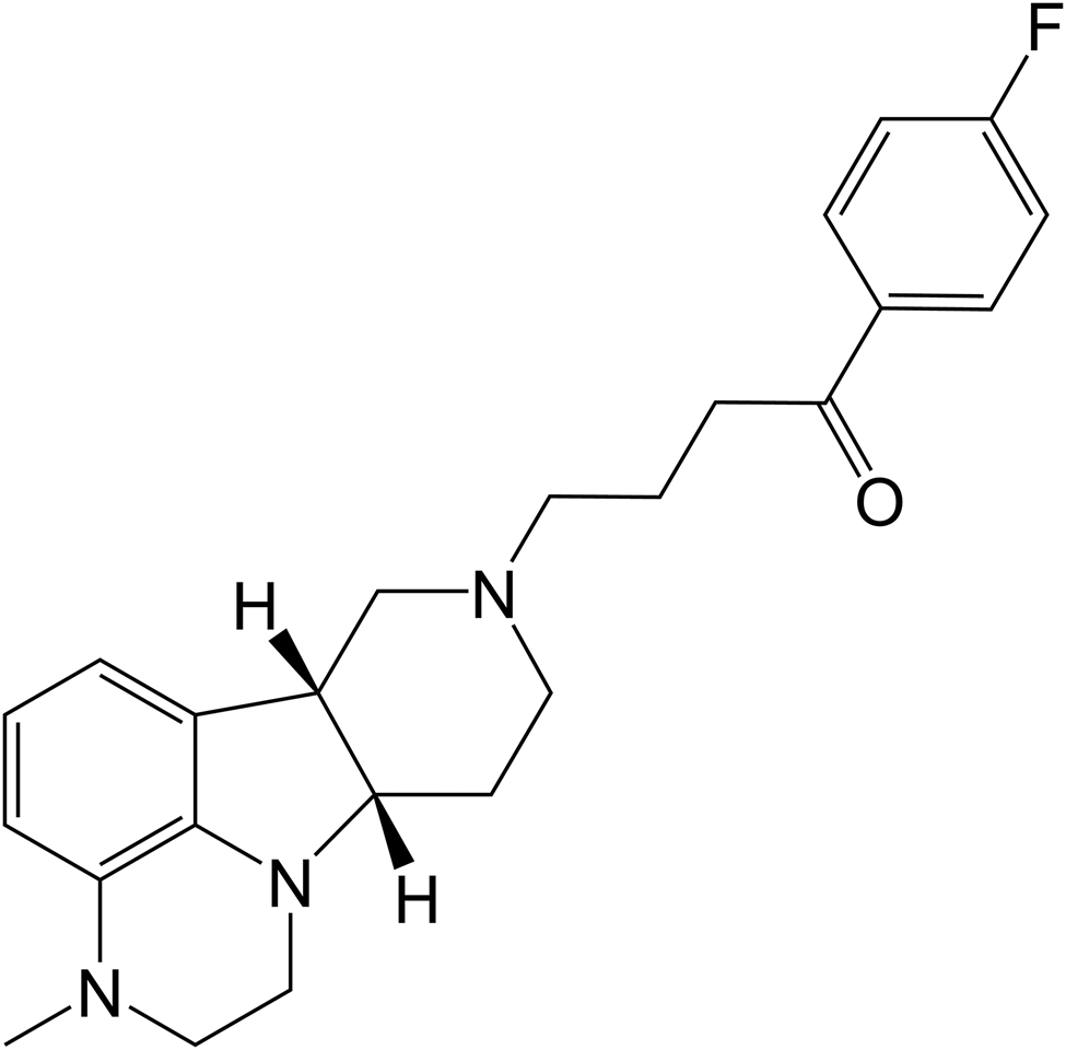
Lumateperone (Lumateperone tosylate, ITI-007, 4-((6bR,10aS)-3-methyl-2,3,6b,9,10,10a-hexahydro-1H,7H-pyrido[3′′,4′:4,5]pyrrolo[1,2,3-de]quinoxalin-8-yl)-1-(4-fluoro-phenyl)-butan-1-one 4-methylbenzenesulfonate) is a novel, orally available agent for the treatment of schizophrenia and other neuropsychiatric and neurological disorders (Blair, Reference Blair2020; Mazza et al., Reference Mazza, Marano, Traversi, Sani and Janiri2020; Maini et al., Reference Maini, Hollier, Gould, Bollich, John LaForge, Cornett, Edinoff, Kaye and Kaye2021; Pahwa et al., Reference Pahwa, Sleem, Elsayed, Good and El-Mallakh2021). Due to its ability to modulate serotonin, dopamine, and glutamate neurotransmission, Lumateperone can be considered a multi-target-directed ligand and a multifunctional modulator of the serotoninergic system with possible precognitive, antipsychotic, antidepressant, and anxiolytic properties, implicated in serious mental illness (Kumar et al., Reference Kumar, Kuhad and Kuhad2018; Frampton, Reference Frampton2020; Greenwood et al., Reference Greenwood, Acharya, Marcellus and Rey2021; Maini et al., Reference Maini, Hollier, Gould, Bollich, John LaForge, Cornett, Edinoff, Kaye and Kaye2021; Syed and Brasic, Reference Syed and Brasic2021; Cao et al., Reference Cao, Yu, Wang, Luo, Liu, He, Qi, Fan, Tang, Chen, Li, Cheng and Wang2022; Titulaer et al., Reference Titulaer, Radhe, Danielsson, Dutheil, Marcus, Jardemark, Svensson, Snyder, Ericson, Davis and Konradsson-Geuken2022). Thus, in December 2019, the U.S. Food and Drug Administration (FDA) approved Lumateperone for the treatment of patients with schizophrenia. The chemical structure of Lumateperone is shown in Figure 1 and its salts, such as Lumateperone tosylate (Wennogle and Tomesch, Reference Wennogle and Tomesch2008), ditosylate (Janton et al., Reference Janton, Cerić, Matečić, Mrsic, Lerman, Momcilovic, Sagud and Jegorov2016), oxalate, 4-aminosalicylate, cyclamate (Wennogle et al., Reference Wennogle, Li and Aret2016), chlorhydrate (Li and Wennogle, Reference Li and Wennogle2017), besylate (Janton et al., Reference Janton, Cerić, Matečić, Mrsic, Lerman, Momcilovic, Sagud and Jegorov2018), mono/di-naphthalenesulfonate (Lengauer et al., Reference Lengauer, Pichler and Margreiter2019), and isonicotinamide and nicotinamide (Wennogle et al., Reference Wennogle, Li and Aret2016) were reported, but their crystal structures have not been reported yet. The current clinically available Lumateperone product ITI-007 is Lumateperone tosylate.
Chemical structure of Lumateperone.

We have not found this compound in the CSD database (Groom et al., Reference Groom, Bruno, Lightfoot and Ward2016) or in the PDF4+ database (Gates-Rector and Blanton, Reference Gates-Rector and Blanton2019). Therefore, we have decided to characterize this compound by X-ray powder diffraction (XRD) technique and X-ray single-crystal diffraction techniques. In our study, we present powder data for Lumateperone tosylate.
II. EXPERIMENTAL
A. Sample preparations
The sample was supplied by Zhejiang Ausun Pharmaceutical Co., LTD (purity >99.9%) and used without further purification. Dissolving Lumateperone tosylate (1.0 g) in the methanol (5 mL) at 50−60°C and slow cooling of the solutions yielded needle crystals of Lumateperone tosylate. Then, the crystals were dried, smashed, and front-loaded into a zero background plate and limited force was used to make the sample surface flat.
B. Powder diffraction data collection
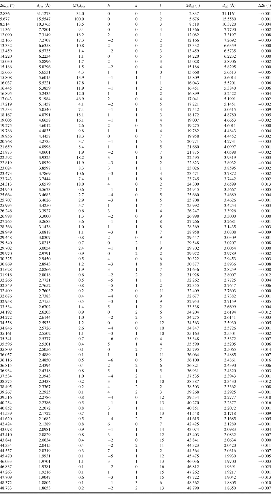
XRD data were collected at room temperature on Bruker D8 Discover diffractometer with parafocusing Bragg–Brentano geometry and one-dimensional LynxEye XE-T detector using a Cu Kα radiation (λ = 1.5418 Å), and operated at 42 kV and 100 mA. The scan 2θ range was from 2° to 50° with a step size of 0.02° and a counting time of 2.0 s/step. In order to reduce the influence of preference orientation, rotating modes with a speed rate of 20 r/min were used. The software package MDI-Jade version 9.4 (Materials Data Inc., USA) (MDI, 2009) was used to smooth the data, fit the background, and eliminate the Kα2 component and the top of the smoothed peaks were used to determine the peak positions and intensities of the diffraction peaks (Table I). The d-spacing was calculated using Cu Kα 1 radiation (λ = 1.5406 Å).
X-ray powder diffraction data of Lumateperone tosylate

C. Single-crystal diffraction data collection
X-ray single-crystal diffraction data were collected at room temperature with a Bruker D8 Venture diffractometer with Ga Kα radiation (λ = 1.34139 Å) for cell determination and subsequent data collection. Data reduction was performed by APEX4 software and multi-scan absorption correction was applied. Using Olex2 (Dolomanov et al., Reference Dolomanov, Bourhis, Gildea, Howard and Puschmann2009), the crystal structure was solved by ShelXT (Sheldrick, Reference Sheldrick2015a) and refined with full-matrix least-squares methods with anisotropic thermal parameters for all non-hydrogen atoms on F2 using SHELXL (Sheldrick, Reference Sheldrick2015b). Diamond (Brandenburg and Putz, Reference Brandenburg and Putz2005) was used to prepare the figures and packing diagrams.
III. RESULTS AND DISCUSSION
Indexing of the experimental XRD patterns and unit-cell refinements was done using MDI-Jade. In the process of refinement, only zero-offset parameter (−0.00102°) was added and refined. The cell refinement results showed that Lumateperone tosylate is monoclinic with space group C2 and unit-cell parameters: a = 15.5848(10) Å, b = 6.0700(4) Å, c = 31.3201(14) Å, β = 96.544(5)°, unit-cell volume V = 2943.58 Å3, Z = 4. The figure of merit is F 30 = 422.3(30) (Smith and Snyder, Reference Smith and Snyder1979). The values of 2θ obs, d obs, I obs, h, k, l, 2θ cal, d cal, and Δ2θ are listed in Table I.
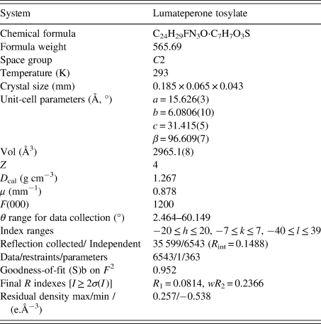
Based on the single-crystal data, the structures of Lumateperone tosylate were solved and refined. The detailed crystallographic information is summarized in Table II and the asymmetric units with the corresponding atom labeling scheme are illustrated in Figure 2(a). In the crystal structure, the expected proton transfer was found between Lumateperone and toluene sulfonic acid, forming Lumateperone cation protonated at the N atom of the hydro-pyridine group. Hydrogen bonds N1–H1⋯O3 linked Lumateperone cations and tosylate anions. In Figure 2(b), two chiral C atoms (C12, C22) of Lumateperone molecules existed “R” and “S” configurations, respectively. The hydrogen bonds (N1–H1⋯O3 and C29–H29C⋯O4) between Lumateperone cations and tosylate anions linked Lumateperone molecules into an infinite straight chain along the a direction in Figure 2(c). Hydrogen bond interactions were listed in Supplementary Table SII.
Crystal and experimental data Lumateperone tosylate

(a) Asymmetric unit of Lumateperone tosylate shown in thermal ellipsoid model with 30% probability. (b) Molecular configuration diagram of Lumateperone. (c) 1D hydrogen bonding chains of Lumateperone tosylate viewed along the b axis.

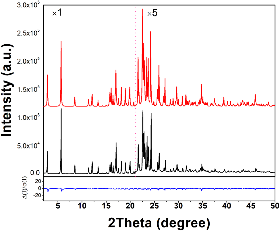
Despite the use of careful grinding and sample rotation during measurement, an evidently preferred orientation was observed in the experimental XRD pattern due to the needlelike crystal morphology of Lumateperone tosylate. An orientation coefficient of 0.75 at the [002] direction (refined by WPF refinement in Jade) was added to the simulated pattern derived from the single-crystal data. The comparison of the experimental powder diffraction pattern and the simulated pattern is shown in Figure 3. Results showed that both single-crystal and powder diffraction methods can get similar structure data and the deviations of the unit-cell parameters and unit-cell volume were between 0.03% and 0.36%.
X-ray powder diffraction pattern (black line) and the simulated pattern (red line) of the crystal structure with preferred orientation in the [002] direction (orientation coefficient = 0.75) (red line) of Lumateperone tosylate. The blue curve is the normalized error plot. The vertical scale has been multiplied by a factor 5× for 2θ >21°.

IV. DEPOSITED DATA
CIF and/or RAW data files were deposited with ICDD. You may request this data from ICDD at pdj@icdd.com.
Supplementary material
The supplementary material for this article can be found at https://doi.org/10.1017/S0885715623000337.
Acknowledgements
This article was financially supported from the Experimental Technology Research Project of Zhejiang University (SYBJS202204) and the Natural Science Exploration Project of Zhejiang Province (LTGC23B050006).