INTRODUCTION
Chagas disease, caused by the haemoflagellate protozoan Trypanosoma cruzi, continues to affects millions of people in Latin America (Pinto-Dias, Reference Pinto-Dias2006). Subjects are infected by fecal matter contact of the insect vector (Hemiptera: Reduviidae), but blood transfusion, congenital transmission and ingestion of contaminated food also cause infection (Moncayo and Silveira, Reference Moncayo and Silveira2009). In the human host, infection is characterized by an acute phase followed by a chronic phase, in which cardiac, digestive or neurological disruptions develop (Coura and Castro, Reference Coura and De Castro2002).
There is no effective treatment once an individual has been infected with T. cruzi, and vaccine development is still in the experimental stage (Maya et al. Reference Maya, Cassels, Iturriaga-Vásquez, Ferreira, Faúndez, Galanti, Ferreira and Morello2007; Gupta et al. Reference Gupta, Wan, Zago, Sellers, Silva, Assiah, Dhiman, Nuñez, Petersen, Vázquez-Chagoyán, Estrada-Franco and Garg2013). Chagas disease treatment is based on the use of benznidazole, a drug with limited use due to its toxicity and minimal benefits during the chronic phase of infection (Urbina and Docampo, Reference Urbina and Docampo2003; Moreno et al. Reference Moreno, D´avoça, Silva, Galvão, Macedo, Chiari, Gontijo and Zingales2010). Currently, Chagas disease has only two pharmaceutical interventions available in clinical trials. Of these, the most advanced is the antifungal posaconazole, which targets parasite sterol synthesis and is undergoing phase II study (Moton et al. Reference Moton, Krishna and Wang2009; Urbina, Reference Urbina2010). The other is the K11777, a peptide which inhibits protease activity, which is in phase I study (Sajid et al. Reference Sajid, Robertson, Brinen and McKerrow2011). Therefore, it is clear that more pharmaceutical options must be developed to improve treatment.
Physalis angulata L. (Solanaceae) is an annual herb widely distributed in tropical and subtropical regions. It is rich in steroid compounds, such as physalins and withanolides. Physalins are a group of seco-steroids found in stem extracts of many Physalis species (Magalhães et al. Reference Magalhães, Veras, Torres, Alves, Pessoa, Silveira, Costa-Lotufo, de Moraes and Pessoa2006; Damu et al. Reference Damu, Kuo, Su, Kuo, Chen, Bastow, Lee and Wu2007; Jin et al. Reference Jin, Mashuta, Stolowich, Vaisberg, Stivers, Bates, Lewis and Hammond2012). Because of the chemical similarity with the glucocorticoid class of steroids, physalins are well-known anti-inflammatory and immunosuppressant agents (Soares et al. Reference Soares, Bellintani, Ribeiro, Tomassini and Ribeiro dos Santos2003, Reference Soares, Brustolin, Santos, Bellintani, Paiva, Ribeiro, Tomassini and Ribeiro Dos Santos2006; Jacobo-Herrera et al. Reference Jacobo-Herrera, Bremner, Marquez, Gupta, Gibbons, Muñoz and Heinrich2006). These compounds regulate many cellular activities, such as differentiation, proliferation, necrosis and apoptosis, adhesion and migration (Brustolim et al. Reference Brustolim, Vasconcelos, Freitas, Teixeira, Farias, Ribeiro, Tomassini, Oliveira, Pontes-de-Carvalho, Ribeiro-dos-Santos and Soares2010; Wu et al. Reference Wu, Leu and Chang2012; Reyes-Reyes et al. Reference Reyes-Reyes, Jin, Vaisberg, Hammond and Bates2013). Based on this, these compounds have been tested for anticancer activity and as inhibitors of pathogenic microbes (Chiang et al. Reference Chiang, Jaw and Chen1992; Januário et al. Reference Januário, Filho, Pietro, Kashima, Sato and França2002; Reyes-Reyes et al. Reference Reyes-Reyes, Jin, Vaisberg, Hammond and Bates2013). Specifically, we observed that physalins B and F inhibit Leishmania major and Leishmania amazonensis infection in macrophages and mice (Guimarães et al. Reference Guimarães, Lima, Santos, Ribeiro, Tomassini, Ribeiro dos Santos, dos Santos and Soares2009). We also determined the antimalarial activity of physalins (Sá et al. Reference Sá, Menezes, Krettli, Ribeiro, Tomassini, Ribeiro dos Santos, de Azevedo and Soares2011). Interestingly, we found that physalin D reduced blood parasitaemia in Plasmodium berghei-infected mice, while physalin F increased parasite load, possibly due to murine immune response interference.
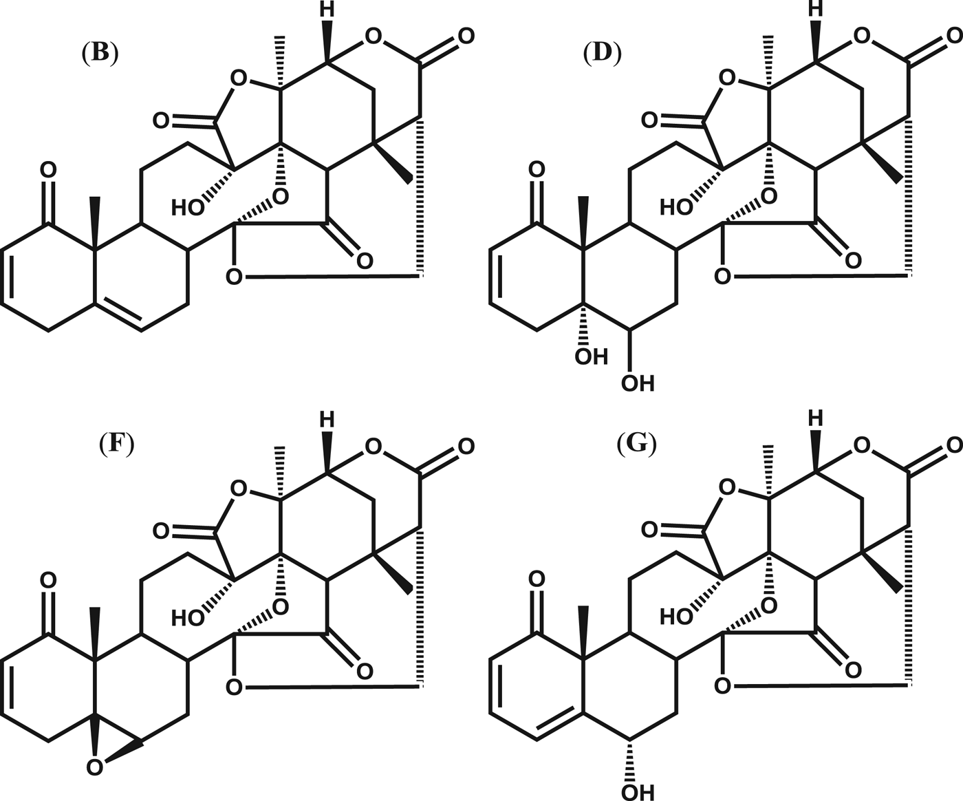
Regarding the anti-T. cruzi activity of physalins, it was previously described that they inhibit epimastigote proliferation in axenic media, as well as in T. cruzi-infected triatomines (Vieira et al. Reference Vieira, Espíndola, Santana, Veras, Pessoa, Pinheiro, de Araújo, Lima and Silveira2008; Castro et al. Reference Castro, Figueiredo, Genta, Ribeiro, Tomassini, Azambuja and Garcia2009, Reference Castro, Moraes, Gonzalez, Ribeiro, Tomassini, Azambuja and Garcia2012). The findings suggest physalins are useful compounds for vector-borne transmission control. Here, we report the activity of physalins B, D, F and G (Fig. 1) against bloodstream trypomastigotes, which is the infectious form in humans. After physalin treatment, we examined ultrastructural alteration and parasite death. Next, the activity of physalins B and F to inhibit the parasite invasion and development in macrophage cell culture was determined.
Structures of physalins B, D, F and G.

MATERIALS AND METHODS
Drugs
Physalins B, D, F and G were isolated from Physalis angulate L. Plants were collected in Belém do Pará (Brazil) and processed as previously described (Soares et al. Reference Soares, Bellintani, Ribeiro, Tomassini and Ribeiro dos Santos2003). Preparations of physalins B, D, F and G (96, 95·6, 97·8 and 95% purity by HPLC, respectively) were dissolved in DMSO and diluted in culture medium for use in the assays. Benznidazole (LAFEPE, Recife, Brazil) was used as a positive control for the anti-T. cruzi assays. Amphotericin B (Gibco Laboratories, Gaithersburg, USA) was used as a positive control in the invasion assay. Gentian violet (Synth, São Paulo, Brazil) was used as a reference drug in the cytotoxicity assays.
Animals
Female BALB/c mice (6–8 weeks old) were supplied by the animal breeding facility at Centro de Pesquisa Gonçalo Moniz (Fundação Oswaldo Cruz, Bahia, Brazil) and maintained in sterilized cages under a controlled environment, receiving a rodent balanced diet and water ad libitum. All experiments were carried out in accordance with the recommendations of Ethical Issues Guidelines, and were approved by the local Animal Ethics Committee.
Parasite and cell cultures
Trypanosoma cruzi epimastigotes (Y strain) were maintained at 26 °C in LIT medium (Liver Infusion Tryptose) supplemented with 10% fetal bovine serum (FBS; Cultilab, Campinas, Brazil), 1% haemin (Sigma Chemical Co., St. Louis, MO, USA), 1% R9 medium (Sigma), and 50 μg mL−1 of gentamycin (Novafarma, Anápolis, Brazil). Bloodstream trypomastigote forms of T. cruzi (Y strain) were obtained from the supernatant of infected LLC-MK2 cells and maintained in RPMI-1640 medium (Sigma Chemical Co., St. Louis, MO, USA) supplemented with 10% FBS and 50 μg mL−1 of gentamycin at 37 °C with 5% CO2.
Cytotoxicity to host cells
Peritoneal exudate macrophages obtained from BALB/c mice were placed on 96-well plates at a cell density of 5×10⁵ cells mL−1 in 200 μL of RPMI-1640 medium (no phenol red) and supplemented with 10% FBS and 50 μg mL−1 of gentamycin and incubated for 24 h at 37 °C and 5% CO2. Compounds were added in a series of eight concentrations (0·13–200 μ m), in triplicate, and incubated for 72 h. 20 μL/well of alamar blue (Invitrogen, Carlsbad, USA) was added to all wells during 10 h. Colorimetric readings were performed at 570 and 600 nm. LC50 values were calculated using data-points gathered from three independent experiments. Gentian violet was used as a positive control, at concentrations ranging from 0·04 to 10 μ m.
Antiproliferative activity against epimastigotes
Epimastigotes were counted in a haemocytometer and dispensed into 96-well plates at a cell density of 5×10⁶ cells mL−1 in 200 μL of LIT medium. Compounds were tested at eight concentrations, in triplicate. The plate was incubated for 5 days at 26 °C, aliquots of each well were collected and the number of viable parasites was counted in a Neubauer chamber, which was compared with the untreated parasite culture. This experiment was performed three times. Benznidazole was used as a positive control.
Cytotoxicity for trypomastigotes
Trypomastigotes collected from the LLC-MK2 cell supernatant were dispensed into 96-well plates at a cell density of 2×10⁶ cells mL−1 in 200 μL of RPMI medium. Compounds were tested at eight concentrations, in triplicate. The plate was incubated for 24 h at 37 °C and 5% CO2. Aliquots from each well were collected and the number of viable parasites, based on parasite viability, was assessed in a Neubauer chamber and compared with untreated parasite culture. This experiment was performed three times.
Trypanosoma cruzi infection assay
Peritoneal exudate macrophages were seeded at a cell density of 106 cells mL−1 in a 24-well plate with round coverslips on the bottom. Cells were cultivated in 1 mL of RPMI-1640 medium, supplemented with 10% FBS and incubated for 24 h. Cells were then infected with trypomastigotes at a ratio of 10 parasites per macrophage for 2 h. Free trypomastigotes were removed by successive washes with saline solution. Cultures were incubated in complete medium, in the presence or absence of physalins B, D, F and G (1 μ m) or benznidazole (10 μ m) for 6 h. After this, the supernatant was removed and fresh medium was added and incubated for 4 days. Cells were fixed in absolute alcohol, stained with haematoxylin and eosin and analysed in an optical microscope (Olympus, Tokyo, Japan). The percentage of infected macrophages and the mean number of amastigotes per 100 macrophages was determined by manual counting.
Trypanosoma cruzi invasion assay
Peritoneal exudate macrophages were cultured at a cell density of 1×10⁵ cells mL−1 in a 24-well plate with rounded coverslips on the bottom in 1 mL of RPMI-1640 supplemented with 10% FBS and incubated for 24 h. Cells were then infected with trypomastigotes at a ratio of 100 parasites per macrophage for 2 h, followed by addition of physalin B or F (at 10 μ m). Amphotericin B (10 μ m) was used as a reference inhibitor. The plate was incubated for 2 h at 37 °C and 5% CO2, followed by successive washes with saline solution to remove extracellular trypomastigotes. Plate was maintained in RPMI-1640 medium supplemented with 10% FBS at 37 °C for 2 h. Cells were fixed in absolute alcohol, stained with haematoxylin and eosin. The percentage of infected macrophages was determined by manual counting.
Propidium iodide and annexin V staining
Trypomastigotes (1×10⁷) in RPMI-1640 medium supplemented with 10% FBS were treated with physalin B (5·0 μ m) and incubated for 24 h at 37 °C. Parasites were labelled with propidium iodide (PI) and annexin V using the annexin V-FITC apoptosis detection kit (Sigma Chemical Co, St. Louis, MO, USA) according to the manufacturer's instructions. Acquisition was performed using a BD FACS Calibur flow cytometer (San Jose, CA, USA), and data were analysed in BD CellQuest software (San Jose, CA, USA).
Electron microscopy analysis
Y strain T. cruzi trypomastigotes (3×10⁷) were treated with physalin B (0·68 or 1·0 μm) and incubated for 24 h at 37 °C. After incubation, parasites were fixed for 1 h at room temperature with 2% formaldehyde and 2·5% glutaraldehyde (Electron Microscopy Sciences, Hatfield, PA, USA) in sodium cacodylate buffer (0·1 m, pH 7·2) for 1 h at room temperature. After fixation, parasites were washed 4 times with sodium cacodylate buffer (0·1 m, pH 7·2), and post-fixed with a 1% solution of osmium tetroxide (Sigma Chemical Co., St. Louis, MO, USA). Cells were subsequently dehydrated in increasing concentrations of acetone (30, 50, 70, 90 and 100%) for 10 min at each step and embedded in Polybed resin (PolyScience family, Warrington, PA, USA). Ultrathin sections were prepared on an ultramicrotome Leica UC7 and sections were collected on 300 mesh copper grids, contrasted with uranyl acetate and lead citrate. Images were collected in a JEOL TEM-1230 transmission electron microscope.
Parasitic vacuole staining
Trypomastigotes (3×10⁷) were treated with physalin B (1·0 μ m) and incubated for 24 h with 5% CO2. After treatment, cells were incubated with 100 μ m of monodansylcadaverine (MDC, Sigma Chemical Co., St. Louis, MO, USA) for 20 min in the absence of light. After MDC staining, parasites were washed with PBS twice. The parasites were analysed in a FV1000 confocal microscope (Olympus). As a positive control, parasites were treated with 0·1 μg mL−1 of rapamycin (Sigma Chemical Co., St. Louis, MO, USA).
Cruzain inhibition
The recombinant cruzain was expressed and purified according to a previously published protocol (Eakin et al. Reference Eakin, MacGrath, McKerrow, Fletterick and Craik1993). Protein was activated in acetate buffer (0·1 m; pH 5·5) containing 5·5 mm of DTT (Invitrogen, Carlsbad, USA) and protein concentration was adjusted to a final concentration of 0·1 μ m. Protein was aliquoted into a 96-well plate, and compounds (previously dissolved in DMSO) in phosphate buffer (in the presence of 0·01% Triton 100) were added to the respective wells. The plate was incubated for 10 min at 35 °C. After this time, a solution containing protease substrate Z-FR-AMC (Sigma Chemical Co., St. Louis, MO, USA) was added, and following incubation periods 1, 5 or 10 min, the plate was read using the EnVision multilabel reader (PerkinElmer, Shelton, CT, USA). The percentage of cruzain inhibition was calculated using the following equation: 100 − (A1/A×100), where A1 represents the cruzain RFU in the presence of the test inhibitor, A refers to the control RFU (cruzain and substrate only). Compound concentration was measured in triplicate. (2S,3S)-trans-epoxysuccinyl-L-leucylamido-3-methylbutane (E-64c) was purchased from Sigma Chemical Co. (catalogue number E0514) and used as a standard cruzain inhibitor.
Statistical analyses
We used a non-linear regression for calculating LC50 and IC50 values. The selectivity index (SI) was defined as the ratio of LC50 by IC50 (trypomastigotes). The one-way ANOVA and Bonferroni multiple comparisons were used to determine the statistical significance of the group comparisons in the in vitro infection studies and cell invasion study. Results were considered statistically significant when P<0·05. All analyses were performed using Graph Pad Prism version 5.01 (Graph Pad Software, San Diego, CA, USA).
RESULTS
Trypanocidal and cytotoxicity activity
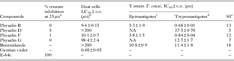
First, we evaluated the trypanocidal activity of physalins B, D, F and G against axenic epimastigotes and bloodstream trypomastigotes from the Y strain of T. cruzi. Cytotoxicity was determined in mouse macrophages. Benznidazole and gentian violet were used as trypanocidal and cytotoxic reference drugs, respectively. Activity was described in term of IC50 or LC50 values (Table 1). Of these, only physalins B and F were able to inhibit epimastigote proliferation, with IC50 values of 5·3±1·9 and 5·8±1·5 μ m, respectively, while benznidazole exhibited an IC50 10·8±0·9 μ m. All physalins exhibited activity against trypomastigotes. Once again, physalins B and F were the most active, presenting IC50 values of 0·68±0·01 and 0·84±0·04 μ m respectively, while an IC50 of 11·4±1·8 μ m was calculated for benznidazole-treated trypomastigotes.
Cruzain activity, cytotoxicity in macrophages and anti-Trypanosoma cruzi activity of purified physalins
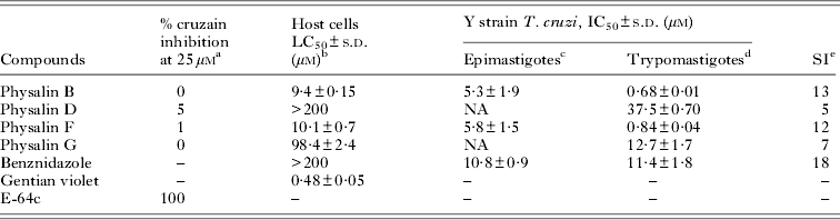
a Determined after 5 min of incubation with enzyme.
b Determined in BALB/c macrophages after 72 h of incubation with the compounds.
c Determined after 5 days of incubation.
d Determined after 24 h of incubation.
e SI = selectivity index. NA = no activity.
s.d. = standard deviation. IC50 and LC50 values are means±s.d. of two or three independent experiments performed in triplicate.
Next, physalin cytotoxicity to host cells (mouse macrophages) was analysed. Compared with gentian violet (LC50 0·48±0·05 μ m), the cytotoxic reference drug in this assay, physalins demonstrated a lower cytotoxic value. Physalins D and G exhibited cytotoxicity similar to each other and they were less cytotoxic than physalins B and F. Regarding the cellular SI, physalins B and F exhibited a selectivity of 13 and 12 respectively, while physalins D and G were two times less selective.
Investigating the mechanism of action
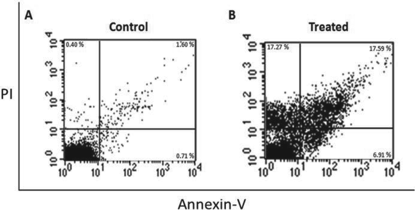
After confirming that physalins were able to kill parasites, our next step was to understand how they affect parasite cells. In the first set of experiments investigating the mechanism of action, we treated Y strain trypomastigotes with 5 μ m of physalin B, which were incubated for 24 h. Cells were then stained with annexin V and PI and examined by flow cytometry. As shown in Fig. 2, most of the parasite cells treated with physalin B were positively stained for PI when compared with untreated cells. Therefore, physalin-based treatment causes parasite cell death through necrosis induction.
Physalin B causes parasite cell death by a necrotic process. Flow cytometry examination of annexin V and propidium iodide staining in Trypanosoma cruzi trypomastigotes treated with 5 μ m of physalin B for 24 h. (A), untreated parasites; (B), physalin B-treated parasites.
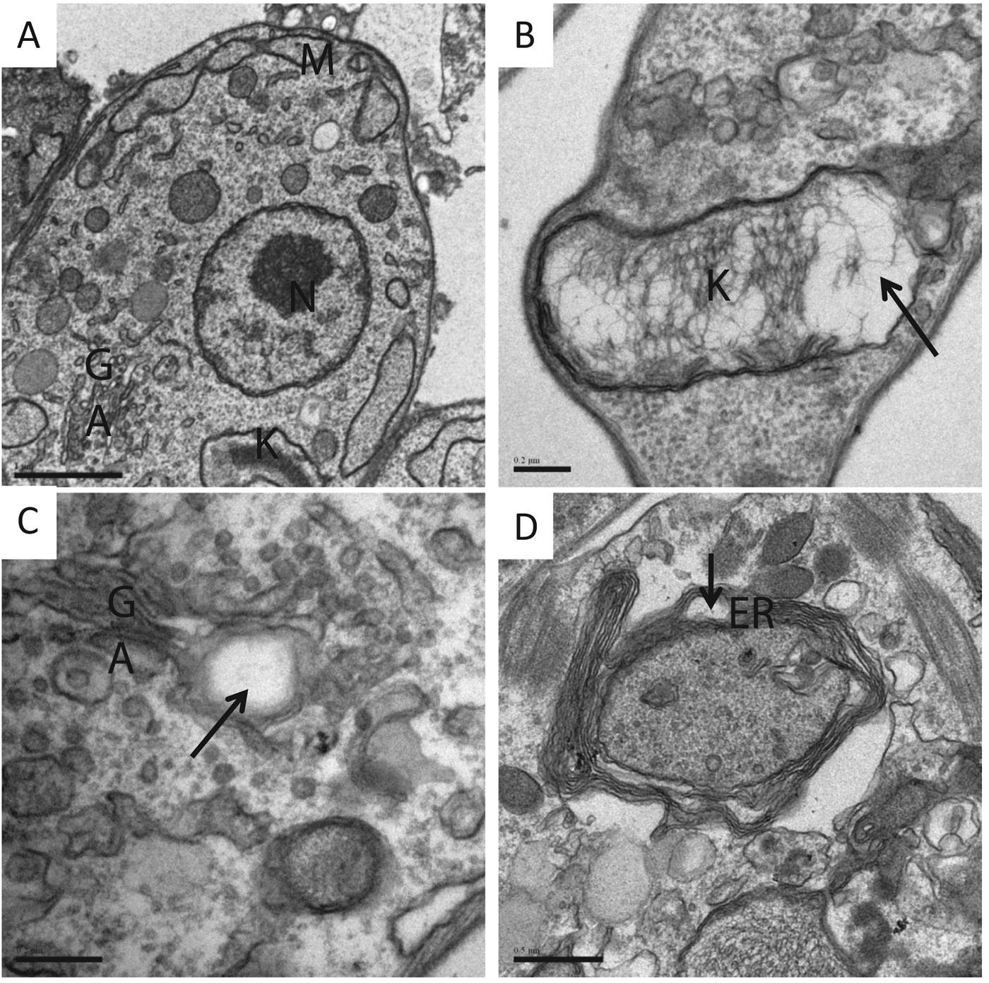
In the second set of experiments, we used electron microscopy to examine the ultrastructural morphology of trypomastigotes treated with physalin B. As we can see in Fig. 3, parasites exhibited kinetoplast enlargement, alterations in the Golgi complex, as well as endoplasmic reticulum morphology. Interestingly, we also observed the presence of myelin-like figures within the cytoplasm. To confirm that these myelin-like figures are parasitic vacuoles, possibly related to autophagy, untreated or treated trypomastigotes were incubated with monodansylcadaverine (MDC) for 20 min and observed by fluorescence microscopy. In a control experiment, untreated cells presented no detectable MDC staining, while rapamycin-treated cells contained stained cytosolic vacuoles. Following this, we tested physalin B (1·0 μ m) which clearly presented stained vacuoles (Fig. 4).
Physalin B induces significant ultrastructural changes in trypomastigotes. (A) shows an image of untreated parasites presenting a typical morphology of the nucleus (N), kinetoplast (K), mitochondria (M) and Golgi apparatus (GA). Treatment for 24 h with physalin B at 0·68 (B and C) or 1·0 μ m (D) causes kinetoplast enlargement (B), Golgi apparatus disorganization (C) and endoplasmic reticulum disorganization (D). Black arrows indicate changes in organelles. Scale bars: A = 1 μm; B and C = 0·2 μm; D = 0·5 μm.
Physalin B induces the formation of cytosolic vacuoles in Trypanosoma cruzi. Trypomastigotes treated with 1·0 μ m of physalin for 24 h and incubated with MDC for 20 min. Left panels are DIC and right panels are fluorescence microscopy. (A) and (B) show untreated trypomastigotes; (C) and (D) show parasite treated with 0·1 μg mL−1 of rapamycin (positive control); (E, F) show treated trypomastigotes with physalin B. Arrows indicate the vacuole structures. Images were captured on confocal microscope with a 60× oil-immersion objective at 3× zoom.
Finally, we tested the ability of physalins to inhibit the activity of recombinant cruzain, the major T. cruzi cysteine protease. As Table 1 demonstrates, none of the physalins tested at a concentration of 25 μ m exhibited inhibitory properties against cruzain.
Physalins inhibit T. cruzi infection in host cells
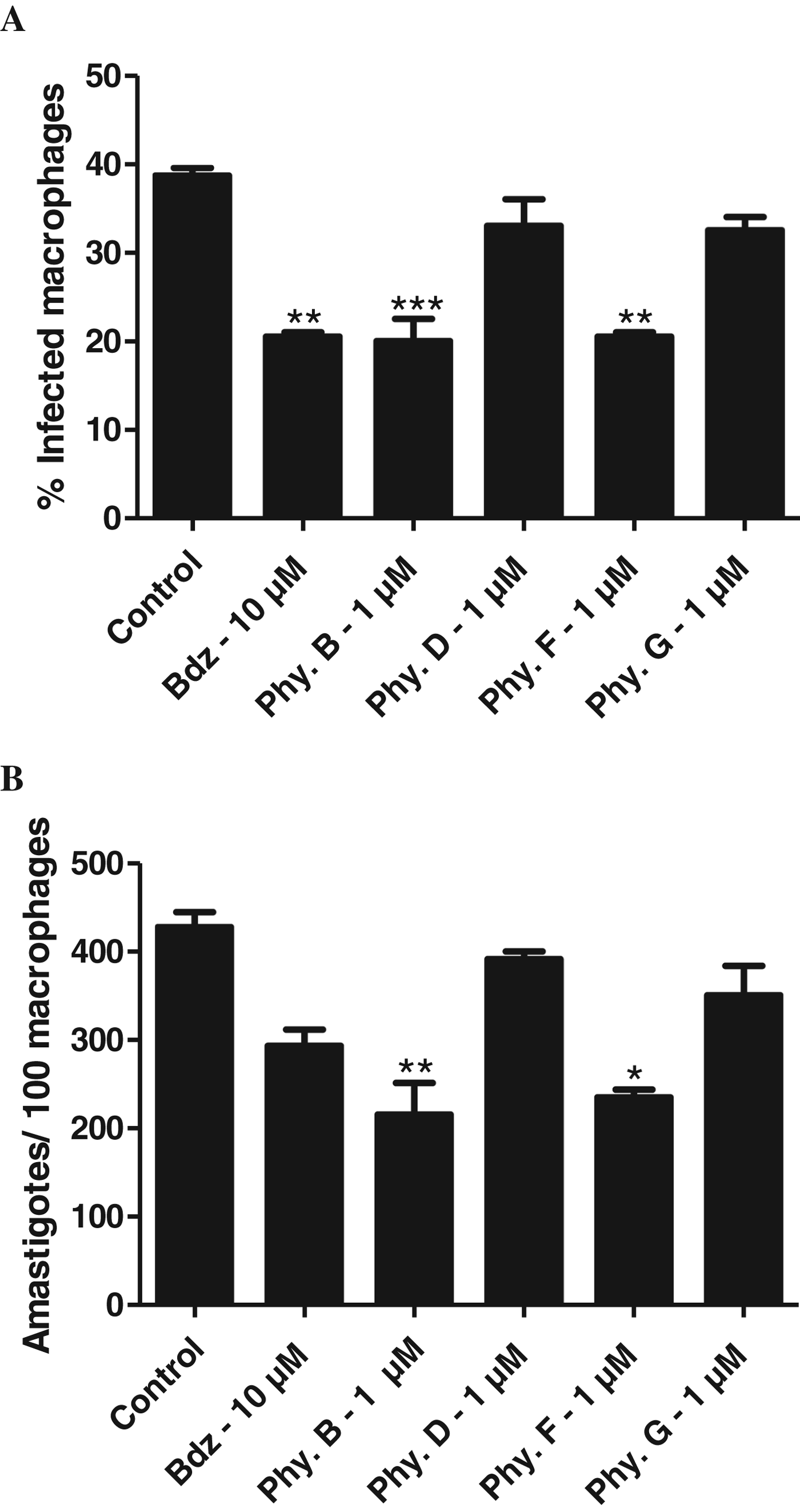
We tested physalins to inhibit the parasite development in host cells. In this assay, mouse macrophages were infected with Y strain trypomastigotes. After infection, the respective physalin (1·0 μ m) was added to the cell culture. Benznidazole was included in this assay as a positive control. Cells were stained with haematoxylin and eosin and analysed by optical microscopy 4 days post-infection. As shown in Fig. 5, physalins B and F significantly reduced the percentage of infected macrophages and the relative numbers of intracellular parasites when compared with control. Interestingly, activity of physalins B and F was quite similar to that observed in benznidazole. In contrast, physalins D and G did not show significant activity in this assay. When physalins B and F were tested in concentrations above 1·0 μ m, they did not inhibit infection in macrophages, probably due to their immunosuppressive properties (data not shown).
Physalins B and F inhibit intracellular amastigotes in host cells. Mouse peritoneal macrophages were infected with Y strain trypomastigotes for 2 h and treated with the respective physalin (1·0 μ m). Infected cells were stained with haematoxylin and eosin and analysed by optical microscopy. (A) shows the percentage of infected macrophages; (B) displays the relative number of amastigotes per 100 macrophages. Bdz = Benznidazole; Phy. = physalin. Values represent the mean±s.e.m. in triplicate. *P<0·05; **P<0·01; ***P<0·001 compared with the control group.
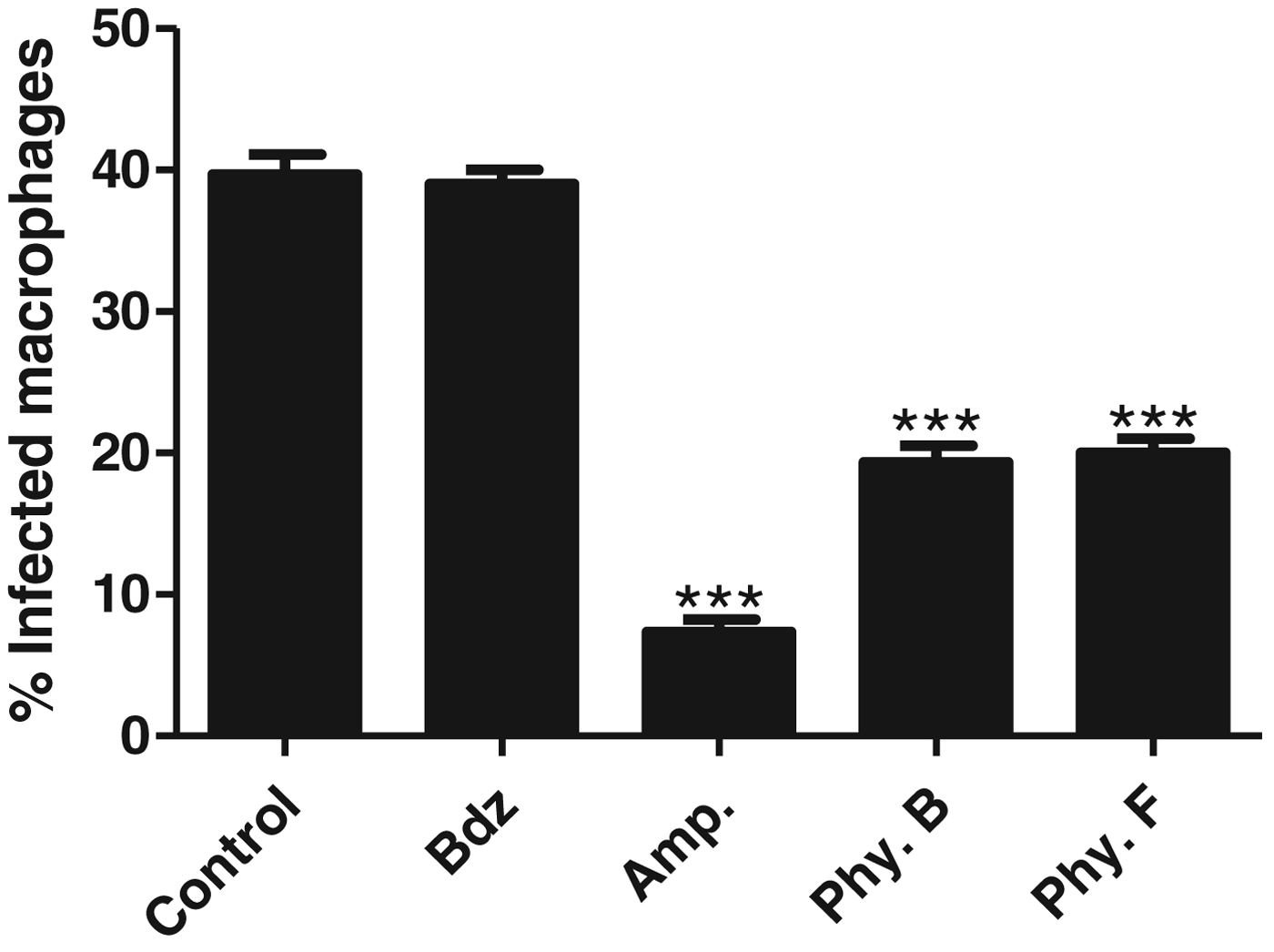
In another experiment, we measure physalins’ ability to impair parasite invasion into host cells. In this assay, mouse macrophages were exposed to trypomastigotes and at the same time treated with physalins (10 μ m). After incubating for 2 h, cell cultures were stained and analysed by optical microscopy. Amphotericin B was used as a positive control. As shown in Fig. 6, physalins B and F significantly inhibited the parasite invasion process in macrophages when compared with untreated cultures. Curiously, while physalins exhibited activity in this assay, equal concentrations of benznidazole did not.
Physalins B and F reduce the parasite invasion process in host cells. Mouse macrophages were infected with Y strain trypomastigotes and at the same time treated with physalins. The percentage of T. cruzi-infected macrophages is shown. Bdz = benznidazole, Amp = amphotericin B. Values represent the mean±s.e.m. in triplicate. ***P<0·001 compared with the control group.
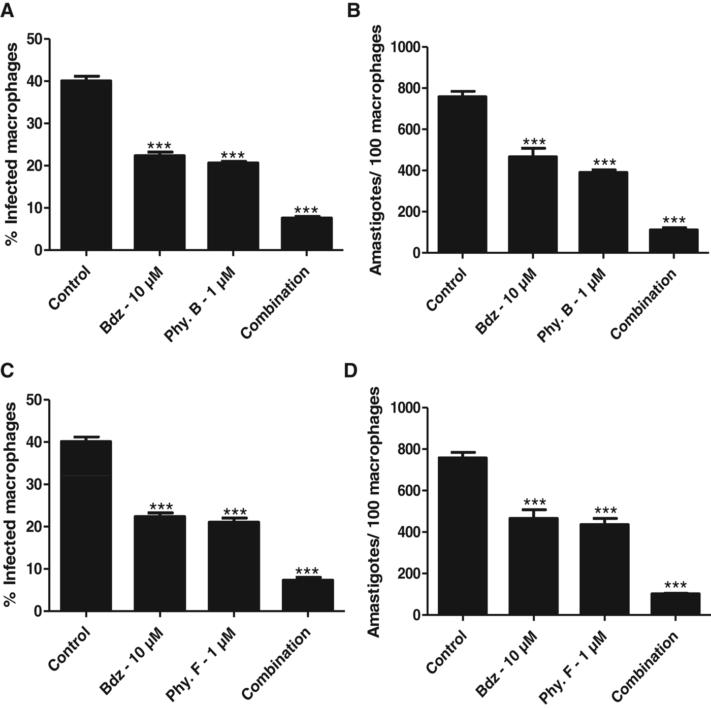
Based on the results described above, a final set of experiments was performed to evaluate the activity in infected macrophages of physalin B or F in combination with benznidazole. As shown in Fig. 7, the combination of physalin and benznidazole reduced the number of infected macrophages as well as the number of amastigotes per macrophage to a greater degree than the respective compound used alone.
A combination of physalin and benznidazole is more potent to inhibit amastigotes than compounds when used alone. Mouse peritoneal macrophages were infected with Y strain trypomastigotes for 2 h. Physalin B or F was added alone or in combination with benznidazole. (A) and (B) show the combination of physalin B and benznidazole, while (C) and (D) display the combination of physalin F and benznidazole. Bdz = Benznidazole. Values represent the mean±s.e.m. in triplicate. ***P<0·001 compared with the control group.
DISCUSSION
Pharmacological treatment of Chagas disease has been limited to benznidazole, which has poor efficacy and causes drug intolerance, as well as adverse effects in many patients. Emergence of parasite resistance to benznidazole is another subject of concern. A large number of small molecules have been screened as anti-T. cruzi agents, however the number is still low when compared with drug discovery efforts for other infectious diseases, such as malaria and AIDS (Goebel et al. Reference Goebel, Ulmer, Projahn, Kloeckner, Heller, Glaser, Ponte-Sucre, Specht, Sarite, Hoerauf, Kaiser, Hauber, Hauber and Holzgrabe2008). Screening of isolated natural compounds is a reliable strategy to identify new anti-T. cruzi agents.
The Physalis genus is a rich source of unique natural compounds. Of these, the seco-steroids physalins are the most investigated in terms of pharmacological property. We previously reported that physalins isolated from P. angulata L. are potent immunomodulatory, antileishmanial and antiplasmodial agents. In 2008, Vieira and co-workers reported that physalin F and withaphysalins inhibit proliferation of T. cruzi epimastigotes in an axenic medium (Vieira et al. Reference Vieira, Espíndola, Santana, Veras, Pessoa, Pinheiro, de Araújo, Lima and Silveira2008). Additionally, Castro and co-workers corroborated these findings, showing that physalin B inhibits T. cruzi development in the gut of triatomines (Castro et al. Reference Castro, Figueiredo, Genta, Ribeiro, Tomassini, Azambuja and Garcia2009, Reference Castro, Moraes, Gonzalez, Ribeiro, Tomassini, Azambuja and Garcia2012). However, the anti-T. cruzi activity of physalins against the infective forms in humans (bloodstream trypomastigotes and amastigotes) have not been described.
In the present study, we determined the anti-T. cruzi activity of physalins B, D, F and G purified from P. angulata. Only physalin F was previously tested against epimastigotes, therefore we performed an extensive examination of all physalins. As expected, we observed that physalins, specifically B and F, are trypanocidal agents. They inhibited epimastigote proliferation, and were toxic against bloodstream trypomastigotes at non-toxic concentrations for mouse macrophages. In addition, they showed a greater potency when compared with benznidazole, the reference anti-T. cruzi drug. Regarding the cellular SI, physalins B and F exhibited some selectivity, albeit low for a desirable anti-T. cruzi drug candidate.
The chemical difference amongst the four physalins is found in the substituents attached to C5 and C6 in the steroid backbone. Physalins D and G are similar because of the hydroxyl groups present and they did not exhibit trypanocidal activity. In contrast, physalin B and F lack hydroxyl groups in C5 and C6, and demonstrated higher activity, exhibiting a pronounced trypanocidal property. In practice, physalin B and F have equipotent trypanocidal activity.
By using trypomastigotes, we observed that physalins cause parasite cell death through necrotic mechanisms. Also, we saw that physalins alter the morphology of the Golgi complex, kinetoplast and endoplasmic reticulum. In the literature, altered Golgi complex morphology is an indication that T. cruzi protease activity was affected (Engel et al. Reference Engel, Doyle, Palmer, Hsieh, Bainton and McKerrow1998). Cruzain is the major cysteine protease present in T. cruzi and has been identified as a drug target (Sajid et al. Reference Sajid, Robertson, Brinen and McKerrow2011). Therefore, we determined the ability of physalins to inhibit the activity of recombinant cruzain; however, physalins did not inhibit it.
Using electron microscopy, formation of myelin-like figures in the parasite treated with physalins was evident. Fluorescence microscopy revealed that parasites treated with physalins were labelled with MDC, a cytosolic vacuole stain (Vanier-Santos and Castro, Reference Vanier-Santos and De Castro2009; Duszenko et al. Reference Duszenko, Ginger, Brennand, Gualdrón-López, Colombo, Coombs, Coppens, Jayabalasingham, Langsley, de Castro, Menna-Barreto, Mottram, Navarro, Rigden, Romano, Stoka, Turk and Michels2011). Therefore, it is possible that the cytosolic vacuoles observed in physalin-treated T. cruzi are due to autophagy. In fact, our results are similar to a very recent finding that physalins exert cellular effects by inducing autophagy (He et al. 2013a, b).
Most importantly, we analysed physalin effects in the in vitro infection. The same order of potency against trypomastigotes was observed when all physalins were tested in T. cruzi-infected macrophages. Physalins B and F at 1·0 μ m were able to reduce intracellular parasite development in macrophages. This was similar to reductions observed in benznidazole-treated infected cells. Physalins B and F are well known for their modulatory property in immune cells (Soares et al. Reference Soares, Bellintani, Ribeiro, Tomassini and Ribeiro dos Santos2003, Reference Soares, Brustolin, Santos, Bellintani, Paiva, Ribeiro, Tomassini and Ribeiro Dos Santos2006). In fact, we observed that physalin B or F at concentrations of 5·0 μ m or higher increased the T. cruzi infection in macrophages, probably because they deactivate host macrophages during the infection development.
The parasite invasion process in macrophages takes approximately 2 h, thereby requiring a drug to work quickly (Matsuo et al. Reference Matsuo, Silva, Torrecilhas, Pascoalino, Ramos, Rodrigues, Schenkman, Caires and Travassos2010). In this assay, benznidazole was not active, while amphotericin B was active to stop the parasite invasion. Interestingly, physalins B and F not only reduced parasite burden in infected macrophages, but they also quickly killed trypomastigotes exposed to macrophages, therefore indicating seco-steroids stop the parasite invasion process in host cells.
An anti-Chagas disease treatment will likely contain a combination of inhibitors to improve the efficacy of the treatment and avoid parasite resistance. In recent years, many efforts have been done to identify optimal drug combinations for Chagas disease (Cencig et al. Reference Cencig, Coltel, Truyens and Carlier2012; Veiga-Santos et al. Reference Veiga-Santos, Barrias, Santos, de Barros Moreira, de Carvalho, Urbina and de Souza2012). In fact, we observed the combination of physalin and benznidazole has a greater activity to reduce T. cruzi infection in macrophages than compounds used alone. The results indicate that the combination of physalin and benznidazole act in an additive fashion. Altogether, these data argue that the screening for new anti-T. cruzi agents based on seco-steroids, specifically those without or with little effects on immune cells, is an attractive line of drug development.
CONCLUSION
Physalins showed potent activity against T. cruzi epimastigotes and trypomastigotes. Of these, physalins B and F exhibited the highest potency and selectivity, and similar to the observations on benznidazole-treated parasites. These compounds achieve trypanocidal activity through autophagy induction, which ultimately results in necrotic parasite death. Regarding the in vitro infection, we observed physalins inhibit parasite development, as well as the invasion process in host cells. Moreover, physalins seem suitable for drug combination with other anti-T. cruzi agents to control infection.
ACKNOWLEDGEMENTS
M. B. P. S. and R. R. S hold a CNPq fellowship, while D. R. M. M. and C. S. M. thank for FAPESB scholarships.
FINANCIAL SUPPORT
This work received funding by Conselho Nacional de Pesquisas Brasileira (CNPq, grant number 502161/2010-8) and Fundação de Amparo as Pesquisas do Estado da Bahia (FAPESB, grant number 6596).
COMPETING INTERESTS
The authors have declared that no competing interests exist.