Introduction
Chronic otitis media with perforation of the tympanic membrane is a common cause of hearing loss and ear discharge in South East Asia.1
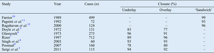
In symptomatic patients, surgical repair of the tympanic membrane, termed a type one tympanoplasty or myringoplasty, is a tried and tested treatment modality. The success of surgery is indicated by the presence of a dry ear with an intact tympanic membrane, an aerated middle-ear space, and improved or maintained hearing.Reference Hirsch and Myers2 There are two popular surgical techniques, the underlay and overlay methods. The underlay technique is quicker and easier to perform, and the creation of a tympano-meatal flap with elevation of the annulus allows inspection of the ossicular chain.Reference Sergi, Galli, De Corso, Parilla and Paludetti3 However, there is a risk of medial displacement of the graft, especially in large and/or anterior perforations, though the risk of this can be minimised by using anterior tagging of the graft.Reference Sharp, Terzis and Robinson4 The overlay technique avoids this pitfall, but there is a risk of keratin pearl formation within the tympanic membrane, and also a risk of blunting of the angle between the drum and the anterior meatal wall.Reference Sergi, Galli, De Corso, Parilla and Paludetti3 Some studies have compared the results of these two techniques (Table I).Reference Sergi, Galli, De Corso, Parilla and Paludetti3, Reference Doyle, Schleuning and Echevarria5–Reference Poonual9 Only minor differences have been found between the outcomes of the two techniques: while the underlay technique produced slightly better outcomes in three studies,Reference Sergi, Galli, De Corso, Parilla and Paludetti3, Reference Doyle, Schleuning and Echevarria5, Reference Glasscock6 the overlay technique produced a higher success rate in another two.Reference Rizer7, Reference Poonual9
Previously reported tympanic membrane closure rates by technique

A number of other techniques of tympanic membrane repair have been described. The term ‘sandwich technique’ was coined by Farrior in 1983 to describe a method in which sheets of temporalis fascia were placed medial and lateral to the drum, with the fibrous layer as the ‘meat’ in the ‘sandwich’.Reference Farrior13 Raghavan et al. used the same term to describe a technique in which a pedicled skin flap is used to partially cover an overlay tympanic membrane graft of temporalis fascia.Reference Raghavan, Malik and Mahmoud12
The technique used in the present study was neither of these, but rather involved the use of temporalis fascia between the epithelial and fibrous layers of the tympanic membrane as described by Pagnini et al. Reference Pagnini, Scarpini, Fanfini and Norberti11 These authors used an endaural approach in their series. To avoid confusion with the techniques of Farrior and Raghavan et al. the author has chosen to term the method used in this series the ‘drum sandwich’ technique. This new technique provides enhanced graft stability because the graft is supported by the fibrous layer of the drum and held in place by the epithelial layer.
Pagnini et al. reported a tympanic membrane closure rate of 93 per cent for their sandwich technique (Table I).Reference Pagnini, Scarpini, Fanfini and Norberti11 However, they reported no data regarding the healing time following surgery.
The present study aimed to describe the author's surgical technique and to investigate the post-operative healing time, success rate and frequency of hearing improvement.
Method
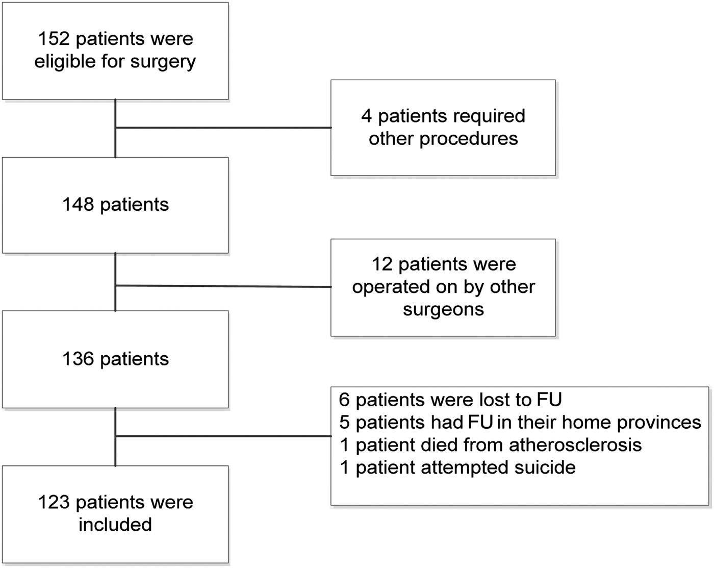
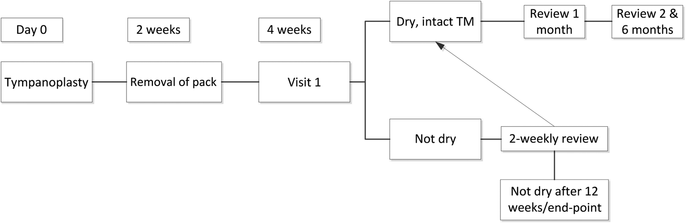
A retrospective chart review was conducted of all type one tympanoplasty operations performed on patients older than 16 years and carried out between January 2009 and December 2011. One hundred and fifty-two patients had originally been scheduled for tympanoplasty during this time period, but only 123 were included in the data analysis (see Figure 1). Demographic data were recorded. The status of the contralateral ear, the duration of aural discharge, and the details of any previous ear surgery were also noted. All patients had received prophylactic antibiotic therapy (oral ciprofloxacin 500 mg twice daily for 5–7 days) following surgery. Audiometric data (i.e. air and bone conduction) had been recorded prior to surgery and, where possible, post-operatively. Pure tone averages had been calculated at 500, 1000 and 2000 Hz. Post-operative air–bone gaps had been calculated using post-operative bone conduction thresholds. Following surgery, the ear canal dressings had been removed after two weeks and the patients reviewed by the operating surgeon according to the schedule shown in Figure 2.
Flow chart showing the procedure for patient inclusion in the study. FU = follow up
Post-operative follow-up schedule. TM = tympanic membrane
The main outcome measures were: healing time, perforation closure success rate and post-operative hearing status.
Surgical technique
All operations had been carried out as day cases under local anaesthesia. A posterior meatal skin flap was raised and the epithelial layer of the tympanic membrane was elevated in continuity with it, exposing the fibrous layer of the tympanic membrane remnant (Figure 3a). It is not difficult to enter the plane between the epithelial and fibrous layers of the tympanic membrane; indeed, trainee surgeons often do so during performance of a tympanotomy. A temporalis fascia graft was placed over the fibrous layer of the tympanic membrane (Figure 3b), and the epithelial layer was replaced over it (Figure 3c). The epithelial layer of the tympanic membrane and the graft were carefully tamped down to avoid anterior blunting of the tympanic membrane during healing. The external auditory canal was dressed with rayon ribbons placed at right angles to each other and cotton wool balls covered in antibiotic ointment.
Diagrams illustrating ‘drum sandwich’ technique for myringoplasty. (a) Elevation of the epithelial layer of the tympanic membrane (TM) in continuity with the tympano-meatal flap. (b) Placement of the fascia graft over the fibrous layer of the tympanic membrane. (c) Replacement of epithelial layer of tympanic membrane and tympano-meatal flap.
Statistical analysis
The hearing data were assessed for normal distribution using the Kolmogorov–Smirnov test. Mean pre- and post-operative hearing thresholds were normally distributed; pre- versus post-operative differences were analysed using Student's t-test. Analyses were carried out using the SPSS version 11 software program (IBM Corporation, Armonk, New York, USA).
Results
One hundred and twenty-three operations meeting the inclusion criteria stated above were carried out during the study period. Of these, 41 (33 per cent) were performed on males and 82 (67 per cent) on females. Patients' ages ranged from 16 to 73 years (mean age, 40.8 years; standard deviation (SD), 11.2 years). One-third of patients had suffered chronic otorrhoea since childhood, and a further one-third for more than 10 years. The mean duration of aural discharge in the group as a whole was 16.4 years (SD, 20.4 years). The tympanic membrane perforation was on the right in 49 patients (40 per cent), on the left in 41 (33 per cent) and on both sides in 33 (28 per cent).
Six patients underwent surgery on both ears during the study period. Eighteen operations were revision procedures. Sixteen of these (89 per cent) were successful. Data on tympanic membrane healing are shown in Figure 4. The perforation was successfully closed in 113 ears (92 per cent of cases). Of those patients with persistent perforations following surgery, eight had dry ears and two had persistent otorrhoea more than 12 weeks after the operation. All patients were followed up for at least 3 months, the average duration being 10.2 months (SD, 7.9 months). Two patients developed small keratin pearls within their tympanic membranes, but these were easily removed in the out-patient clinic.
Patients' tympanic membrane healing times (n = 121).
Table II presents the mean pre- and post-operative air and bone hearing thresholds and air–bone gaps, and the mean pre- versus post-operative air–bone gap difference, which was statistically significant. The air–bone gap was closed to within 10 dB in 50 cases (41 per cent). Thirty-five patients (28 per cent) spontaneously reported subjective improvement in their hearing.
Differences between pre- and post-operative audiograms

*Pre- versus post-operative mean. Pre-op = pre-operative; post-op = post-operative; SD = standard deviation; CI = confidence interval; AC = air conduction; BC = bone conduction; ABG = air–bone gap
Discussion
In this study, the success rate for tympanic membrane closure (92 per cent) was consistent with Pagnini and colleagues' findings.Reference Pagnini, Scarpini, Fanfini and Norberti11 However, the follow up in the present study was relatively short, so the occurrence of late re-perforation could not be excluded. Patients who were lost to follow up or who died (13 cases) were excluded from analysis. In the unlikely event that all of these operations failed, the success rate would have fallen to 83 per cent. In any case, the results suggest that the outcomes of the drum sandwich technique are at least as good as those of other surgeons' methods, and that this technique can be recommended for general use.
The average air–bone gap, which gives an indication of the degree of hearing loss due to the perforation, was 20 dB (±10.54 dB SD) before surgery and 11 dB (±9.8 dB SD) post-operatively. This implies that many patients had a useful improvement in their hearing and obtained a dry ear. Previous authors have reported mean hearing gains of between 3.1 and 16 dB following myringoplasty, with less improvement in older patients.Reference Gyo, Yanagihara, Saiki and Hinohira14–Reference Ribeiro, Rui, Natercia, Jose and Antonio17 The comparable figure for the present study was 10.6 dB. Pagnini et al. reported air–bone gap closure to within 10 dB in 57 per cent of their series, compared with 41 per cent of the present series.Reference Pagnini, Scarpini, Fanfini and Norberti11 This difference in outcomes may be due to the long history of chronic otitis media in the majority of patients in the present study group.
By four weeks after pack removal, the ears of 87 per cent of the current study patients had healed; this is a relatively short time. Rizer reported a healing time of four to eight weeks for overlay operations, but only two to four weeks when the underlay technique was used.Reference Rizer7 Singh et al. reported a healing time of four to six weeks for the underlay technique and six to eight weeks for the overlay method.Reference Singh, Rai, Bandyopadhyay and Gupta8 It is not clear from these papers whether the healing time was measured from the day of surgery or the time when the dressing was removed from the external auditory canal. The drum sandwich technique, using placement of the residual epithelial layer of the tympanic membrane over the graft, may have contributed to the rapid healing observed in the present series.
However, this result could also be explained by the pressure effect of cotton wool balls adjacent to the rayon ribbons next to the ear canal and tympanic membrane. Rayon ribbons do not stick to soft tissue and can therefore be removed without trauma. There is relatively little in the literature on the effects of different ear canal dressings on healing. Zeitoun et al. compared bismuth iodoform paraffin paste dressings, Pope Otowicks (Xomed-Treace, Cabo Rojo, Puerto Rico), Silastic sheeting (Dow Corning, Midland, Michigan, USA) and Tri-adcortyl ointment following middle-ear surgery, and found no difference between them in terms of discharge, granulation tissue formation or the development of canal stenosis.Reference Zeitoun, Sandhu, Kuo and Macnamara18 Nakhula et al. compared bismuth iodoform paraffin impregnated ribbon gauze and Tri-adcortyl ointment and found no difference between the two methods.Reference Nakhula, Tawoingi and Sinha19 Neither of these studies reported the healing times which resulted from the different packing methods.
In the current series, another factor which might have influenced the healing time was the use of prophylactic oral antibiotics. Carlin et al. investigated the use of systemic antibiotic prophylaxis for myringoplasty in 71 patients but failed to demonstrate any benefit from its use.Reference Carlin, Lesser, John, Fielder, Carrick and Thomas20 Bacteria present in the ear pre-operatively were not eliminated and the appearance of organisms post-operatively was not prevented. In addition, there was no association between ear discharge detection and the presence of pathogenic organisms on culture. John et al. carried out a randomised trial of antibiotic prophylaxis versus no antibiotics in 130 patients, and found no difference in the success rate of surgery between the 2 groups (although they assessed the status of the tympanic membrane at 8 weeks rather than the rate of healing).Reference John, Carlin, Lesser, Carrick and Fielder21 These findings suggest that the use of antibiotics in the present study probably did not influence the speed of healing.
Rapid healing is beneficial, because patients can return to their normal activities sooner and require fewer out-patient visits.
• ‘Drum sandwich’ tympanoplasty involves placement of a temporalis fascia graft between the fibrous and epithelial layers of the tympanic membrane remnant
• This provides enhanced graft stability and may reduce healing time
• In this study, perforation closure rates for this technique were comparable with previous reports for other techniques
• Hearing results were also comparable
A larger series of cases with longer follow up will be required to confirm the efficacy of the drum sandwich technique for myringoplasty.
Conclusion
The drum sandwich technique for myringoplasty produces results comparable with those reported in the literature for other techniques. It appears to be associated with rapid post-operative healing of the ear.
Acknowledgement
The author thanks Professor R P Mills for his help with the English language editing of this paper.