
Introduction
Porphyrins are essential materials in nature.[Reference Penon, Patino, Barrios, Nogues, Amabilino, Wurst and Perez-Garcia1] They are part of many physiological processes, including metal chelation, catalysis,[Reference Barona-Castaño, Carmona-Vargas, Brocksom and De Oliveira2] gas transport, photosynthesis,[Reference Allison and Moghissi3] as well as disease eradication via the production of reactive oxygen species. As a result, many synthetic porphyrins have been developed and employed in many fields, including environmental engineering,[Reference La, Bhosale, Jones and Bhosale4] organic synthesis, electronic processing,[Reference Paolesse, Nardis, Monti, Stefanelli and Di Natale5] and biomedics.[Reference Gao, Liu, Yin, Li, Bai and Zhang6] In clinical applications, they are a major contribution to the essential materials for photodynamic therapy,[Reference Winkler, Simon, Finke, Bleses, Birke, Szentmáry, Hüttenberger, Eppig, Stachon, Langenbucher, Foth, Herrmann, Seitz and Bischoff7,Reference Li, Wang, Cheng, Gong, Yin, Gong, Li and Liu8] biosensors, and photoluminescence-based bio-imaging.[Reference Dou, Teng, Ye and Loh9] For example, many porphyrins have been used for the treatment of cancers,[Reference Fakayode, Kruger, Songca, Abrahamse and Oluwafemi10,Reference Ormond and Freeman11] as sensing agents for volatile organic compounds[Reference Paolesse, Nardis, Monti, Stefanelli and Di Natale5] and fluorescence imaging probes for drug and disease site localizations.[Reference Li, Wang, Cheng, Gong, Yin, Gong, Li and Liu8] However, despite all these attributes, many porphyrins are limited in the bio-application due to a high degree of hydrophobicity and low absorption within the biological window (regions where the absorption of the biological matrices are low).[Reference Fakayode, Tsolekile, Songca and Oluwafemi12–Reference Vieira, Castilho, Ferreira, Ferreira-Strixino, Hewitt and Raniero14] The former can cause them to aggregate in aqueous solutions and thus cause an embolism (blockage of blood vessels). This, in turn, may lead to cellular complications such as inflammation or cell death. As a result, efforts are being made to circumvent these challenges. These include conjugation with water-soluble functionalized nanoparticles, interaction with hydrophilic polymers, and conversion to chlorins or bacteriochlorins.[Reference Mccarthy, Bhaumik, Merbouh and Weissleder15,Reference Aravindu, Mass, Vairaprakash, Springer, Yang, Niedzwiedzki, Kirmaier, Bocian, Holten and Lindsey16] Among these methods, conjugation to nanoparticles seems to have gained considerable attention over the years due to the effects of some of the properties of the nanomaterials on porphyrin's chemistry such as attraction to an external magnetic field, site-specific nano-delivery, and improved water solubility.[Reference Gao, Liu, Yin, Li, Bai and Zhang6,Reference Fakayode, Kruger, Songca, Abrahamse and Oluwafemi10,Reference Muresan and Lindsey17,Reference Huan, Fei, Shan, Wang, Xu and Feng18] Superparamagnetic iron oxide nanoparticles (SPIONs) are especially recognized for specific site-delivery without embolism due to their unique magnetic properties (re-dispersibility into the biological fluid after removal of an applied external magnetic field).[Reference Fakayode, Songca and Oluwafemi19] More often than not, the SPIONs are usually covered with gold nanoparticles to prevent leaching of Fe(III) ions, provide a platform for linking thiol-compounds, and enhance the hyperthermia characteristics of the SPIONs.[Reference Tintoré, Mazzini, Polito, Marelli, Latorre, Somoza, Aviñó, Fàbrega and Eritja20,Reference Mohammad, Balaji, Weber, Uppu and Kumar21] Polyethylene glycols are recognized materials that exhibit excellent hydrophilicity. Thus, many conjugates of water-soluble SPIONs, gold nanoparticles, and porphyrins have been synthesized via their interactions with polyethylene glycol system.[Reference Penon, Patino, Barrios, Nogues, Amabilino, Wurst and Perez-Garcia1,Reference Li, Wang, Cheng, Gong, Yin, Gong, Li and Liu8,Reference Anbarasu, Anandan, Chinnasamy, Gopinath and Balamurugan22,Reference Silva, Lima, Mansilla, Zysler, Troiani, Pisciotti, Locatelli, Benech, Oddone, Zoldan, Winter, Pasa and Creczynski-Pasa23] However, despite all these advantages, some porphyrins conjugated to SPIONs or gold nanoparticles[Reference Penon, Patino, Barrios, Nogues, Amabilino, Wurst and Perez-Garcia1,Reference Li, Wang, Cheng, Gong, Yin, Gong, Li and Liu8] show some deformities in their absorption properties. These effects can lower their efficiencies for biomedical applications. To circumvent this challenge, a new method is required for the development of nondeformed water-soluble SPIONs/gold porphyrin conjugates.
Nosocomial bacteria are the causative agents responsible for most common types of hospital-acquired infections (HAIs) such as pneumonia, urinary tract infections (UTIs), diarrhoa, bedsores, bacteremia, and surgical wound infections.[Reference Murray, Rosenthal and Pfaller24] These microorganisms may already be present in the patient's body or may come from the environment, contaminated hospital equipment, health care workers, or other patients.[Reference Murray, Rosenthal and Pfaller24] The most frequently found nosocomial bacteria include: Escherichia coli which accounts for 23% of nosocomial infections, Staphylococcus aureus (20%), Pseudomonas aeruginosa (11%), and Enterococcus species (6%).[Reference Khan, Ahmad and Mehboob25]
In this report, a simple, low-cost method, which utilizes the advantage of the solubility of certain porphyrin molecules in two miscible polar solvents of different dipole moments, is employed for the syntheses of four water-soluble thiolated polyethylene glycol gold-coated SPIONs (SPIONs@Au)–porphyrin conjugates. The first solvent, organic alcohol, allows for the easy dissolution of the hydrophobic porphyrins and generates a conducive environment for the dispersibility of the porphyrins in the second solvent (water). Furthermore, the antimicrobial evaluation of the potentials of the as-synthesized magnetic porphyrin conjugates against four common nosocomial bacterial pathogens was carried out to explore their efficacy as antibacterial agents.
Experimental
Materials
All reagents were purchased from Sigma-Aldrich (Steinem, Germany) and used without further purifications, except for the pyrrole, which was vacuum distilled before utilization. For the bacterial assays, Gram-positive bacterial strains such as S. aureus (ATCC# 25923) and Enterococcus faecalis (ATCC# 29212) as well as Gram-negative strains such as P. aeruginosa (ATCC# 27853) and E. (ATCC# 8739) were obtained from Davies Diagnostics, Johannesburg, South Africa, and retained viable in the Department of Pharmacy and Pharmacology, University of the Witwatersrand where all microbiological tests were carried out. Detailed descriptions of the syntheses and characterizations of porphyrins (5,10,15,20-tetrakis(4-hydroxyphenyl)porphyrin(m-THPP),5,10,15,20-tetrakis(3,5-dimethoxyphenyl) porphyrin (m-TdMPP), 5,10,15,20-tetrakis(3-pyridyl)porphyrin(m-T3-PyP), and 5,10,15,20-tetrakis(1-methylpyridinium-3-yl)porphyrin tetra-iodide (m-T3-Py+P4I−) are given in the Supplementary Material.
Synthesis of SPIONs
A mixture of ferric chloride hexahydrate solution (50 mL, 0.300 M) and α-D-glucose solution (50 mL, 0.100 M) were heated at 95 °C for 40 min without stirring until the total volume was reduced to 15 mL. This was followed by cooling for 5 min and subsequent addition to a preheated ammonium hydroxide solution (50 mL, 1.000 M; 55 ± 2 °C) dropwise without stirring to form a black colloidal solution. Thereafter, the resulting solution was continuously heated to boil for 10 min until a black precipitate of SPION was formed. The latter was separated out from the solution with the aid of a permanent magnet leaving out a clear supernatant, which was removed. At this time, the colloidal solution was allowed to cool to room temperature and finally dried in the oven at 88 °C for 15 h. Thereafter, the as-synthesized SPIONs were washed with deionized water (4 × 50 mL) and dried under ambient conditions until analysis.
Synthesis of SPIONs@Au
A small amount of the as-synthesized SPIONs (0.02101 g) was sonicated in 30 mL of deionized water for 15 min to obtain a dark chocolate color. This was followed by the addition of 20 mL gold (III) chloride monohydrate solution (0.02152 g/20 mL), and the resulting solution subsequently heated to 74 °C without stirring for 4 h to obtain an antique gold reddish-brown color solution. The resulting gold-coated SPION solution was cooled, washed with deionized water, and harvested from the supernatant using a magnet. This was followed by drying at 60 ± 2 °C for 18 h. The material was stored under ambient conditions until analysis.
Synthesis of thiol-PEGylated SPIONs@Au–TdMPP conjugate
An aqueous solution of SPIONs@Au (10 mL, 0.00075 g/mL) was sonicated for 10 min. This was followed by the addition of methoxy-polyethylene glycol-thiol (mPEG-SH) (0.250 g), and the resulting solution sonicated for another 5 min. A TdMPP solution in methanol (15 mL, 0.00052 g/mL) was then added to the gold-coated SPIONs-mPEG-SH mixture and subsequently shaken for 10 min. Thereafter, the resulting solution was evaporated to dryness under ambient conditions to give a brown color SPIONs@Au–TdMPP conjugate.
Synthesis of thiol-PEGylated SPIONs@Au–THPP conjugate (brown)
An aqueous solution of SPIONs@Au (10 mL, 0.00075 g/mL) was sonicated for 10 min. This was followed by the addition of mPEG-SH (0.250 g) and sonication for another 5 min. A THPP solution in methanol (15 mL, 0.00052 g/mL) was then added to the gold-coated SPION-mPEG-SH mixture, and the resulting solution shaken for 10 min. Thereafter, the mixture was evaporated to dryness under the ambient condition to give a brown color SPIONs@Au–THPP conjugate.
Synthesis of thiol-PEGylated SPIONs@Au–THPP conjugate (green)
An aqueous solution of SPIONs@Au (10 mL, 0.00075 g/mL) was sonicated for 10 min. This was followed by the addition of 0.125 g mPEG-SH and subsequently sonication of the resulting solution for another 5 min. The resulting mixture was added to a THPP solution in methanol (15 mL, 0.00052 g/mL), and the resulting solution subsequently was shaken for 10 min. The final solution was then evaporated to dryness under ambient conditions to obtain a green color SPIONs@Au–THPP conjugate.
Synthesis of thiol-PEGylated SPIONs@Au–T3-PyP and SPIONs@Au–T3-Py+P4I− conjugates
The procedure for SPIONs@Au–TdMPPC was repeated using T3-PyP and T3-Py+P4I− instead of TdMPP to form SPIONs@Au–T3-PyPC and SPIONs@Au–T3-Py+P4I−C, respectively.
Characterizations
Absorption and fluorescence spectra were recorded at room temperature using 1 cm path length cells on a UV/VIS Perkin-Elmer Lambda 25 Spectrophotometer (Buckinghamshire, UK) and Shimadzu RF-6000 Spectro-fluorophotometer (Kyoto, Japan), respectively. The Fourier transform-infrared spectroscopy (FTIR) spectra were obtained on a Perkin-Elmer Universal Attenuated Total Reflection UATR Spectrum Two (Buckinghamshire, UK). Proton (1H) and carbon-13 (13C) nuclear magnetic resonance (NMR) spectra were recorded on a Bruker 500-MHz Advance III HD spectrometer (Billerica, MA, USA) at 298 K with the CDCl3 solvent. Saturation magnetism and magnetization curves of SPIONs were measured using the SQUID magnetometer PPMS, Quantum Design Inc. (San Diego, CA, USA). For zeta potential studies, the aqueous solutions of the as-synthesized conjugates were run using the Anton Paar Litesizer 500 (Graz, Austria).
Antibacterial assays
The quantitative microplate bioassay method[Reference Eloff26] was used to determine the minimum inhibitory concentrations (MICs) of the as-synthesized materials against four nosocomial bacterial strains. The as-synthesized materials (5.0 mg/mL) were dissolved in an appropriate volume of solvent (water for injection was used for the conjugates, cationic porphyrin, gold-coated SPIONs, and ciprofloxacin, while acetone was used for THPP, TdMPP, and T3-PyP), followed by sonication for 10 min to aid dissolution before use. Ciprofloxacin (starting concentration 0.01 mg/mL), a broad-spectrum antibiotic, was used as a reference standard and positive control, while acetone was used as a negative control. The experiments were performed in triplicates. The detail descriptions are summarized in Supplementary Table S1.
Results and discussion
Synthesis and characterization of gold-coated SPIONs–porphyrin conjugates
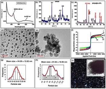
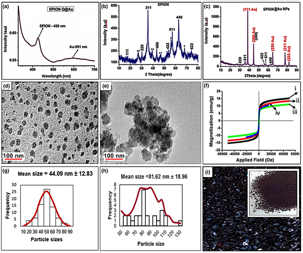
The gold-coated SPIONs emerged as a dark red wine magnetic material when compared to the dark brownish-black of the pristine SPIONs. The red wine color indicated the presence of Au (0) obtained from the reduction of Au(III). The presence of gold on SPIONs was further confirmed using UV–Vis, transmission electron microscope (TEM), and X-ray powder diffraction (XRD) [Figs. 1(a)–1(c)]. According to Fig. 1(a), the SPIONs' absorption peak was at 426 nm while gold peaked at 591 nm. The XRD showed that both the SPIONs and the gold-coated SPIONs materials were crystalline in nature [Figs. 1(b) and 1(c)] by virtue of their narrow, sharp Bragg's reflection peaks at (220), (311), (400), (511), (440) and (111), (200), (220), (311), (222), corresponding to a cubic iron oxide,[Reference Fakayode, Songca and Oluwafemi19,Reference He, Miao, Li, Wang, Cao, Xie, Yang, Zou and Burda27] and face-centered cubic gold planes, respectively.[Reference Krishnamurthy, Esterle, Sharma and Sahi28] The morphology of the SPIONs was spherical [Fig. 1(d)], while the presence of the gold on the SPIONs was seen as shell structures surrounding the spherical core [Fig. 1(e)].
Characterization of SPIONs@Au. (a) UV–Vis; (b) XRD for SPIONs; (c) XRD for SPIONs@Au; (d) TEM image of SPIONs; (e) TEM image of SPIONs@Au; (f) SQUID of SPIONs@Au at (i) 10 K, (ii) 150 K, and (iii) 300 K; and of SPIONs at (iv) 300 K; (g) size distribution of SPIONs; (h) size distribution of SPIONs@Au; and (i) digital image of SPIONs. Inset: digital image of SPIONs@Au.

The results from the evaluation of the magnetic properties of the gold-coated SPIONs showed that the coating of gold on SPIONs did not significantly affect the magnetic properties of the SPIONs since the SPIONs maintained its superparamagnetic property, and there was not much change in the magnetization (Ms) value after coating [Fig. 1(f)]. This agreed with the reports in the literature that stated that passivating SPIONs' surfaces with Au had a negligible effect on their magnetic behavior.[Reference Wu, He and Jiang29,Reference Sun, Reddy, Marquez, Jena, Gonzalez and Wang30] The size distribution curves of the SPIONs and the gold-coated SPIONs are given in Figs. 1(g) and 1(h). According to Figs. 1(g) and 1(h), the average size of the magnetic nanomaterials increased by a factor of about 1.85 after coating with gold. Figure 1(i), together with the inset, represents the digital images of the SPIONs and the gold-coated SPIONs, respectively.
Moreover, the decrease in Ms values as the temperature increases was due to the enhancement of the effect of the thermal energy on the nanoparticles, which promotes the transition from ferromagnetism to superparamagnetism.
All porphyrins evolved as purple except T3-Py+P4I− which appeared as red-violet. However, all conjugates appeared as brown materials except the THPP conjugate synthesized with a lower amount of mPEG-SH, which emerged as a green material. All conjugates were soluble in water and exhibited attraction to the external magnetic field. The presence of the gluconic acid on the SPIONs@Au coupled with the presence of thiolated polyethylene glycol contributed to the enhancement of the water solubility of the porphyrin conjugates. Thiol-terminated polyethylene glycol is known to enhance the solubility of the gold matrix in water because of the high affinity of the thiol group to gold surfaces.[Reference Xue, Li, Li and Zhang31] The results from the zeta potential analysis of the conjugates in aqueous solution revealed overall negative charges of −15.7, −19.6, and −22.5 mV for SPIONs@Au–T3-PyPC, SPIONs@Au–THPPCbr, and SPIONs@Au–TdMPPC and positive charges of +3.3 and +13.8 mV for SPIONs@Au–T3-Py+P4I−C and SPIONs@Au–THPPCgr, respectively (Supplementary Table S2). The large negative or positive values of the zeta potentials of the conjugates indicate large colloidal stability, while a small positive charge may promote the prevention of protein adsorption or sequestration in the aqueous biomedical environment.[Reference Honary and Zahir32,Reference Hirsch, Kinnear, Moniatte, Rothen-Rutishauser, Clift and Fink33]
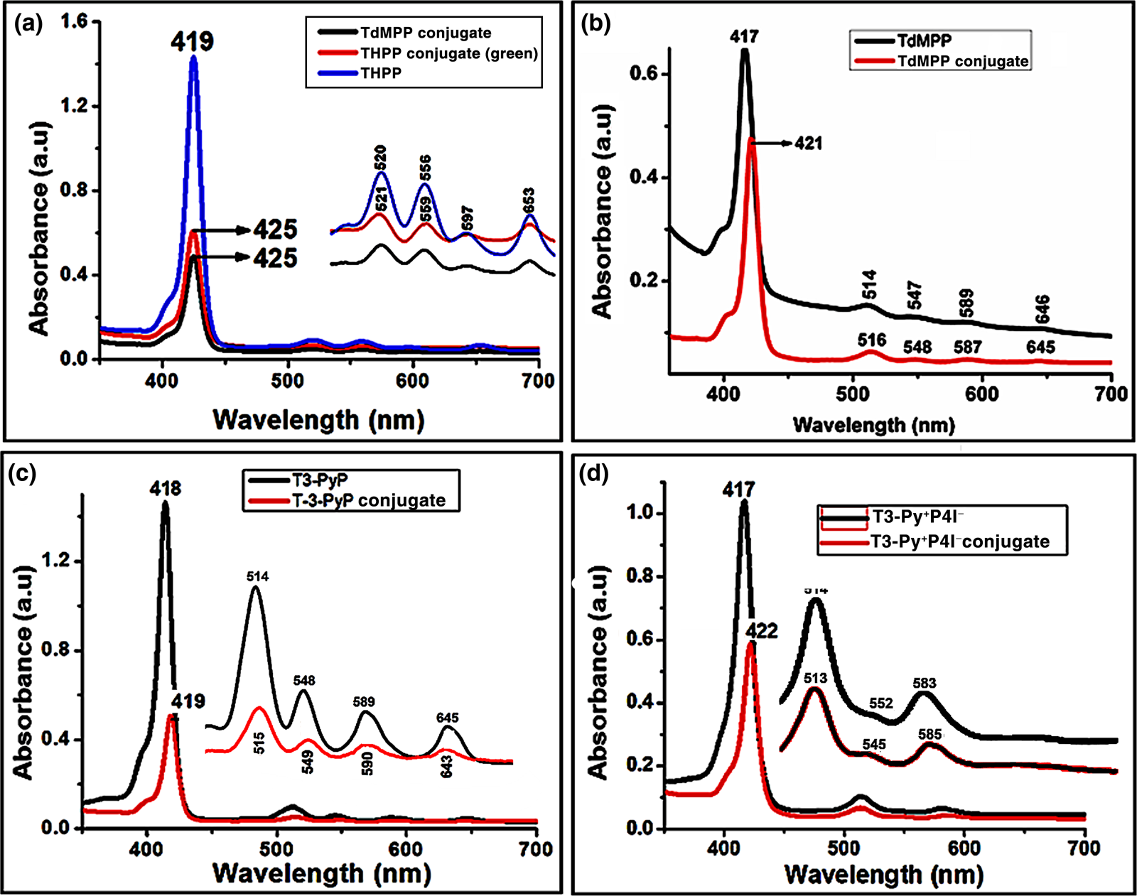
The UV–Visible absorptions evaluation of the conjugates showed the characteristic features of free-base porphyrins without distortion of the Soret and four Q-bands [Figs. 2(a)–2(d)]. This means that the porphyrins were not metallated during synthesis. This shows that the method employed in this study for the conjugation of SPIONs@Au to porphyrins provides porphyrins with better absorption properties compared with some methods reported in the literature.[Reference Penon, Patino, Barrios, Nogues, Amabilino, Wurst and Perez-Garcia1,Reference Li, Wang, Cheng, Gong, Yin, Gong, Li and Liu8] Moreover, bathochromic shifts of bands were evident in the conjugates relative to the pristine porphyrins, suggesting better absorption of longer wavelengths [Figs. 2(a)–2(d)].
Absorption spectra of the pristine porphyrins and their corresponding conjugates. (a) THPP and conjugate; (b) TdMPP and conjugate; (c) T3-PyP and conjugate; and (d) T3-Py+P4I− and conjugate.

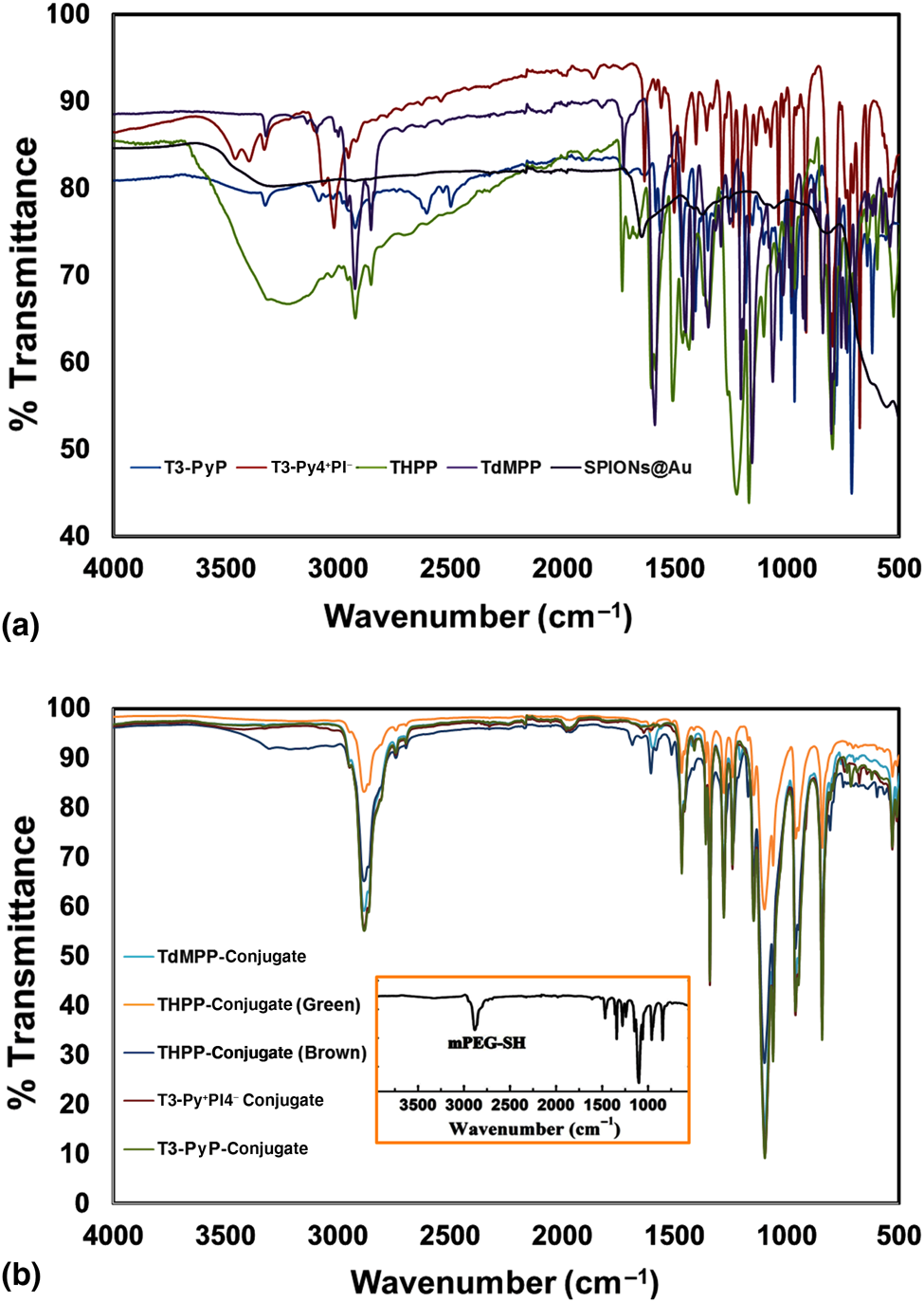
The results of the FTIR analyses of the conjugates showed the characteristic presence of C–Hstr at 2884 cm−1 and C–Ostr at 1096 cm−1 similar to the functional groups of the mPEG-SH except for the conjugates containing T-3-PyP and T3-Py+P4I− which did not have C–O bond within their molecular structures [Figs. 3(a) and 3(b)]. However, significant differences between the spectra of the conjugates and those of the precursors were observed. For example, a blueshift occurred for THPP and TdMPP after conjugation from 2853 and 2854 to 2884 cm−1 for C–Hstr absorption, respectively, while a redshift was experienced by the T3-Py+P4I− from 2923 to 2884 cm−1. In contrast, a redshift was observed for the THPP and TdMPP for C–Ostr absorption from 1170 and 1155 to 1096 cm−1, respectively. The absence of some characteristic features of the precursors between 4000–3000 and 1700–500 cm−1 in the conjugates' absorptions suggested a possible interaction of the functional groups within these regions with the functional groups of the mPEG-SH or encapsulation of these functional groups within the polymer matrix [Figs. 3(a) and 3(b)].
FTIR characterization of the precursors and conjugates. (a) The precursors before conjugation and (b) conjugates. Inset: FTIR spectrum of the mPEG-SH.

Antibacterial activities of the conjugates
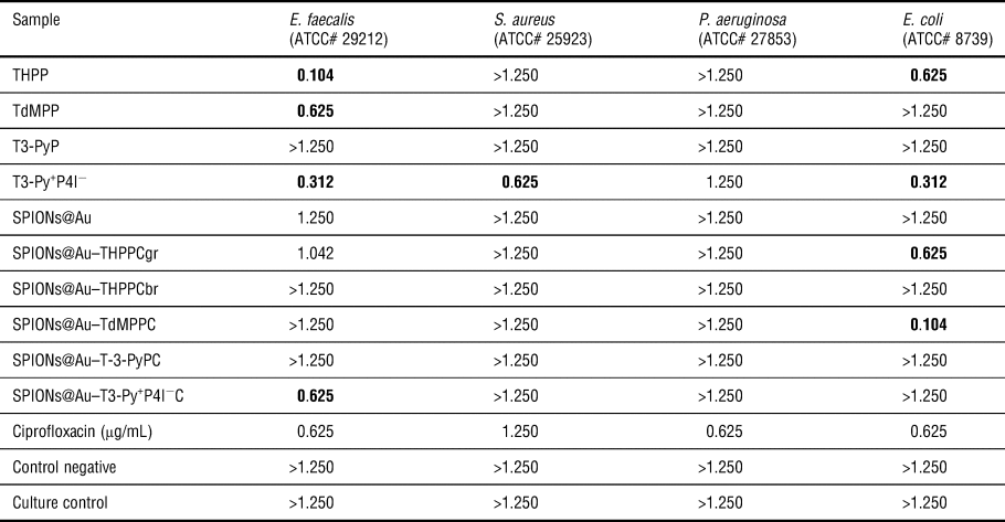
The results of the antibacterial activities of the as-synthesized conjugates together with their precursors, against four pathogenic bacterial strains, are shown in Table I. SPIONs@Au–TdMPPC with a MIC value of 0.104 mg/mL showed the most noteworthy inhibitory effects against E. coli. THPP also demonstrated the same noteworthy activity (0.104 mg/mL) against E. faecalis. Other noteworthy activities are indicated in bold in Table I. Furthermore, while the T3-Py+P4I− and THPPgr conjugates showed equal activity in comparison with the commercially available antibiotic Ciprofloxacin against E. faecalis and E. coli, respectively (Table I), the TdMPP conjugate exhibited a better activity (about 16%) than the antibiotic agent against E. coli. Also, the TdMPP conjugate showed greater activity than all of the pristine porphyrin precursors evaluated against E. coli. For reasons, yet unknown, the THPPbr and T-3-PyP conjugates did not show any worthwhile activities against all the nosocomial agents evaluated in this work. This simply means that the inhibition activities of the conjugates reported in this work might go beyond just the structure, surface charge, and interaction between the biomolecules at the outer membrane of microbial cells as conventionally perceived. For example, the distinguishable MIC value displayed by the SPIONs@Au–TdMPPC (0.104 mg/mL) against the E. coli compared to the other conjugates might be described as being electrostatic interaction dependent. However, the average zeta potential results showed that SPIONs@Au–TdMPPC had the highest negative surface charge value of −22.5 mV (Supplementary Table S2), meaning it should be the one to face the highest cellular repulsion since the cell surface is generally negatively charged at the physiological pH. In terms of structure, SPIONs@Au–TdMPPC has lesser attributes (–O–CH3) to form hydrogen bonds with the proteins, lipids, and the carbohydrates at the cellular surface than the SPION@Au–THPPCgr (–O–H), yet it showed higher inhibition activity than the latter. Thus, elucidating the mechanism of the inhibition activities by these conjugates against the tested organisms will be a source of attractive research as information from this may provide a better understanding on how these potential molecules carry out their activities against the understudied pathogenic organisms. Nonetheless, the amount of mPEG-SH present within a conjugate matrix may have a significant influence on the biological activity of such a material against a given pathogen as seen in the activities of SPION@Au–THPPCgr and SPION@Au–THPPCbr.
MIC (mg/mL) of as-synthesized samples against bacterial pathogens.

Note: Culture control (bacterial suspension only); Control negative: acetone; values in bold indicate noteworthy activity.
E. coli, a Gram-negative bacterium, is accountable for many food and water-borne diseases and deaths in some cases,[Reference Murray, Rosenthal and Pfaller24,Reference Khan, Ahmad and Mehboob25] while E. faecalis, a Gram-positive bacterium, is considered the predominant microorganism in secondary infections such as bacteremia, wound infections, and chronic periodontitis.[Reference Murray, Rosenthal and Pfaller24,Reference Khan, Ahmad and Mehboob25] Both E. coli and E. faecalis are prevalent in all mammalian feces where they are introduced into water sources such as bore-hole, wells, tap water, and drinking troughs.[Reference Ahmed, Neller and Katouli34,Reference Byappanahalli, Nevers, Korajkic, Staley and Harwood35] Hence, these pathogens are chosen as the biological indicators of water treatment safety.[Reference Byappanahalli, Nevers, Korajkic, Staley and Harwood35,Reference Edberg, Rice, Karlin and Allen36] The presence of these pathogens in potable water sources, as well as in human body, poses a great risk to health. Thus, stringent regulation of absolute zero concentration of E. coli and E. faecalis is currently required in ready-to-drink portable water. This means that the as-synthesized conjugates that exhibited excellent activity against these pathogens possessed some potential to be applied as antibacterial inhibition agents in water treatment plants and clinical environment for the prevention of the existence of these pathogens.
Conclusion
A facile approach to the synthesis of non-distorted visible light-absorbing gold-coated SPIONs–porphyrin conjugates has been established for four porphyrins of different structures (three neutral and one cationic). The synthesis involved a simple transfer of an organic hydrophobic molecule into an aqueous phase containing gluconic acid-capped gold-coated SPIONs and the hydrophilic agent, thiolated polyethylene glycol. The Soret and the Q-bands of these porphyrin conjugates were intact after conjugation, together with shifts to higher wavelengths. The results of the evaluation of the antibacterial inhibition effects of these conjugates together with their porphyrin precursors against four nosocomial pathogens showed that two of the conjugates, SPIONs@Au–TdMPPC and SPIONs@Au–T3-Py+P4I−C, showed high noteworthy activities against E. coli and E. faecalis, respectively. Interestingly, the conjugates containing T3-Py+P4I− and THPPgr molecules showed equal activity in comparison with Ciprofloxacin, a commercially available antibiotic agent against E. faecalis and E. coli, respectively, while the TdMPP conjugate exhibited better activity than the antibiotic agent (about 16%) and all the pristine porphyrin against E. coli. All these findings suggested that these new porphyrin conjugates have the potential to be used as alternative antibacterial inhibition agents against E. coli and E. faecalis in the environmental and biomedical fields.
Supplementary material
The supplementary material for this article can be found at https://doi.org/10.1557/mrc.2019.141.
Acknowledgments
The authors are grateful to the National Research Foundation (NRF), South Africa, under the Nanotechnology Flagship Programme (Grant no. 97983), and the Collaborative Postgraduate Training Grant (Grant no. 105262) for financial support. B.M.A.-T. appreciates Niger Delta University, Wilberforce Island, Bayelsa State, Nigeria, for granting him study leave to accomplish this work.