I. INTRODUCTION
Wear is usually minimized by applying lubrication. However, in some situations, such as high temperature, longer operating time, vacuum environment etc., oil or grease lubrications alone are not sufficient to resolve the surface wear issues.Reference Zygmunt1 Abrasive resistant coatings provide a better option for replacing the lubrications since this method is simple and economic.Reference Wu, Guo and Zhang2
Nowadays, coating has been used incrementally for engineering applications especially in oil and gas and heavy machineries industries. Generally, the capability of the surface without coating in terms of wear and corrosion resistivity is much lower compared to the surface of the coated material. Applications of coating are potential to prolong the lifespan of material and machineries besides reducing the maintenance cost in industrial sector.Reference Marita and Yaacob3 In practice, coatings confer characteristics, such as wear and corrosion resistance, hardness, high melting temperature, high density, good adhesion, low permeability and diffusion, for oxidation.Reference Kennedy and Hashmi4
Throughout the research and study made recently, there are lots of claims that addition of nanoparticle coating on the surface of the material can enhance and improve the capability of surface in terms of corrosion and wear. Nanocrystalline coatings have attracted great attention potential due to their superior characteristics which are not typically found in conventional coatings. These materials demonstrate the superior improved performance and sometimes completely new performance as a result of the grain size reduction.
Electrodeposition is an easy, economic, and simple method in coating preparation nanocrystalline particles.Reference Marita and Yaacob3 The deposition can be done on various types of substrates. This method also can be conducted approximately at room temperature without the need of high vacuum or high temperature conditions. Besides, high surface finish and pore free coating can be produced at high rates using electrodeposition if compared with other coating methods.Reference Pellicer, Varea, Pane, Sivaraman, Nelson, Surinach, Baro and Sort5 The samples can be grown at high deposition rates and there are no limitations on substrate geometry.
Over the years, cobalt coatings as well as cobalt-based alloys have been explored mainly because of their unique physicochemical properties and wide applications. They have gained much attention when they are found to be the most potential candidates to replace hazardous hexavalent chromium. Alloying with Ni and Fe enhances the properties of Co coatings. It is reported that both Co–Ni and Ni–Fe provide good magnetic properties, high hardness, thermal stability, corrosion, and wear resistances.Reference Marita and Yaacob3,Reference Pellicer, Varea, Pane, Sivaraman, Nelson, Surinach, Baro and Sort5 However, there is still lack of investigation on wear and surface roughness for Co, Ni and Fe nanoparticles combined as a coating. Thus, the idea of investigating the wear properties and surface roughness of the Co–Ni–Fe nanoparticle coating prepared at different time depositions is proposed in this paper. Mild steel substrate was selected for the testing since it is a common steel type used in industry but easy to corrode. Hence, Co–Ni–Fe nanocrystalline coatings will be coated on the mild steel to protect it from the corrosion.
The physical behaviors of electrodeposited coating such as crystal phase, grain size, and phase composition are strongly dependent on deposition parameters. The deposition parameters involved pH bath solution, time deposition, current density, electrode potentials, working temperature and amount of additives included in the electrolyte.Reference Franczak, Levesque, Bohr, Douglade and Chopart6
This paper presents the effect of deposition time on the slurry erosion wear and corrosion performance of Co–Ni–Fe nanoparticles electrodeposited on mild steel.
II. EXPERIMENTAL WORK
A. Electrodeposition of Co–Ni–Fe nanoparticles
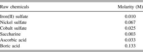
Co–Ni–Fe nanocrystalline coatings were electrodeposited onto mild steel (AISI 1018 Steel). Mild steel substrate was cut into dimensions of 76 mm × 25 mm × 6 mm from the mild steel bar. The substrate was cleaned from the oxide layers using a 120 grit flat disk. It was then polished using emery papers of 240 and 320 grit sand paper flat disk, followed by mirror polished using a cloth polishing wheel machine. Mild steel and platinum were used as cathode and anode, respectively in the electrodeposition process. Co–Ni–Fe nano-coatings were prepared by electrodeposition from a sulfate bath. The electrolyte bath was a mixture of CoSO4, NiSO4, FeSO4, H3BO3, and saccharin using required amount of mole as shown in Table I. The chemical composition of Co–Ni–Fe electrolyte bath was prepared using optimum composition obtained from previous work.Reference Resali, Hyie, Berhan, Salleh and Kasolang7 Cobalt sulfate, nickel sulfate, and iron sulfate were used as a source of soluble metal ions to produce a coating layer on the anodic surface. As for boric acid, it was added to regulate the pH value from abrupt fluctuation. On the other hand, saccharin was performed as a grain refining agent to reduce the tension and compression stress deposit besides producing a smaller grain size.
Chemical composition for electrolyte preparation.

The pH of electrolyte solution was fixed at about 3, whereas the working temperature was set at about 50 °C. Coating used in conventional bath at high pH may induce significant hydrogen evolution at the cathode surface. Hydrogen evolution is not favorable in the coating synthesis as it can result in void formation.Reference Zhang and Ivey8 Voids can degenerate coating uniformity and thus affect the performance of coating in mechanical, wear, and corrosion. Therefore, the development of stable baths with relatively low pH is practical. The deposition was performed at a range of current densities from 0.01796 to 0.001995, at different deposition times of 15, 30, and 45 min. After electrodeposition completed, the coated samples were removed from the solution immediately and then thoroughly rinsed with distilled water. The wet samples were dried overnight for further analysis.
B. Characterization of Co–Ni–Fe nanoparticles coated on mild steel
The phase formation of nanoparticles was characterized via ULTIMA IV FD 3668N, x-ray diffraction (XRD; Rigaku Corporation, Tokyo, Japan) analysis. The samples were measured using a Cu target. The scan region (2θ) was ranged from 30° to 100° at a scan rate of 4°/min. The morphology of the nanoparticles was analyzed using Quanta 450 FEG, field emission scanning electron microscope (FESEM; FEI Corporation, Tokyo, Japan). FESEM images were taken using electron beam energy of 10 kV. The elemental composition of the nanocrystalline coating was detected using a JEOL JSM-6701 model energy dispersed x-ray spectrometry machine (JEOL Ltd., Tokyo, Japan). The thickness of coating was measured by FESEM.
C. Hardness and surface roughness measurement
The micro hardness of the sample was measured by a MITUTOYO MVK-H1 (Mitutoyo, Tokyo, Japan), Vickers Micro hardness technique by applying 1000 g load. The final micro hardness values quoted were an average of five measurements performed on different locations in the sample. A standard specimen (uncoated mild steel) was prepared to compare the improvement of hardness due to Co–Ni–Fe nanocrystalline coating.
A surface measuring instrument (Mitutoyo SURFTEST SV-600) was used to obtain the surface roughness of Co–Ni–Fe nanoparticle deposit on the samples. The average values were taken from three measurements.
D. Slurry erosion wear test
Wear tests were conducted by using a slurry erosion tester machine (Model TR-40, Ducom, Bangalore, India) inconformity with ASTM G40 Standard. This is done by immersing the samples in slurry of the desired sand composition and concentration and rotating the samples (rectangular shape) for certain speed and operation time. As a result, erosion of the samples and loss of weight can be determined. The standard dimension of specimen needed for this test is 76 mm × 25 mm × 6 mm. Rotation speed of 300 and 1200 rpm were applied and individually operated for constant 12 h. Mining sands having an average particle size of 710 µm were used in the slurry erosion test. About 60 wt% of sands have been utilized throughout the testing. This type of wear test was chosen due to the application of Co–Ni–Fe nanoparticles usually involved in corrosive environment such as marine.
E. Salt fog test
Salt fog test is an accelerated corrosion test that replicates the condition of corrosive attack to the surface of coated samples to verify the capability of the coating produced as a protective finish even under corrosive environment. The testing was conducted according to ASTM B117. The samples were exposed to a salt spray generated from a 5% sodium chloride solution with a pH between 6.5 and 7.2. All the samples were placed in the salt spray chamber for 24 h. Surface micrographs of the corroded samples were taken using a camera. A mild steel without coating was tested for comparison purpose.
III. RESULTS AND DISCUSSION
A. Characterization of Co–Ni–Fe nanocoating electrodeposited on mild steel
Co–Ni–Fe nanoparticle deposits were seen to have similar XRD patterns regardless the effect of deposition time. It can be seen in Fig. 1 that all Co–Ni–Fe nanocrystalline coatings show the same crystallographic structure. The nanocrystalline coating was proven to have a mixture phase of body centered cubic (bcc) and face centered cubic (fcc). The characteristic peaks correspond to fcc(111), bcc(200), and fcc(220) planes at 2θ of 44.3°, 51.3°, and 92.4°, respectively. The peaks were identified with JCPDS No. 10745694 and JCPDS No. 3-1208. The peaks show that the coating is crystalline but not amorphous. No other contamination phase appeared in the coating.
XRD patterns of Co–Ni–Fe nanocoating deposited mild steel at different deposition times.

It is known that Co–Ni–Fe alloy existed in three distinct phases namely ε (hexagonal closed-packed, HCP), γ (fcc), and α (bcc). The phase transformation was affected by pH bath as reported in Ref. Reference Nakahara and Mahajan9 on pure cobalt nanoparticles. The fcc phase was produced in a solution of pH less than 3 due to the simultaneous incorporation of hydrogen. The phase was transformed from fcc to HCP at pH more than 3 as a result of the out-diffusion process of hydrogen.Reference Nakahara and Mahajan9 However, Sundaram and other researchers10 found a mixture phase of bcc + fcc in their fabrication of Co–Ni–Fe coatings at pH less than 3.5. On the other hand, other researchers reported the effect of pH bath on the Co–Ni–Fe nanocoating deposited on stainless steel.Reference Sundaram, Dhanasekaran and Mahalingam10 It is found that only the nanocoating prepared at pH 7 showed a single fcc structure. The Co–Ni–Fe nanocoating deposited at pH 3 and 9 formed fcc + bcc dual phase. In this study, there is no the effect of deposition time on phase transformation on Co–Ni–Fe nanocoating deposits. The coating was prepared at about a pH of 3, hence the coating exhibited the fcc and bcc mixture phase.
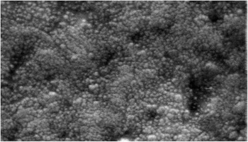
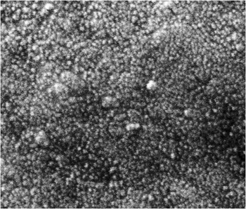
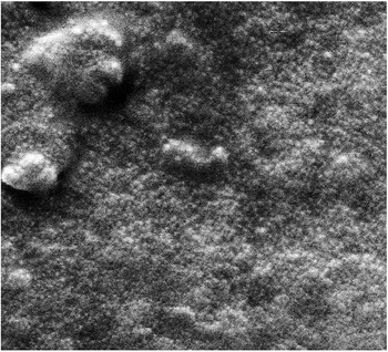
Figures 2–4 illustrate typical FESEM images of CoNiFe nanocrystalline coating coated sample at three different deposition times. Although all the samples show the same spherical morphologies, the uniformity and homogeneous were differently affected by time deposition. Voids existed on the sample coated at 15 min as shown in Fig. 2. This may be due to the insufficient coating time to fully coat the surface of the substrate. As a result, the uncoated surface led to the void formation. The Co–Ni–Fe nanocrystalline coatings coated at 30 min time deposition revealed a uniform and smooth coating. This is due to the orderly arrangement of atoms in sufficient deposition time. As for the Co–Ni–Fe nanocrystalline coatings coated at 45 min time deposition, some agglomerates were visible in Fig. 4. Since 30 min time is sufficient to fully coat the substrate, the extra time for the electrodeposition may cause excess nanoparticles deposited on the surface of coating. The particles were intended to combine and coalesce with each other to form agglomeration. The excess deposits may also produce thicker coating.
FESEM image of Co–Ni–Fe nanocoating deposited mild steel at 15 min deposition time.

FESEM image of Co–Ni–Fe nanocoating deposited mild steel at 30 min deposition time.

FESEM image of Co–Ni–Fe nanocoating deposited mild steel at 90 min deposition time.

It was observed that all the particles sizes of the nanocrystalline coating were less than 100 nm excludes the agglomerates. However, the particle size was difficult to be measured due to poor image condition. Overall, the coating is still considered uniform and homogeneity of particles can be viewed in Figs. 3 and 4. This evidently shows that electrodeposition can produce uniform and pore free coating by controlling the process parameters.
Longer deposition time allowed more Co–Ni–Fe nanocrystalline coatings deposited on the mild steel substrate and resulted in higher thickness of the coating.Reference Resali, Hyie, Abdullah, Ghani and Kalam11 One problem of the coating is the adhesive between coating materials and substrate. If the adhesive is poor among them, the mechanical, wear, and corrosion properties of the coated mild steel will be definitely affected. Thus, further analysis on the adhesive contact between substrate and Co–Ni–Fe nanocrystalline coatings was observed by FESEM.
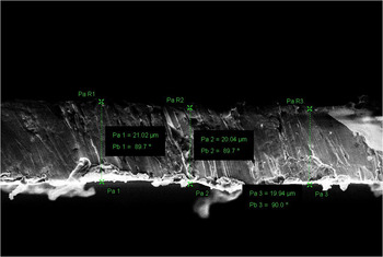
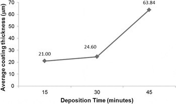
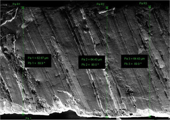
The coating thickness was measured at the cross section of coated samples. Figure 5 shows the example of how the thickness was measured in the coated sample prepared at 15 min time deposition. In Fig. 5, a good adhesion between substrate and coating materials was observed. The average coating thickness measured for all three samples deposited at different times is plotted in Fig. 6. It is observed that the thickness was increased by increasing the deposition time. The thickness of coating was not much different (about 17%) when compared between the deposits at 15 and 30 min. This is because the coating was not fully coated and voids were visible at the substrate prepared at 15 min time deposition. This was confirmed by the FESEM images as shown in Fig. 2. By extending the time, the voids were filled with electrodeposited Co–Ni–Fe until the whole substrate was fully covered with the nanocrystalline coatings. At 30 min, the coating was fully electrodeposited. After that, the excess particles started to combine and stack with each other to create layer coating if the deposition time was prolonged. The thickness was increased almost 72% for coating prepared at 45 min if compared to samples deposited at 30 min. The multiple stacking layers of deposition were believed to form in the sample due to agglomeration. Anyway, the adhesive was observed in good condition without cracking as shown in Fig. 7. Hence, 30 min time deposition is considered sufficient for complete uniform electrodeposition of Co–Ni–Fe nanocrystalline coatings on the mild steel.
FESEM image of cross section of Co–Ni–Fe nanocoating deposited mild steel at 15 min deposition time.

Average coating thickness as a function of deposition time of Co–Ni–Fe nanocrystalline coatings coated mild steel.

FESEM image of cross section of Co–Ni–Fe nanocoating deposited mild steel at 45 min deposition time.

B. Hardness and surface roughness of Co–Ni–Fe nanocrystalline electrodeposited on mild steel
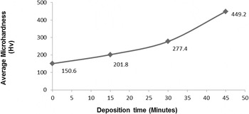
The microhardness of the sample was measured with average 5 measurements for each nanoparticle coating samples by using a Vickers Micro hardness tester. The application of Co–Ni–Fe nanocoating was proven to improve the hardness of mild steel, as seen in Fig. 8. The micro hardness of Co–Ni–Fe nanocrystalline coating coated for 45 min deposition was found to be the highest whereas 15 min coating sample shows the lowest hardness. The factor affecting the hardness may be attributed to grain size effect, phase presence, effect of porosity, adhesion, and solid solution hardening mechanism.Reference Portinha, Teixeire, Carneiro, Dup, Shmegera and Tavares12 Nano grain size has resulted in high volume fraction of the boundary atoms inside the grain boundaries.Reference Marita and Yaacob3 The dislocation motion was blocked by these grain boundaries and resulted in hardness improvement with the application of nanocoating. The presence of porosity found in the coating deposited at 15 min was the main reason contributed to low microhardness. Uniformity over the surface of the coating could also increase the hardness of the sample. The compact and full spherical structure resulted in the reduced quantity of void and agglomerate formation consequently improved the surface morphology and hardness of the Co–Ni–Fe nanoparticle sample. Due to the drastic increase of thickness in the sample electrodeposited at 45 min, the resistance of the materials to the force was increasing provided the adhesive coating is continuous. Previous workReference Hyie, Resali and Abdullah13 confirmed that the Co–Ni–Fe nanocoating was harder than Co–Ni and pure Co nanocoating due to the presence of bcc and fcc dual phase.
Average microhardness of Co–Ni–Fe nanocoated mild steel prepared at different deposition times compared to uncoated mild steel.

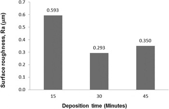
Figure 9 shows the relationship of surface roughness, Ra with respect to different coating deposition times. Lower surface roughness is preferable for better surface finishing of working parts. It was found that the surface roughness varied for all samples. The lowest surface roughness of 0.293 μm was recorded from 30 min coating sample while the highest surface roughness of 0.593 μm was found in 15 min coating sample. The decrement of surface roughness at 30 min deposition time may be due to the uniform particle formation with suitable particle size and dense population as a result of the rise in the nucleation density.Reference Lee and So14 Based on the FESEM morphologies, the surface roughness value might differ due to the void and agglomeration that existed in the 15 and 45 min coating samples, respectively. The existence of void on the coating sample prepared at 15 min time deposition exhibited higher surface roughness compared to the agglomeration that formed in the 45 min coating sample. It was noted that the agglomerated particle is one of the reason for high roughness of deposits. These particles were combined and coalesced with each other to form agglomerates and bigger particles.Reference Chinnasamy, Narayanasamy, Ponpandian and Chattopadhyay15
Surface roughness of Co–Ni–Fe nanocrystalline coating electrodeposited on the mild steel at different times.

Although both hardness and coating thickness were increased when the deposition time was longer. The resultant surface roughness did not express the same trend. In fact, there is no direct relation between coating thickness, hardness, and surface roughness of the coating. Previous study proved that the hardness of coating was independent of the coating thickness and interfacial roughness.Reference Kato, Nazul, Itti, Akebono, Sugeta and Mitani16
C. Slurry erosion wear and corrosion resistance of Co–Ni–Fe nanocrytalline electrodeposited on mild steel
Co–Ni–Fe nanocoating is a potential material to be used in marine application due to their corrosion resistance. Slurry erosion condition may become an important issue to be encountered when this coating was in contact with aqueous solutions containing hard particles that impact against the surface coating. Due to this reason, this testing was carried out to study the slurry erosion wear performance of Co–Ni–Fe nanocrytalline electrodeposited on mild steel.
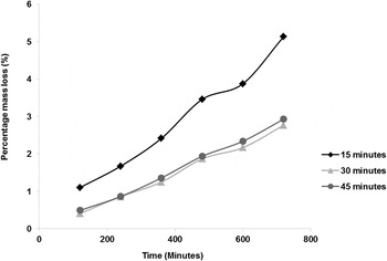
Figures 10 and 11 represent the results of progressive mass loss obtained from the wear test at 300 and 1200 rpm. The erosion wear rate (mass loss) was increased with respect to operation time.Reference Satish Kumar and Mohapatra17 The coating samples prepared at 30 min time deposition seems to have the best erosion wear resistance as the sample produced the least mass loss. The result was further supported by previous work.Reference Siu and Li19 The effect of the coating deposition time can be seen clearly at higher rotation speed (1200 rpm) compared to the lower rotation speed (300 rpm). At 1200 rpm, the material removal is excessive due to higher rpm.Reference Patel and Sejkar18 A wear map philosophy has been created by Siu and Li.Reference Siu and Li19 It is stated that the effect of surface roughness is not significant when Ra is below 0.1 mm. However, in this case, the wear erosion of Co–Ni–Fe nanocoated mild steel was significantly related to the surface roughness. The lowest surface roughness produced the lowest progressive percentage of mass loss.
Mass loss (percentage) after low speed slurry erosion test.

Mass loss (percentage) after high speed slurry erosion test.

Figure 12 shows the samples after the salt fog test. In this experiment, salt fog test had been conducted for 24 h operation time to observe the effect of the corrosion on the materials and surface coatings. From the observation, the coating sample prepared at 15 min time deposition shows several corrosion spots on the surface. The corrosion reaction took place particularly at voids area in the sample. On the other hand, the coating samples prepared at 30 min time deposition show less corrosion whereas the coating sample having the highest thickness was not corroded. The purpose of this testing was to verify the function of Co–Ni–Fe nanocrystalline coatings as a corrosion resistance coating. It is shown that Co–Ni–Fe nanocrystalline coating was successfully used to minimize the corrosion of mild steel when compared to corroded mild steel without coating.
The physical appearance of CoNiFe coating samples after salt fog test compared to uncoated mild steel (a) 15 min time deposition (b) 30 min time deposition (c) 45 min time deposition (d) uncoated mild steel.

IV. CONCLUSIONS
The paper focused on the effect of deposition time on the physical, wear, and corrosion properties of Co–Ni–Fe nanocrystalline coating coated on the mild steel. It is concluded that the deposition time played an important role to determine the phase formation, homogeneity of coating and coating thickness which may directly influence the performance of Co–Ni–Fe nanocrystalline coating in terms of hardness, surface roughness, slurry erosion wear, and corrosion protection.
30 min of deposition time is sufficient to produce a full coating of Co–Ni–Fe nanocrystalline coating. The existence of agglomeration and void could deteriorate the quality of surface coating. Anyway, the spherical shape with uniform nanocrystalline coating distribution can only be achieved within 30 min deposition time. FESEM showed the evidence of good adhesion between Co–Ni–Fe nanocrystalline coating and mild steel substrate can be obtained using electrodeposition method. The thickness of coating is in range of 20–65 μm. The coating became thicker with increasing deposition time. Likewise, the adhesion was still in good condition after 90 min time deposition.
XRD analysis indicated that the Co–Ni–Fe nanoparticles have fcc and bcc crystallite structure. This dual phase formation was believed to enhance the adhesive coating and resulted in better hardness if compared to uncoated mild steel.
Surface hardness of the coated mild steel was improved after being coated with Co–Ni–Fe nanocrystalline coating. The increment of the deposition time increased the surface hardness of the coated mild steel. The lowest surface roughness value, Ra recorded was 0.293 μm in the coated mild steel prepared at 30 min time deposition. On the other hand, less coating mass loss was produced from the lower surface roughness of the coated sample during slurry erosion wear test. The existence of void on coating surface showed the higher mass loss compared to the agglomerate coated surface. Furthermore, higher rotation speed or velocity applied caused the progressive increment of coating mass loss. This may reduce the effectiveness of coating as a protection layer to the erosion wear and increase the possibilities of coated mild steel being rusted in shorter time.
ACKNOWLEDGMENTS
The authors would like to thank Research Management Institute (RMI) UiTM and Ministry of Higher Education Malaysia for financial supports. The research was conducted at Faculty of Mechanical Engineering, Universiti Teknologi MARA (UiTM), Malaysia under support Grant 600-RMI/ERGS 5/3 (24/2013).