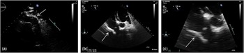
A 32-year-old man with double-outlet right ventricle associated with a 22q11 microdeletion underwent percutaneous pulmonary valve replacement with a Melody valve (Medtronic Inc., Minneapolis, Minnesota, United States of America) in January, 2014. The replacement was performed because of stenosis and regurgitation in his right ventricle-to-pulmonary artery conduit. A Hancock tube was installed upon full correction of the cardiopathy at 5 years of age, and “opened” at age 13. In August of 2014, the patient was hospitalised for suspected infective endocarditis with a fever and night chills. His blood cultures were positive for Rothia dentocariosa, and echocardiography showed dilation of the right cavities and an increase in right ventricular pressure. Images of his chest CT-scan indicated distal bilateral septic emboli, as well as in the right proximal pulmonary artery. The diagnosis of infective endocarditis was considered possible on the basis of the modified Duke criteriaReference Li, Sexton and Mick 1 – one main criterion and two minor criteria – but not certain because no vegetation could be seen on the Melody valve despite repetitive transthoracic (Fig 1a) and transoesophageal echocardiography (Fig 1b and c).
Transthoracic and transoesophageal echocardiographic images of the Melody valve. ( a ) A transthoracic echocardiographic image, parasternal short-axis view, focussed on the right ventricle-to-pulmonary artery conduit without any vegetation visualised on the prosthesis. ( b ) A transoesophageal echocardiographic image, transgastric view, showing the right ventricle-to-pulmonary artery conduit. ( c ) A closer view of the right ventricle-to-pulmonary artery conduit in transoesophageal echocardiography, transgastric view, showing a slight thickening of the Melody valve without any clear vegetation. Arrow: Melody valve.

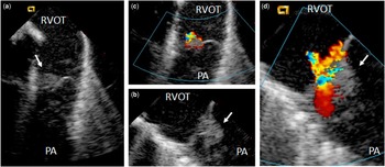
In November, 2015, persistence of increased right ventricular pressure prompted a haemodynamic study and angiography that ruled out a stent fracture type of mechanical complication. An intracardiac echocardiography was performed at the same time with the AcuNav™ 8-F catheter (Biosense Webster Inc., Diamond Bar, California, United States of America) through the right femoral vein, allowing for a posteriori confirmation of the infective endocarditis diagnosis, by revealing the presence of a sessile vegetation on one of the cusps of the Melody valve (Fig 2a and b) with moderate pulmonary regurgitation (Fig 2c), two jets (Fig 2d), and no stenosis (Supplementary videos 1a and 1b).
Intracardiac echocardiography imaging of a mobile vegetation within a percutaneously implanted bioprosthetic pulmonary valve; intracardiac echocardiographic images with the transducer placed in the right ventricular outflow tract (RVOT) ( a ). A mobile vegetation (arrow) attached to the Melody valve ( b ). A closer view of the vegetation ( c ). A pulmonary regurgitation due to the vegetation, with two jets ( d ). PA=pulmonary artery.

Infective endocarditis is a serious complication of percutaneous Melody valves, estimated to occur in 2–3.9% of patients per year,Reference Patel, Malekzadeh-Milani, Ladouceur, Iserin and Boudjemline 2 although its incidence remains uncertain due to difficulties in confirming the diagnosis.
Definitive infective endocarditis diagnosis is based on the modified Duke criteria;Reference Li, Sexton and Mick 1 however, true valve involvement may be difficult to demonstrate in the case of a Melody valve. Melody valves are made of bovine jugular vein valves that are sutured into an iridium and platinum stent. The anterior position of the right ventricular outflow tract and artefacts generated by the stent make it difficult to analyse the valve and to visualise vegetations with transthoracic and transoesophageal echocardiography.
Infective endocarditis with a Melody valve is characterised by the appearance or sudden worsening of the right ventricular outflow tract pressure gradient in the absence of a mechanical explanation; therefore, some groups consider this sudden stenosing development as a major diagnostic criterion.Reference Buber, Bergersen and Lock 3
The usefulness of intracardiac echocardiography has been shown for anatomical and functional assessment of the Melody valve immediately following its implantation or to guide the procedure.Reference Awad, Masood and Gonzalez 4 It has also been reported for the diagnosis of infective endocarditis related to pacemaker leads. With transoesophageal echocardiography as the gold standard, intracardiac echocardiography had a sensitivity and specificity of 100 and 82.8%, respectively.Reference Narducci, Pelargonio and Russo 5
Previously, two cases of infective endocarditis on a Melody valve have been confirmed by intracardiac echocardiography, reported in the literature by Cheung et al.Reference Cheung, Vejlstrup and Ihlemann 6 Intracardiac echocardiography provides high-quality images; however, it remains an invasive and arduous examination with a steep learning curve that limits its routine use. Intracardiac echocardiography uses a catheter of 8 or 10 F that is 90 cm in length, equipped at one end with a 64-phased array, high-frequency ultrasound captor allowing 90° sectorial sections, with up to 15 cm of penetration (AcuNav™). The tip of the catheter can be moved in the anterior, posterior, and lateral planes and maintained in the desired position.
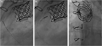
The intracardiac echocardiography catheter is introduced through femoral venous access, and then inserted with fluoroscopic monitoring up to the right atrium. It is then “bent” and pushed through the tricuspid valve in the right ventricle where a clockwise rotation allows the right ventricular outflow tract to be viewed. The tip of the intracardiac echocardiography catheter can be positioned directly under the pulmonary valve, thus providing excellent visualisation (Fig 3). A second method is to cross the pulmonary valve with the intracardiac echocardiography catheter.
Fluoroscopic images during the intracardiac echocardiography procedure. Lateral ( a and b ) and anterior–posterior ( c ) fluoroscopic projections showing the intracardiac echocardiography catheter positioned in the right ventricular outflow tract, directly under the Melody valve, allowing an excellent analysis of the prosthesis.

An increase in the right ventricular outflow tract gradient – unexplained by a mechanical complication – must be considered as a sign of pulmonary prosthetic valve involvement. This could become a major diagnostic criterion in suspected cases of infective endocarditis of a Melody valve. By being as close as possible to the cardiac structure of interest, intracardiac echocardiography provides high-resolution, artefact-free images, and allows a firm diagnosis, which confirms the existence of valve vegetations. This technique could be used at an early stage for patients with suspected prosthetic pulmonary valve infective endocarditis when transthoracic and transoesophageal echocardiography are inconclusive. This may confirm prosthesis involvement, allow for tailored medical or surgical therapy, as well as help establish the actual frequency of Melody valve endocarditis.
Acknowledgement
None.
Financial Support
This research received no specific grant from any funding agency or from commercial or not-for-profit sectors.
Conflicts of Interest
None.
Ethical Standards
Informed written consent was obtained from the patient, whose data were anonymised. The procedures were reviewed and approved by the institution’s ethics committee and comply with the Helsinki Declaration of 1975, as revised in 2008.
Supplementary Material
To view supplementary material for this article, please visit https://doi.org/10.1017/S1047951117000403