


Highlights
-
• Non-motor symptoms are common across hereditary and sporadic adult-onset cerebellar ataxia.
-
• Mood disorders and cognitive impairment are among the most prevalent non-motor features.
-
• Adult-onset cerebellar ataxia extends beyond the cerebellar motor syndrome.
Introduction
Adult-onset cerebellar ataxia (AOCA) encompasses a heterogeneous group of hereditary and sporadic neurodegenerative disorders characterized by progressive cerebellar dysfunction. The clinical phenotype typically includes impaired balance, incoordination, dysarthria and oculomotor abnormalities.Reference Kemani1 Presentation may be subacute or chronic depending on etiology, and affected patients are commonly evaluated in tertiary movement disorders clinics. Diagnostic frameworks for sporadic adult- or late-onset ataxia have evolved over the past decade, driven by advances in genetic testing and refined diagnostic criteria.Reference Wirth, Faber and Depienne2 Recent genetic discoveries, including SCA27B caused by FGF14 repeat expansions, now account for a substantial proportion of cases previously classified as sporadic adult-onset ataxia of unknown etiology (SAOA).Reference Pellerin, Danzi and Wilke3
The motor manifestations of cerebellar degeneration, collectively referred to as the cerebellar motor syndrome, are well described and typically dominate clinical assessment and research outcomes.Reference Manto4 Growing evidence, however, indicates that non-motor symptoms (NMS) – including cognitive impairment, mood disorders (depression and anxiety), sleep abnormalities, pain and autonomic symptoms – contribute substantially to disease burden in cerebellar disorders.Reference Malek, Makawita, Al-Sami, Aslanyan and de Silva5 Moreover, additional motor features beyond gait and limb ataxia, including oculomotor abnormalities, dysphagia, parkinsonism, dystonia and abnormalities of tone or reflexes, are also common and inconsistently captured in phenotypic descriptions of adult-onset ataxia.Reference Villanueva6 These features may profoundly affect quality of life, functional independence and caregiver burden, yet they remain under-recognized and inconsistently assessed in routine clinical practice.
Although NMS have been reported in individual hereditary ataxias, including specific spinocerebellar ataxias (SCAs) and selected genetically defined cohorts,Reference Malek, Makawita, Al-Sami, Aslanyan and de Silva5,Reference Lin, Kuo and Cognitive7 systematic characterization of NMS and non-ataxia motor features across the broader spectrum of AOCA remains limited. Consequently, the prevalence and distribution of NMS across diagnostic categories are poorly defined, limiting comprehensive phenotypic characterization and the development of targeted therapeutic approaches.
Movement disorders clinics are uniquely positioned to identify and longitudinally follow patients with progressive hereditary and sporadic ataxias, enabling comprehensive phenotypic characterization beyond motor features alone. In this study, we sought to characterize NMS and non-ataxia motor features in patients with genetically confirmed hereditary ataxias and SAOA at a tertiary Canadian movement disorders center.
Methods
Study design and patient identification
We conducted a retrospective observational study based on a comprehensive review of paper and electronic medical records at a tertiary movement disorders clinic. Institutional review board approval was obtained with a waiver of informed consent due to the retrospective nature of the study (REB21-0012). Clinical visits and investigations conducted between January 1, 2000, and December 31, 2020, served as primary data sources. We identified consecutive patients presenting with a primary complaint of ataxia using the International Classification of Diseases, Tenth Revision, Clinical Modification diagnostic codes.
The primary objective was to describe the prevalence and distribution of non-ataxia motor and non-motor symptoms across diagnostic subgroups of AOCA.
Inclusion criteria comprised (1) progressive cerebellar ataxia with diagnosis in adulthood (age ≥ 18 years) and (2) chronic cerebellar symptoms persisting for more than 3 months with documented clinical progression across serial assessments. Patients were included regardless of whether a genetic diagnosis was ultimately identified.
We excluded patients with acquired ataxias due to non-genetic causes, those meeting diagnostic criteria for multiple system atrophy during follow-up and those with a confirmed alternative diagnosis such as paraneoplastic or alcoholic cerebellar degeneration. Patients with incomplete diagnostic evaluations were also excluded.
Clinical evaluation and investigations
All patients underwent initial assessment by a movement disorders specialist, including a detailed medical history with emphasis on family history and ethnic background when available and comprehensive neurological examination. Brain MRI was reviewed in all patients primarily to assess for the presence of cerebellar atrophy, as this was the most consistently documented imaging feature across the cohort. Although extra-cerebellar imaging abnormalities may be relevant in specific hereditary ataxias, these findings were not uniformly or systematically reported and therefore could not be reliably analyzed retrospectively.
Laboratory investigations were performed to exclude alternative etiologies, including vitamin B12, vitamin E, thyroid-stimulating hormone, syphilis serology, anti-gliadin antibodies, tissue transglutaminase antibodies and anti-neuronal antibodies. Additional investigations were ordered at the discretion of the treating neurologist and considered diagnostically relevant if they confirmed an alternative diagnosis.
We reviewed all follow-up visits for the duration of care within the movement disorders clinic and included new symptoms or examination findings documented at any point during follow-up in the final analysis.
Genetic testing and diagnostic classification
Genetic testing was performed in a clinical setting and evolved over the study period according to availability and clinical suspicion. All patients underwent molecular genetic testing for common autosomal dominant SCAs, including ATXN1 (SCA1), ATXN2 (SCA2), ATXN3 (SCA3), CACNA1A (SCA6), ATXN7 (SCA7), ATXN8 (SCA8) and TBP (SCA17). All patients were tested for FXN mutations associated with Friedreich ataxia. FMR1 testing for fragile X-associated tremor-ataxia syndrome was performed selectively based on clinical phenotype.
Testing availability evolved over the study period. Patients initially classified as SAOA were subsequently tested for SCA17 when testing became available and reclassified accordingly. Selected patients with negative repeat expansion testing underwent commercially available next-generation sequencing ataxia panels, including genes associated with autosomal dominant SCAs, autosomal recessive ataxias, mitochondrial disorders and syndromic neurodegenerative conditions (including SETX, SYNE1, ANO10, POLG, SPG7, NPC1 and OPA1, among others). Whole-exome sequencing was not performed in this cohort. Patients in whom no pathogenic or likely pathogenic variant was identified after panel testing, and where applicable, whole-exome sequencing, were classified as SAOA. Importantly, testing for recently recognized genetic causes of adult-onset ataxia, including RFC1 repeat expansions associated with Cerebellar Ataxia, Neuropathy, and Vestibular Areflexia Syndrome (CANVAS) and FGF14-related SCA27B, was not routinely available during much of the study period. As a result, some patients classified as sporadic adult-onset ataxia may have harbored currently identifiable genetic etiologies.
Motor and non-motor symptom assessment
We extracted data from all documented clinical encounters, including initial and follow-up examinations, using a standardized data abstraction framework. NMS were identified through routine clinical documentation across longitudinal visits and were not systematically assessed using standardized instruments. Data abstraction was performed by two investigators with discrepancies resolved by consensus.
NMS were identified through review of clinical documentation, neurophysiological studies and psychiatric assessments throughout follow-up. These included pain, symptoms or objective evidence of peripheral neuropathy, unprovoked seizures or epilepsy, restless legs syndrome (RLS), rapid eye movement (REM) sleep behavior disorder (confirmed by polysomnography or suggested by clinical history), excessive daytime sleepiness, narcolepsy and insomnia. Cognitive status was assessed through review of serial mental status examinations, formal neuropsychological assessments and bedside cognitive screening tools, including the Mini-Mental Status Examination and Montreal Cognitive Assessment. Depressive and anxiety symptoms were identified through documented clinical diagnoses and routine clinical assessments recorded in the medical chart. Standardized psychiatric rating scales were not systematically used. Subjective fatigue, autonomic symptoms (e.g., orthostatic hypotension and bowel or bladder dysfunction) and visual symptoms were also recorded.
Non-ataxia motor features recorded included weakness, muscle atrophy, fasciculations, spasticity, hyperreflexia or hyporeflexia and additional movement disorders such as chorea, myoclonus, parkinsonism or tremor when deemed disproportionate to the degree of cerebellar ataxia. Peripheral neuropathy was defined by documented clinical examination findings and/or confirmatory electrodiagnostic studies or by clearly documented neuropathic symptoms (e.g., distal numbness, paresthesia or burning pain) judged by the treating neurologist to be consistent with a peripheral neuropathic process.
Oculomotor dysfunction was defined by documented complaints or examination findings of diplopia with or without dysconjugate gaze, nystagmus, saccadic hyper- or hypometria or saccadic intrusions. Dysphagia was recorded based on clinical documentation and speech–language pathology assessments when available.
Statistical analysis
We performed statistical analysis using Stata version 16. Categorical variables are presented as frequencies and percentages and continuous variables as means ± standard deviations. No formal sample size calculation was performed due to the retrospective and exploratory design. Distribution normality was assessed using visual inspection and Shapiro–Wilk testing. We compared groups using chi-square tests for categorical variables and Wilcoxon rank-sum or Kruskal–Wallis tests for non-normally distributed continuous variables. Pearson correlation coefficients were calculated to assess associations between NMS and demographic or clinical features. Correlation analyses were explored with stratification by sex and inheritance pattern. Analyses were conducted using available-case data. Given the exploratory nature of the study, no correction for multiple comparisons was applied. Statistical significance was set at p < 0.05 (two-sided).
Results
Cohort characteristics
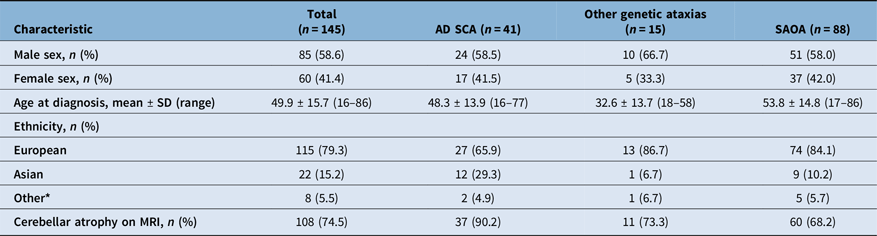
We identified 145 patients with AOCA (Table 1). The cohort included 85 males (58.6%) and 60 females (41.4%) with a mean age at diagnosis of 49.9 ± 15.7 years (range: 16–86). Two patients reported symptom onset at ages 16 and 17, respectively, but were diagnosed and entered longitudinal follow-up in adulthood. Most patients were of European ancestry (79.3%), followed by those of Asian ancestry (15.2%), with other or mixed ethnicities accounting for 5.5%.
Clinical characteristics and demographics by diagnostic group

Table 1. Long description
Clinical characteristics are demographics are categorized in rows and include male sex, female sex, age at diagnosis, ethnicity (european, asian, other), and cerebellar atrophy on MRI. The rows are diagnostic groups, including Autosomal Dominant Spinocerebellar Ataxia, other genetic ataxias, and Sporadic Adult-Onset Ataxia of Unknown Etiology.
AD = autosomal dominant; SCA = spinocerebellar ataxia; SAOA = sporadic adult-onset ataxia of unknown etiology; SD = standard deviation.
One X-linked case (fragile X–associated tremor/ataxia syndrome) was excluded from subgroup comparisons due to small sample size. Percentages are calculated within each diagnostic subgroup.
*“Other” ethnicity includes African, Portuguese and mixed ethnicities. MRI percentages reflect the proportion of patients within each subgroup with documented cerebellar atrophy.
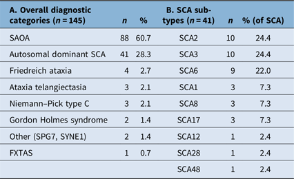
Diagnostic categories included SAOA in 60.7% (n = 88), autosomal dominant SCAs in 28.3% (n = 41) and other genetic ataxias in 10.3% (n = 15). Diagnoses in the other genetic ataxias category included Friedreich’s ataxia (n = 4), ataxia-telangiectasia (n = 3), FXTAS (n = 1), Niemann–Pick type C (n = 3), SPG7 (n = 1), SYNE1 mutation (n = 1) and Gordon Holmes syndrome (n = 2) (Table 2a). The most common SCA subtypes were SCA2, SCA3 and SCA6 (Table 2b). Cerebellar atrophy on MRI was present in 108 individuals (74.5%).
Diagnostic distribution and spinocerebellar ataxia (SCA) subtypes in adult-onset cerebellar ataxia

Table 2. Long description
Overall diagnostic categories included are Sporadic Adult-Onset Ataxia of Unknown Etiology, Autosomal Dominant Spinocerebellar Ataxia, Freidreich Ataxia, Ataxia Telangiectasia, Niemann-Pick type C, Gordon Holmes syndrome, Other (SPG7, SYNE1), and Fragile X Tremor Ataxia Syndrome. In this category, the Sporadic Adult Onset Ataxia of unknown etiology groups was the largest. Within the spinocerebellar ataxia group, the subtypes are listed as SCA2, 3, 6, 1, 8, 17, 12, 28, and 48. The SCA2, 3, and 6 groups were the largest.
FXTAS = fragile X–associated tremor/ataxia syndrome; SAOA = sporadic adult-onset ataxia of unknown etiology; SPG7 = Spastic Paraplegia 7; SYNE1 = Spectrin Repeat Containing Nuclear Envelope Protein 1. Percentages in Column A are calculated using the total cohort (n = 145). Percentages in Column B are calculated using the SCA subgroup (n = 41).
Non-motor symptoms
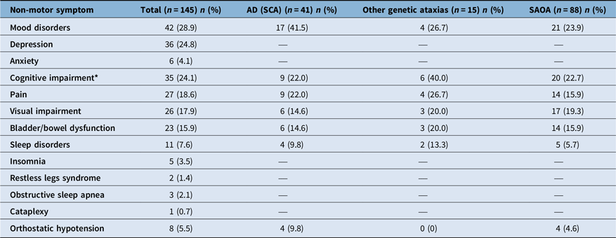
NMS were common across all diagnostic groups (Table 3). The most frequently documented NMS were mood disorders (28.9%), cognitive impairment (24.1%) and chronic pain (18.6%). Visual disturbances (17.9%), bladder or bowel dysfunction (15.9%) and sleep-related symptoms (7.6%) were also observed.
Non-motor symptoms overall and by diagnostic group

Table 3. Long description
The non motor symptoms catergorized in the header row are mood disroders, depression, anxiety, cognitive impairment, pain, visual impairment, bladder/bowel dysfunction, sleep disroders, insomnia, restless legs syndrome, obstructive sleep apnea, cataplexy,and orthostaic hypotension. Across all categories, mood disorders were most common overall, with cognitive impairment, pain and visual impairment following closely.
AD = autosomal dominant; SCA = spinocerebellar ataxia; SAOA = sporadic adult-onset ataxia of unknown etiology.
Percentages are calculated within each diagnostic subgroup.
One X-linked case was excluded from subgroup comparisons due to small sample size.
*Cognitive assessments were only formally performed in 23 patients (15.9% of total cohort).
Among mood disorders, depression (24.8%) was more common than anxiety (4.1%). Formal cognitive testing was performed in 23 patients (15.9% of the cohort).
Among sleep-related symptoms, insomnia (3.5%) and obstructive sleep apnea (2.1%) were most frequently reported, with fewer patients reporting restless legs symptoms (1.4%).
Non-ataxia motor and other neurological features
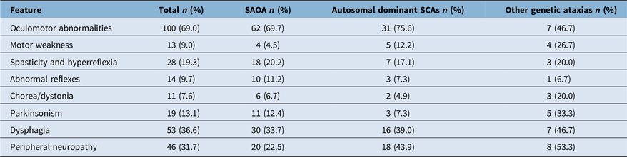
Motor features beyond gait and limb ataxia are summarized in Table 4. Oculomotor abnormalities were the most frequent finding, present in 68.9% of patients. Dysphagia was documented in 36.6% of patients. Additional non-ataxia motor features included spasticity and hyperreflexia (19.3%), parkinsonism (13.1%), hyporeflexia (9.7%), weakness (9.0%) and chorea or dystonia (7.6%). Peripheral neuropathy was reported in 31.7% of patients based on clinical and/or electrodiagnostic assessment. Epilepsy was present in 4.9% of cases.
Non-ataxia motor features overall and by diagnostic group

Table 4. Long description
The non ataxia motor symtpoms in the header row are oculomotor abnormalities, motor weakness, spasticity/hyperreflexia, abnormal reflexes, chorea/dystonia, parkinsonism, dysphagia, and peripheral neuropathy. Oculomotor abnormalities were most common at 69 percent, followed by motor weakness and hyperreflexia.
SCA = spinocerebellar ataxia; SAOA = sporadic adult-onset ataxia of unknown etiology.
Percentages are calculated within each diagnostic subgroup.
Exploratory comparison by diagnostic classification
Rates of NMS by diagnostic subgroup are shown in Table 3. Given the evolving genetic classification of adult-onset ataxia, subgroup analyses were considered exploratory. Depression and anxiety were significantly more frequent in autosomal dominant SCAs compared to SAOA (41.5% vs. 23.9%, p = 0.03). Other NMS, including pain, bladder or bowel dysfunction and cognitive impairment, did not differ significantly between these groups.
No statistically significant differences in overall NMS frequency were observed between autosomal dominant and autosomal recessive ataxias. However, nonsignificant trends toward higher rates of cognitive impairment (40.0% vs. 21.9%, p = 0.18) and pain (26.7% vs. 22.0%, p = 0.71) were observed in autosomal recessive ataxias.
Among autosomal dominant SCAs (n = 41), mood disorders were present in 41.5%, cognitive impairment in 22.0% and pain in 22.0% of patients. Subtype-level analyses were descriptive given small sample sizes, but higher rates of mood disorders were observed in SCA2, SCA1 and SCA17, while pain was most frequent in SCA3 (Supplemental Table S1).
Correlation analyses
An inverse correlation was observed between age at diagnosis and the presence of depression or anxiety (r = –0.22, p = 0.0066). This association remained significant after stratification by sex and inheritance pattern.
A positive correlation was observed between sleep-related symptoms and depression/anxiety (r = 0.30, p = 0.002).
Discussion
While AOCA is most often characterized by deficits in motor coordination and balance, our findings demonstrate that hereditary and sporadic forms frequently involve substantial non-motor and non-ataxia motor manifestations. Although some of these features implicate structures and pathways outside the cerebellum, accumulating evidence suggests that NMS may also arise from cerebellar dysfunction itself. In this study, we characterized NMS as a primary focus, together with non-ataxia motor features, in a clinically heterogeneous AOCA cohort encompassing autosomal dominant SCAs, autosomal recessive conditions, an X-linked disorder and SAOAs.
The high frequency of NMS across diagnostic categories reinforces the concept of AOCA as a multisystem network disorder rather than a purely cerebellar motor syndrome.
Mood disorders and cognitive impairment were among the most frequently identified NMS in this cohort, consistent with prior reports in AOCA. Depression and anxiety were the most documented mood disorders; however, neuropsychiatric manifestations in ataxia syndromes are broader and may include apathy, agitation and psychosis.Reference Karamazovova, Matuskova, Ismail and Vyhnalek8 A systematic review of neuropsychiatric symptoms of SCAs and Friedreich ataxia reported depression prevalence ranging from 17–26% in SCAs and 36–51% in Friedreich ataxia, with SCA3 being the most commonly affected subtype.Reference Karamazovova, Matuskova, Ismail and Vyhnalek8 The prevalence of depression observed in our cohort (24.8%) aligns with these estimates. Although mood disorders may partially reflect the psychological impact of progressive neurological disease, prior studies demonstrate that the relationship between depression and motor severity is not linear, supporting a neurobiological contribution beyond reactive psychological distress.Reference Fancellu, Paridi and Tomasello9,Reference Lo, Figueroa and Pulst10 Current pathophysiological models implicate cerebellar vermis involvement and disrupted connectivity between the posterior vermis and limbic regions, including the cingulate cortex.Reference Villanueva6
We observed an inverse correlation between age at diagnosis and the presence of depression or anxiety. This suggests that earlier-onset disease may be associated with greater affective burden, independent of inheritance pattern. This is in contrast to prior studies that showed no association between mood disorders and repeat length in SCAs or Friedreich’s ataxia, despite the established relationship between repeat length and earlier disease onset.Reference Karamazovova, Matuskova, Ismail and Vyhnalek8 The broader composition of our cohort may partly account for this discrepancy. It is also possible that older-onset patients had under-recognition of affective symptoms, particularly in the context of cognitive decline, although this could not be directly assessed.Reference Moriarty, Cook, Hunt, Adams, Cipolotti and Giunti11 Cognitive impairment was documented in 24.1% of patients. Given that formal neuropsychological testing was performed in only a minority of cases, this likely represents a conservative estimate. Cognitive deficits in cerebellar ataxia are thought to arise from disruption of cerebellar-cortical networks, particularly those between the posterior cerebellar lobes and association cortices. The resulting constellation of deficits has been termed the cerebellar cognitive affective syndrome, characterized by impairments in executive function, visuospatial processing and language.Reference Argyropoulos, van Dun and Adamaszek12 Our findings are consistent with involvement of cerebellar-cortical networks implicated in cognitive and affective processing across both hereditary and sporadic adult-onset ataxias. Pain was the third most frequently identified NMS (18.6%). Pain in cerebellar ataxia may arise from spasticity, musculoskeletal strain or coexisting peripheral neuropathy. Certain ataxia subtypes may be associated with scoliosis or headache syndromes, further contributing to pain burden. Despite this, pain remains under-characterized as a core feature of AOCA. Recognition of pain as a prevalent and potentially modifiable symptom has important implications for supportive management. Sleep disturbances, visual impairment and bladder or bowel dysfunction were also frequently observed. Although insomnia, RLS and obstructive sleep apnea were documented, sleep symptoms were likely substantially underestimated in this retrospective cohort.Reference Malek, Al-Sami, Aslanyan and Desilva13 Prior studies report substantially higher prevalence of REM sleep behavior disorder (up to 55%) and RLS (20–30%) in specific ataxia subtypes, particularly SCA3.Reference Iranzo, Muñoz, Santamaria, Vilaseca, Milà and Tolosa14,Reference Abele, Bürk, Laccone, Dichgans and Klockgether15 The relatively low prevalence observed here for some symptoms likely reflects reliance on routine clinical documentation rather than systematic screening.
Non-ataxia motor features were common. Oculomotor abnormalities and dysphagia were frequent findings as expected, forming part of the broader cerebellar syndrome.
Parkinsonism was present in over 13% of patients, supporting involvement of basal ganglia–cerebellar network interactions. It has been described in association with several forms of AOCA, including SCAs, FXTAS and DNA polymerase gamma catalytic subunit (POLG)-related disorders,Reference Manto4 and in some cases, may precede the onset of ataxia.Reference Lin, Kuo and Cognitive7 Some reports suggest that an akinetic-rigid phenotype represents the most common movement disorder seen in SCAs and POLG disorders as a whole.Reference Karamazovova, Matuskova, Ismail and Vyhnalek8,Reference Fancellu, Paridi and Tomasello9 Spasticity and hyperreflexia likely reflect associated corticospinal tract involvement. On the other hand, hyporeflexia may represent a combination of primary cerebellar dysfunction and coexisting peripheral neuropathic processes. Overall, these findings highlight that motor phenotyping in AOCA extends beyond gait and limb ataxia and requires systematic examination.
This study has several strengths. First, it includes a relatively large, clinically well-characterized cohort of AOCA evaluated within a single tertiary movement disorders center over two decades. Second, the inclusion of both genetically confirmed hereditary ataxias and SAOA allows for comparison across a broad and clinically representative disease spectrum. Third, the systematic review of longitudinal clinical documentation enabled capture of both motor and non-motor features beyond initial presentation, reflecting real-world disease evolution. Finally, the focus on NMS alongside non-ataxia motor manifestations provides a more comprehensive phenotypic description than many prior subtype-specific studies.
Several limitations merit consideration. First, because symptom ascertainment relied on routine clinical documentation rather than standardized non-motor assessments, the prevalence of symptoms not systematically screened in routine care, particularly cognitive, psychiatric and sleep-related symptoms, was likely underestimated. Second, formal cognitive and psychiatric assessments were not uniformly performed, potentially contributing to underestimation of these symptoms. A third important limitation relates to the evolution of genetic diagnostics over the study period. Since completion of data collection, several common genetic causes of adult-onset ataxia have been identified, including RFC1 repeat expansions associated with CANVAS and FGF14-related SCA27B.Reference Pellerin, Danzi and Wilke3,Reference Rafehi, Read and Szmulewicz16 A proportion of patients classified as sporadic adult-onset ataxia in this cohort may therefore carry variants that were not identifiable at the time of evaluation. This limitation may have influenced subgroup classification and reduces the ability to draw genotype-specific conclusions, particularly in comparisons involving the SAOA group.
Taken together, our findings should therefore be interpreted as reflecting the broader clinical spectrum of AOCA rather than genotype-specific phenotypes.
In addition, the heterogeneous mix of etiologies enhances generalizability but may obscure genotype-specific patterns and limit statistical power for subgroup analyses. This is particularly true as it relates to the subgroup analysis of the “other genetic ataxias” group, which is highly heterogeneous. Small sample sizes within individual genetic subtypes further restrict the detection of subtype-specific differences. As a tertiary referral cohort, findings may reflect referral bias and greater disease severity. Finally, the absence of standardized severity scales and validated non-motor instruments limits precise quantification of symptom burden and its relationship to motor severity.
Conclusion
AOCA encompasses heterogeneous disorders that extend beyond the cerebellar motor syndrome. In this cohort, NMS were common across both hereditary and sporadic forms, with mood disorders and cognitive impairment among the most frequent features. These findings support routine assessment and multidisciplinary management of NMS, particularly in younger-onset patients. They also support the importance of using validated assessment tools for future prospective work. Greater recognition of non-motor and non-ataxic motor burden may inform symptom-directed management and improve comprehensive clinical care.
Supplementary material
The supplementary material for this article can be found at https://doi.org/10.1017/cjn.2026.10653.
Acknowledgments
We sincerely thank the physicians of the Movement Disorders Program for their dedicated clinical care of the patients included in this study.
Author contributions
DA collected the data, wrote the original draft of the introduction and methods of the manuscript and reviewed/edited the final manuscript. EW compiled information the introduction, methods, original data and statistical analysis to prepare the original complete draft of the manuscript. VK provided oversight for the research planning and execution and reviewed/edited the manuscript. VB provided oversight for the research planning and execution, completed the statistical analysis of the raw data and reviewed/edited the manuscript. SF provided oversight for the research planning and execution and reviewed/edited the manuscript.
Funding statement
This research received no specific grant from any funding agency in the public, commercial or not-for-profit sectors.
Competing interests
DA, EW, SF and VK declare that they have no known competing financial interests or personal relationships that could have appeared to influence the work reported in this paper. In the last 3 years, VB has received research grants from Parkinson Canada, Parkinson Foundation and Calgary Health Foundation. None of these have been paid to her personally and were paid to specific institutions (Alberta Health Services, Allergan and Paladin Labs, respectively). She has also received honoraria from the International Parkinson and Movement Disorders Society for educational activities.


 Open access


Target article
The Spectrum of Motor and Non-Motor Symptoms of Adult-Onset Genetic and Sporadic Cerebellar Ataxia
Related commentaries (2)
Reviewer Comment on Abo AlSamh et al. “The Spectrum of Motor and Non-Motor Symptoms of Adult-Onset Genetic and Sporadic Cerebellar Ataxia”
Reviewer Comment on Abo AlSamh et al. “The Spectrum of Motor and Non-Motor Symptoms of Adult-Onset Genetic and Sporadic Cerebellar Ataxia”