Introduction
Far-advanced otosclerosis as a clinical diagnosis of otosclerosis associated with an air-conduction threshold more than 85 dB without a measurable bone-conduction threshold.Reference Marx, Lagleyre, Escudé, Demeslay, Elhadi and Deguine1 In 2011, Merkus et al.Reference Merkus, van Loon, Smit, Smits, de Cock and Hensen2 recommended the requirement of a speech reception threshold decrease. At this stage, management of the patients includes stapedotomy and an efficient hearing aid or cochlear implantation.Reference Merkus, van Loon, Smit, Smits, de Cock and Hensen2 When sensorineural hearing loss (SNHL) is profound with an aided word recognition score less than 50 per cent at 60 dB, the patient is a clinical candidate for cochlear. Changes in the cochlear structure during surgery present challenges to the surgeon. These range from round window obliteration to obliteration of the cochlear lumen or loss of scalar definition.Reference Sainz, Garcia-Valdecasas and Ballesteros3 The aim of our study is to report on surgical difficulties encountered in patients with advanced otosclerosis of various stages. Their audiological outcomes were also compared to a matching group of non-otosclerosis patients.
Patients and methods
In this retrospective study, we included 17 patients with bilateral profound SNHL due to otosclerosis. All patients underwent cochlear implantation in the period from January 2020 until January 2023. Patients were selected according to clinical history of progressive hearing loss diagnosed as otosclerosis, normal otoscopic examination, severe to profound hearing loss in audiometry and typical computed tomography (CT) findings of inner ear affected by otosclerosis. Patients with previous stapes surgery were included. We compared this group with a group of 17 non-otosclerotic post-lingually deaf patients. Both groups were matched for age at implantation, sex and duration of hearing deprivation.
The study was done in accordance with the ethical standards the 1964 Helsinki declaration and its later amendments or comparable ethical standards. This study was approved by the Institutional Review board of Ain Shams University, Faculty of Medicine (FMASU R185/2023).
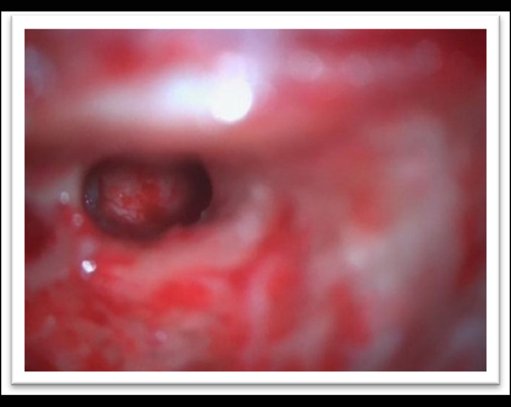
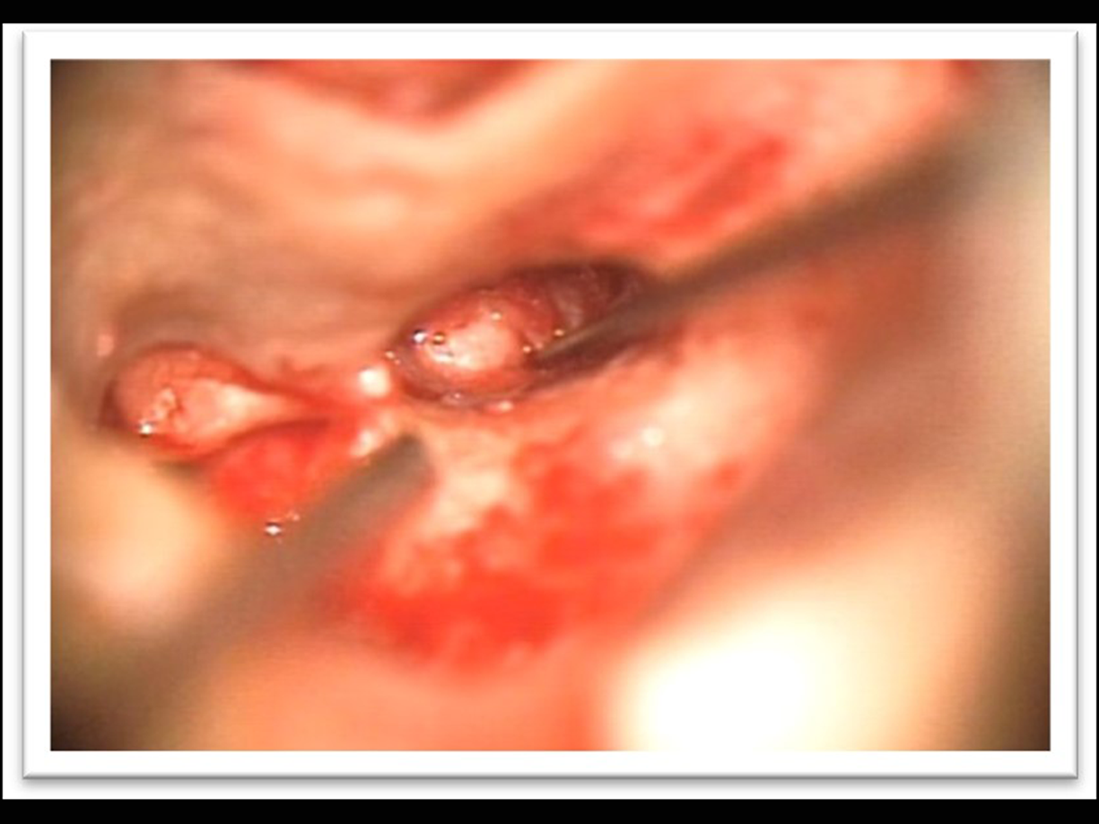
Surgical technique (Figures 1, 2)
Two experienced consultants from our team performed all the operations under general anaesthesia, using standard facial recess approach in all cases with a round window or extended round window insertion. If basal turn was partly ossified, a progressive drill-out was performed until the lumen was identified. More ossification necessitated either a scala vestibuli insertion or a middle turn approach with retrograde insertion.
Devices from different companies were used. Documentation of all adverse effects was done with a follow-up period of at least one year post surgery. The mean PTA (pure tone audiometry) average at 500,1000 and 2000 Hz, speech reception threshold, and speech discrimination score were documented.
Right side promontorial focus.

A big otosclerotic red focus was seen obscuring the round window and extending to the hypotympanum.

Imaging
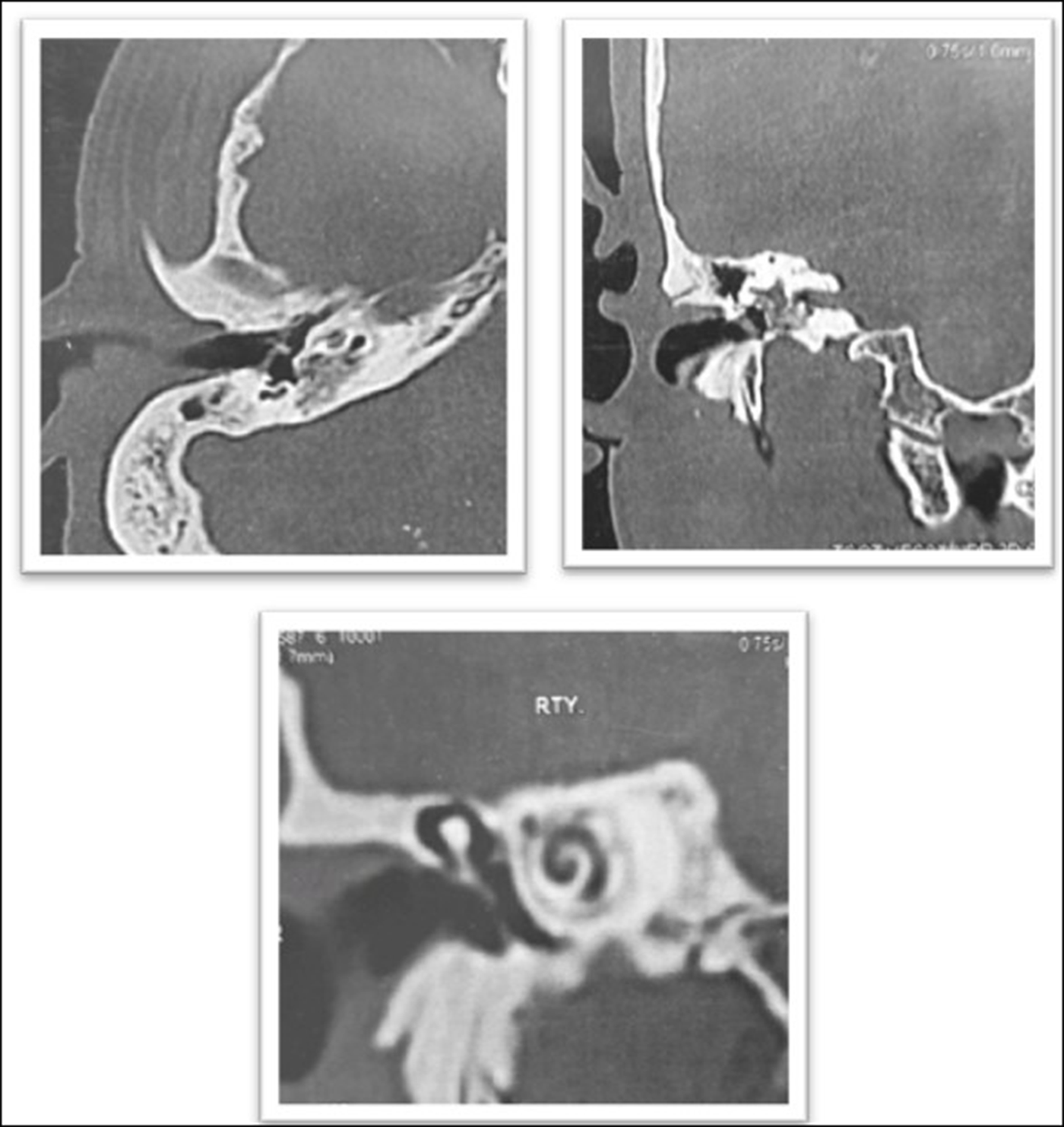
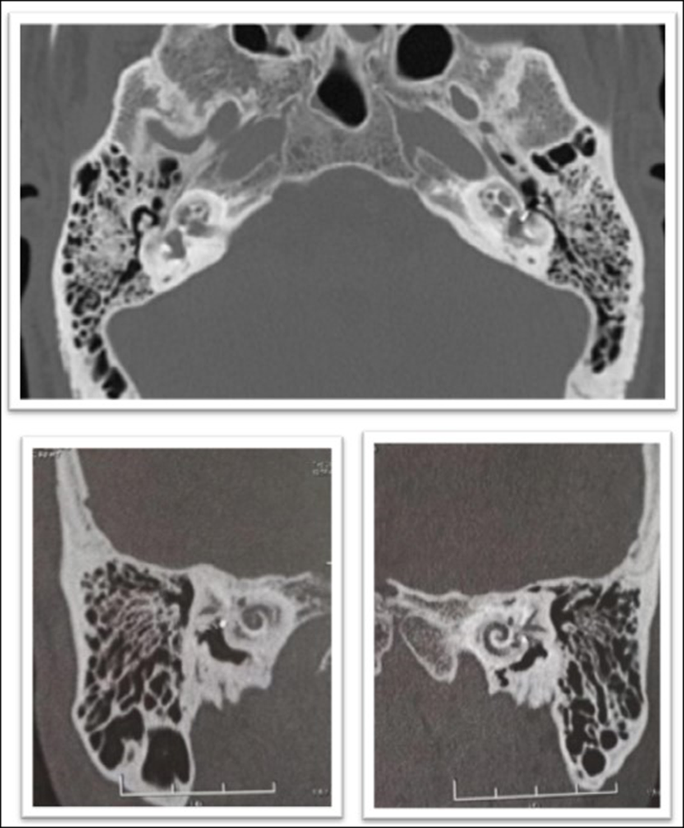
High resolution CT scans of petrous bone were done and analysed by two surgeons and a radiologist. Accordingly, the advanced otosclerosis group patients were classified into grades according to the Rotteveel classificationReference Rotteveel, Snik, Cooper, Mawman, van Olphen and Mylanus4 (Figures 3, 4).
47-year-old male patient with grade 2A retrofenestral otosclerosis with double ring appearance. An otosclerotic focus was obstructing round window and scala tympani.

Bilateral retrofenestral otosclerosis: right: diffuse grade 3 otosclerosis, left: grade 2c double ring appearance with narrowed basal turn. Patient underwent stapedectomy bilaterally with prostheses seen in place.

Results
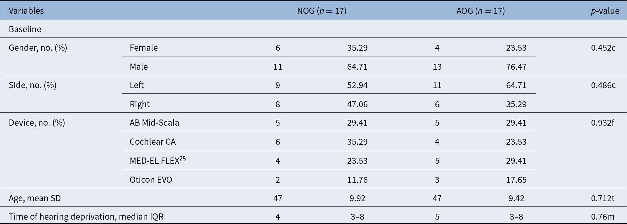
The two groups did not differ with regard to gender, age at implantation and duration of hearing deprivation. Electrodes from different companies were used equally in both groups (Table 1).
Demographic and outcome data analysis; NOG = non-otosclerosis group; AOG = advanced otosclerosis group; IQR = interquartile range; SD = standard deviation; chi-square test (c), Fisher’s exact test (f), independent t-test (t) and Mann–Whitney U test (m) were used for the analyses; * = significant p-value (p ≤ 0.05). AB Mid-Scala (Advanced Bionics, Valencia, California); Cochlear CA (Dubai, UAE); MED-EL FLEX28 (MED-EL, Innsbruck, Austria); Oticon EVO (Oticon Medical, Smørum, Denmark)

CT grading and difficulties (Table 2)
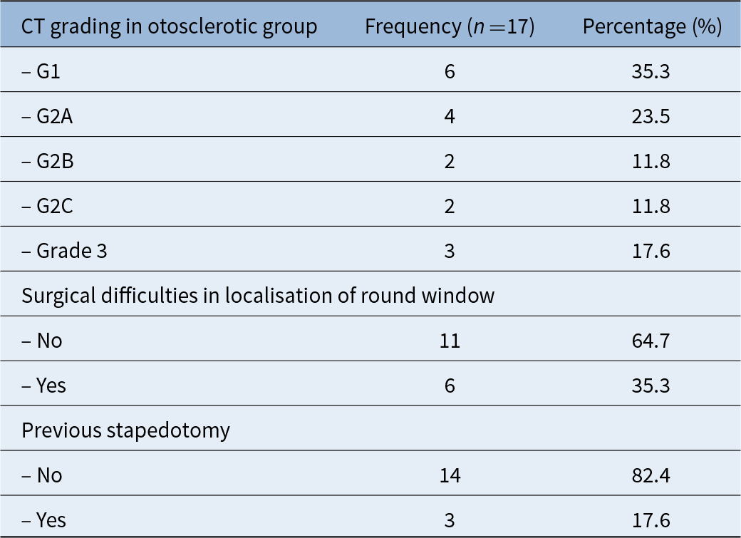
The CT scans in six patients (35.3 per cent) show fenestral affection only (grade 1), while four patients (23.5 per cent) had retro-fenestral and double ring (grade 2A), two patients had narrowed basal turn (grade 2B), two patients had double ring and narrowed basal turn (grade 2C) and three patients (17.6 per cent) had diffuse confluent cochlea (grade 3). Three patients had previous stapedotomy.
Computed tomography (CT) grading and difficulties

Surgical challenges
All cases had a very brittle otic capsule which was easily fragmented. During drilling of the niche tegmen soft debris tended to block the access to the RW membrane. Difficulty in localisation of round window during implantation with excessive bleeding was encountered in six patients (35.3 per cent). In three patients a false passage through the soft bone lateral to the scala tympani was encountered. In two of these patients, this was immediately remedied intra-operatively and in one of these patients post-operative imaging revealed the misplacement, and the patient was revised and the electrode properly reinserted. Incomplete insertion mainly due to endo-luminal fibrosis was seen in only one case in our series.
In two patients, a scala vestibuli insertion was performed. In two other patients a middle turn retrograde insertion was the only available option.
Electrode selection was based on the radiological grading and expected permeability and accessibility of the scala tympani. In cases with expected or documented fibrosis, a more rigid electrode was selected (Cochlear contour advance or Advanced Bionics MS) to give some rigidity to the electrode, but care must be taken to avoid the creation of a false passage. In less severe cases with a well-defined lumen, more flexible electrodes are preferable to follow the lumen without dislocation or false passages.
Hearing outcomes
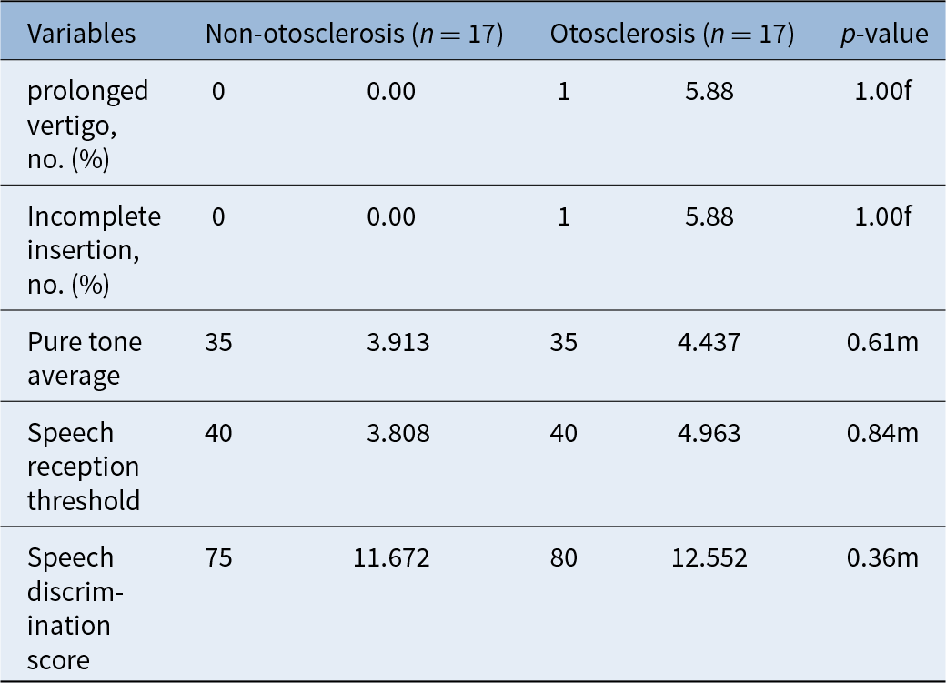
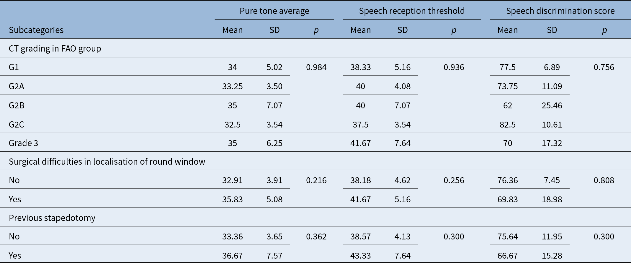
Hearing outcomes were evaluated one year after surgery. In the otosclerotic group the mean PTA was 35 dB ± 4.43, speech reception threshold was 40 ± 4.96, and speech discrimination score was 80 per cent ± 12.55. No significant difference was found between the two groups. The linear regression models assessing PTA, speech reception threshold, and speech discrimination score indicate that the adjusted effect of otosclerosis on these variables was not statistically significant (regression coefficient = 1.34, 0.937, and 1.613, respectively), after controlling for sex, age, and duration of hearing deprivation (Tables 3, 4). Hearing outcomes were evaluated in different otosclerotic grades (Table 5).
Hearing outcome; Fisher’s exact test (f) and Mann–Whitney U test (m) were used for the analyses

Multivariate analyses of hearing outcomes; B = regression coefficient; SE = standard error; * = significant p-value (p ≤ 0.05)

Subgroup analysis for the otosclerosis group; CT = computed tomography; FAO = far-advanced otosclerosis; Kruskal–Wallis test was used for CT grading analysis; Mann–Witney U test was used for binary variable analyses; * = significant p-value (p ≤ 0.05)

Discussion
Otosclerosis is an osteodystrophy disease affecting the otic capsule. It is associated with an unstoppable process of osteolysis and osteogenesis leading to an unusual histology including vascular proliferation, new creation of connective tissue stroma and bone resorption.Reference Marx, Lagleyre, Escudé, Demeslay, Elhadi and Deguine1 Many authors have documented that, if the inner ear is affected by otosclerosis, hearing loss will continue to propagate, even when stapedotomy has closed the air–bone gap.Reference Maxwell, Shokry, Master and Slattery5, Reference Lee, JB and Eddington6 According to a recent meta-analysis on stapedotomy in candidates for implantation with advanced otosclerosis, some authors recommend stapedotomy and hearing aid in advanced otosclerosis with a mean speech discrimination score of 59 per cent.Reference van Loon, Merkus, Smit, Smits, Witte and Hensen7 In more advanced cases, cochlear implantation is warranted. Radiologic evaluation of and otosclerosis-affected inner ear is helpful in pre-operative staging of the extent of the disease.Reference Rotteveel, Snik, Cooper, Mawman, van Olphen and Mylanus4, Reference Castillo, Polo, Gutiérrez, Reyes, Royuela and Alonso8 It also helps in building a strategy for operative access through either scalae. Among all stages requiring implantation, advanced otosclerosis suffers from a relatively high percentage of complications, which occur as facial nerve stimulation and electrode dislocation. Facial nerve stimulation may result from current flow from the electrode to the labyrinthine part of the nerve due to the low impedance of the otosclerotic bone between the nerve canal and upper part of the basal turn of the cochlea.Reference Seyyedi, Herrmann, Eddington and JB9, Reference Dumas, Schwalje, Franco-Vidal, Bébéar, Darrouzet and Bonnard10 This is a fairly rare complication, as few cases were documented in the literature.Reference Battmer, Pesch, Stöver, Lesinski-Schiedat, Lenarz and Lenarz11 In a recent meta-analysis, the pooled rate of facial nerve stimulation was found to be 18 per cent.Reference Kondo, Vasan, Jufas and Patel12 It occurred in 8.6 per cent of the study group of Dumas et al., 2018, with one patient requiring revision surgery due to persistent facial stimulation.Reference Dumas, Schwalje, Franco-Vidal, Bébéar, Darrouzet and Bonnard10 In a recent meta-analysis done by Teaima et al., facial nerve stimulation was 12.4 per cent.Reference Teaima, Elnashar, Hakim and Hadaey13
In the present study we document results of cochlear implantation in 17 patients with different stages of advanced otosclerosis and in matched non-otosclerosis patients. Our results showed that cochlear implantation is an effective solution in advanced otosclerosis. Indeed, our study revealed no significant difference between the otosclerosis and non-otosclerosis groups regarding PTA, speech reception threshold, and speech discrimination score, and results were parallel to those found in the literature.Reference Kabbara, Gauche, Calmels, Lepage, Escude and Deguine14–Reference Marshall, Fanning, Symons, Shipp, Chen and Nedzelski16 In follow-up audiological evaluations one year after surgery, the mean PTA and speech discrimination scores were similar in both groups with no statistical difference. No facial nerve affection or stimulation occurred.
Surgical difficulties in the advanced otosclerosis group were encountered in six cases in the form of difficulty in localisation of the round window, excessive bleeding and different degrees of labyrinthitis ossificans. Two patients had complete occlusion of round window, one underwent cochleostomy for scala tympani insertion and the second underwent scala vestibuli insertion. Incomplete insertion mainly due to endo-luminal fibrosis was seen in only one case in our series. In two patients, a scala vestibuli insertion was performed. In two other patients a middle turn retrograde insertion was the only available option. Meticulous pre-operative imaging interpretation by the surgeon, is helpful in surgical planning and electrode selection.Reference Ricci, Lapenna, Gambacorta, Della Volpe, Faralli and Di Stadio17 The rate of partial insertion in the literature is around 10 per cent.Reference Kondo, Vasan, Jufas and Patel12 In the literature, 8.1 per cent cases of cochlear implantation in advanced otosclerosis needed surgical revision.Reference Teaima, Elnashar, Hakim and Hadaey13
In the current study we had three patients with previous stapedotomy and the hearing results of those patients were similar to the hearing outcomes of patients with no history of stapedotomy. Our results and those of Skarzynski et al.,Reference Skarzynski, Pastuszak, Gos, Lorens, Kolodziejak and Obrycka18 Kabbara et al.,Reference Kabbara, Gauche, Calmels, Lepage, Escude and Deguine14 and Teaima et al.,Reference Teaima, Elnashar, Hakim and Hadaey13 indicate that cochlear implantation is a very effective treatment for advanced otosclerosis, even after stapes surgery.
The findings we obtained in advanced otosclerosis after cochlear implantation were satisfactory, with good hearing results and speech discrimination. Cochlear implantation in advanced otosclerosis presents some challenges, which can be predicted before or during surgery. Judicious planning and flexibility in the access to the inner ear and choice of electrode raise the chances of a successful implantation. Surgical difficulties were directly related to the advanced stage of the disease, which can be detected by proper interpretation of CT. In properly implanted patients the audiological outcomes are satisfactory and comparable to non-otosclerotic patients.
• Patients with far-advanced otosclerosis with poor cochlear reserve and a low speech discrimination score (<50 per cent) present a challenge in the management of their associated hearing loss
• Cochlear implantation is one of the available options but presents number of specific challenges and difficulties related to the advanced stage of the disease
• Difficulties were overcome by proper interpretation of computed tomography scan, special precautions during surgery and the judicious selection of electrodes
• There was no significant difference in audiological outcome between otosclerotic and non-otosclerotic groups after cochlear implantation after one year
• There was no difference in audiological outcomes in different radiological stages of advanced otosclerosis
• Cochlear implantation is a safe and effective management for advanced otosclerosis patients even after stapes surgery
Conclusion
Cochlear implantation is a safe and effective management for advanced otosclerosis patients even after stapes surgery. Cochlear implantation should be suggested to all patients who fail to achieve satisfactory results with stapedectomy with or without a hearing aid. There is no difference in audiological outcomes in different radiological stages of advanced otosclerosis.
Funding
The authors have no funding to disclose.
Conflicts of interest
The authors have no conflicts of interest to disclose.