We reported a rare case of hypothetical double aortic arch with interruption between left and right common carotid arteries.
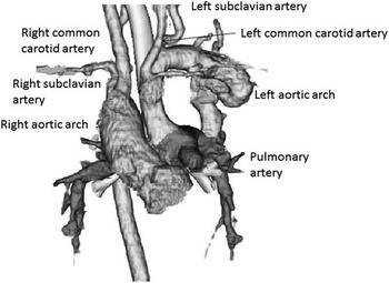
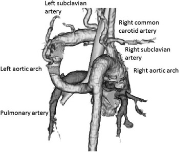
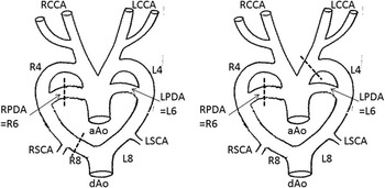
Echocardiography indicated pulmonary atresia with ventricular septal defect in a male infant born at 38 weeks of gestation weighing 2130 g, although the arborisation of neck vessels was unclear. His oxygen saturation measured by pulse oximeter was about 90% without continuous prostaglandin infusion. Furthermore, three-dimensional computed tomography revealed double aortic arch with interruption between the left and right common carotid arteries, in which the left arch flowing into the pulmonary artery branched the left subclavian and common carotid arteries and the right arch branched the right subclavian and common carotid arteries (Figs 1and 2). We hypothesised that involution of left 4th branchial arch and the remnant of right 8th intersegmental artery are combined simultaneously (Fig 3). G-banding stain and the target site of the fluorescent in situ hybridisation on chromosome 22 detected no chromosomal disorder.
Front three-dimensional view of computed tomography angiogram.

Back three-dimensional view of computed tomography angiogram.

Development of aortic arch: dotted lines are the points of involution in the development. Left, normal development; right, this case. aAo=ascending aorta; dAo=descending aorta; RSCA=right subclavian artery; RCCA=right common carotid artery; RPDA=right patent ductus arteriosus; LSCA=left subclavian artery; LCCA=left common carotid artery; LPDA=left patent ductus arteriosus; R4=right 4th branchial arch; R6=right 6th branchial arch; R8=right 8th intersegmental artery; L4=left 4th branchial arch; L6=left 6th branchial arch; L8=left 8th intersegmental artery.

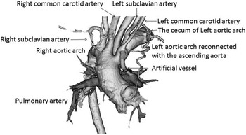
Excision of both ends of the left aortic arch was performed when the patient was one-year old. The pulmonary arterial side was connected to the ascending aorta and the descending aortic side was connected to the caecum. To facilitate pulmonary circulation, an artificial vessel was connected from the left common carotid artery to the pulmonary artery (Fig 4). One year after this palliative surgery, he underwent the Rastelli procedure, and biventricular circulation was established.
Front three-dimensional view of computed tomography angiogram after first surgery.

Although we found few papers showing the double aortic arch with interruption in the left arch proximal to the common carotid artery,Reference Nakajima, Satomi and Kawamura 1 no report has described the cardiovascular anatomy of our patient.
Acknowledgements
None.
Financial Support
This research received no specific grant from any funding agency, commercial, or not-for-profit sectors.
Conflicts of Interest
None.